

SKU:EA1-1017868
Brain model that also shows arteries. Can be separated into 9 parts
Brain model that also shows arteries. Can be separated into 9 parts
Out of stock
ATTENTION! This item ships separately. The delivery time may vary.
Couldn't load pickup availability
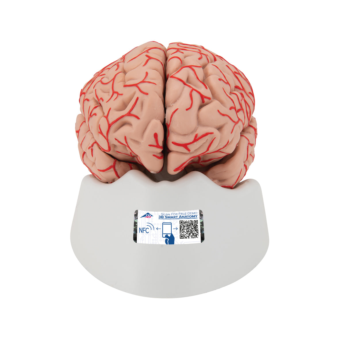
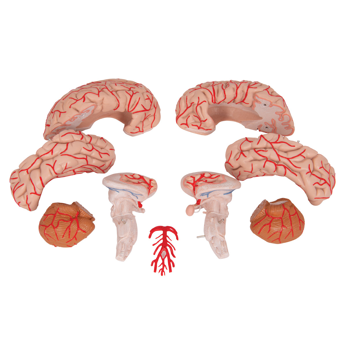
This brain model shows arteries in the brain, many internal structures and can be separated into 9 parts.
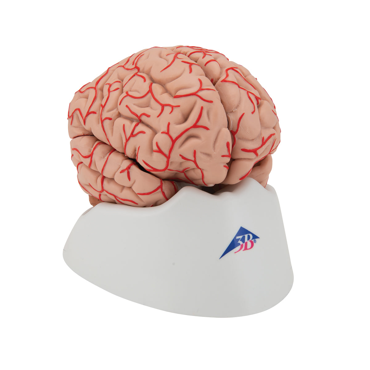
The brain model is cast in hollow and solid plastic, which makes the material flexible because it can both be squeezed quite a bit and moved a bit. This makes the model pleasant to touch and work with when it needs to be taken apart and studied.
The model's 9 parts are held together via metal pins. Different areas of the brain appear in different shades, and below you can read more about the anatomical details such as the arteries and the limbic system. The size of the model corresponds to the brain of an adult person. The dimensions are 15 x 14 x 16 cm. The weight is approximately 850 grams and it comes on a white removable stand.
Anatomically speaking
Anatomically speaking
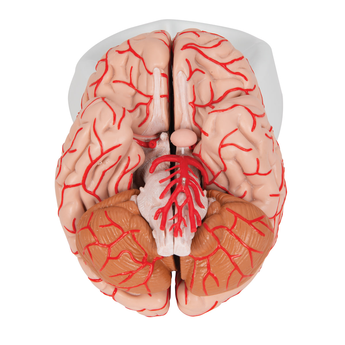
Anatomically, the model shows the human brain, which can generally be divided into the cerebrum (cerebrum), the cerebellum (cerebellum) and the brain stem (truncus encephali).
These 3 structures are clearly separated via different color tones, and the difference between gray and white matter can be clearly seen on this model.
In the cerebrum (telencephalon and diencephalon), the lobes of the brain, as well as the thalamus and hypothalamus (and the pituitary gland) are primarily seen
In the cerebellum, the vermis cerebelli and the cerebellar hemispheres (hemisperium cerebelli) are seen
In the brainstem you can see its 3 parts (the midbrain, the pons and the medulla oblongata) as well as the apparent origin of the cranial nerves (also called the cranial nerves)
Other structures such as the brain stem, fornix, ventricular system and the first 2 cranial nerves (the olfactory and optic nerves) are also seen, which do not originate from the brainstem
The pictures on the left show how the model can be separated into 9 parts. This makes it possible to study the internal structures of the brain, and several of these are seen in 3 dimensions.
Arterial vascular supply of the brain
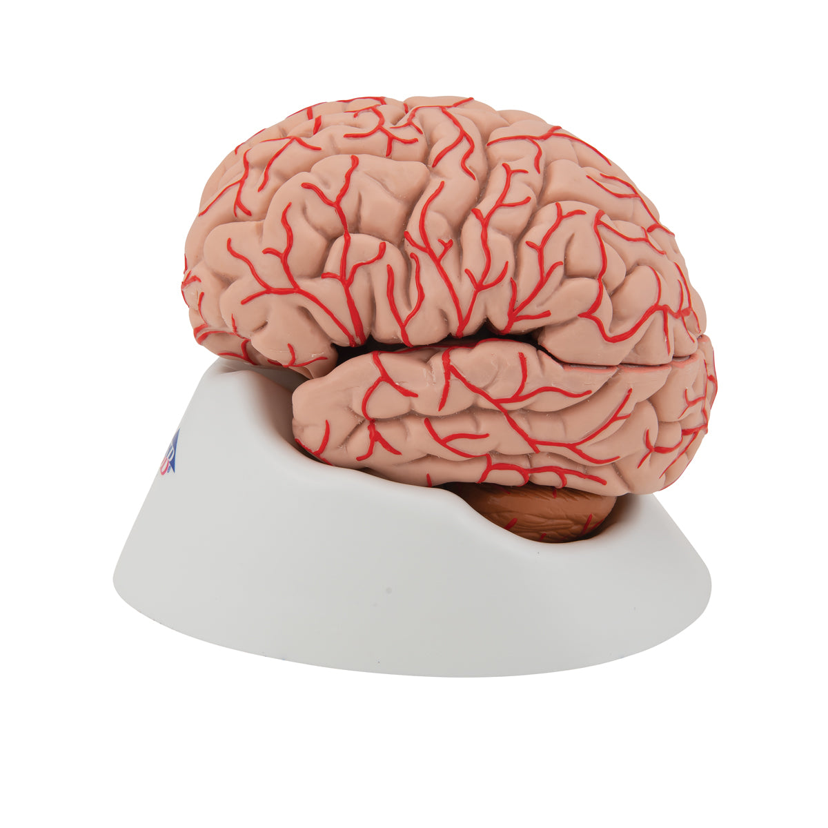
The two paired arteries that supply the brain with blood are called the internal carotid artery and the vertebral artery. They are illustrated on both sides of the model.
A. carotid interna (the carotid system): It has an S-shaped course, which is called the carotid siphon and is very clearly visible. In addition, the medial cerebral artery and the anterior cerebral artery are seen, which are the two main branches that the internal carotid artery divides into.
A. vertebralis (basilaris system): It joins the opposite vertebral artery to form a. basilaris (on the front of the pons/pons). This is seen very clearly, just as you see other smaller ramifications.
NB: As the brain model can be separated into 9 parts, the manufacturer has chosen to show several of the arteries separated/disconnected. It has the following meaning:
A. carotis interna: A. cerebri media and anterior are not shown in their uninterrupted course to the lateral part of the brain's surface and the medial part of the hemisphere, respectively, so that you can follow them "all the way" with the index finger. However, it must be emphasized that numerous arteries corresponding to their branches are seen in these regions.
A. vertebralis: As something special, a collateral vessel mouth is formed in the human brain, because smaller arteries from the basilar system are anastomosed (connected) with smaller arteries from the carotid system. This connection is not visible because the manufacturer has chosen to separate the carotid system from the basilar system, so that the latter can be taken out of the brain model completely (see also the images on the left).
The limbic system
Many of our customers ask about the limbic system in connection with the purchase of brain models. Hence this description.
The limbic system includes various anatomical structures in the central nervous system (CNS), and is primarily responsible for emotional functions such as anxiety, aggressiveness, mood, memory and social adaptability. Clinically, it is therefore often related to psychiatric disorders.
The limbic system includes, among other things amygdala, hippocampus, gyrus parahippocampalis, hypothalamus, fornix, corpus mammillare, the prefrontal cerebral cortex and the monoaminergic systems of the brainstem. The list is quite a bit longer - especially because numerous fiber connections connect the limbic structures. Many customers ask in particular about the amygdala and hippocampus (which is why they are mentioned first in this section).
NB: In this brain model, the hippocampus can be seen as well as some of the other limbic structures such as the fornix - but not the amygdala.
The amygdala is involved in anxiety and emotional coloring of sensory impressions. It lies as an almond-shaped nucleus IN FRONT of the hippocampus in the anterior pole of the temporal lobe (amygdala and hippocampus are therefore separate).
The hippocampus is involved in memory. It lies as an irregular twisted structure in the medial part of the temporal lobe.
Along with the amygdala, the hippocampus lies IN FRONT of the hippocampus (roughly speaking further "toward the forehead"), both of these structures can only be seen on a brain model if the model includes at least 2 frontal/coronal sections through the temporal lobe - or if the brain model is partially transparent (frontal/coronal cut roughly corresponds to the cut direction "from ear to ear").
We have not yet seen a brain model that shows 2 cuts through the temporal lobe, so that both the amygdala and the hippocampus are seen. In our range, on the other hand, we have a partially see-through brain model in the highest price range, which shows both structures.
All brain models in our range can be separated into different parts. All models (both with and without educational colors) that can be separated into 4 or more parts show the hippocampus. On almost all of these models, the hippocampus is also numbered and named on an overview that can be downloaded from the product descriptions of the brain models. This also applies to this brain model.
Flexibility
Flexibility
Clinically speaking
Clinically speaking
Clinically, the model is ideal for understanding apoplexy (stroke). The model can also be used to understand many other lesions and disorders such as epilepsy, brain tumors, lesions involving cranial nerves and sclerosis (multiple sclerosis).
Share a link to this product


A safe transaction
For 19 years I have been managing eAnatomi and sold anatomical models and posters to 'almost everyone' who has anything to do with anatomi in Scandinavia and abroad. When you place your order with eAnatomi, you place your order with me and I personally guarantee a safe transaction.
Christian Birksø
Owner and founder of eAnatomi and Anatomic Aesthetics