Skip to product information
1 of 3

SKU:EA1-1010005

Detailed ear model that shows both the entire cochlea and a cross-section with three-dimensional details

Detailed ear model that shows both the entire cochlea and a cross-section with three-dimensional details

Regular price 3 784 SEK
Regular price Sale price 3 784 SEK
Sale Sold out
Not including taxes if ordering from outside EU. Shipping calculated at checkout.

Out of stock

this product is made to order. To place an order please call or write us.

This impressive model shows both the entire cochlea (the cochlea) and a cross-section from the cochlea with three-dimensional details of, among other things, the cortical organ (organum spiral).


The ear model is enlarged, weighs approx. 1.2 kg and the dimensions are 26 x 19 x 26 cm. It consists of both a small part that shows the cochlea and a larger part that primarily shows the cortical organ on the basilar membrane. It is all delivered together on a stand (white plastic sheet).

Anatomical features

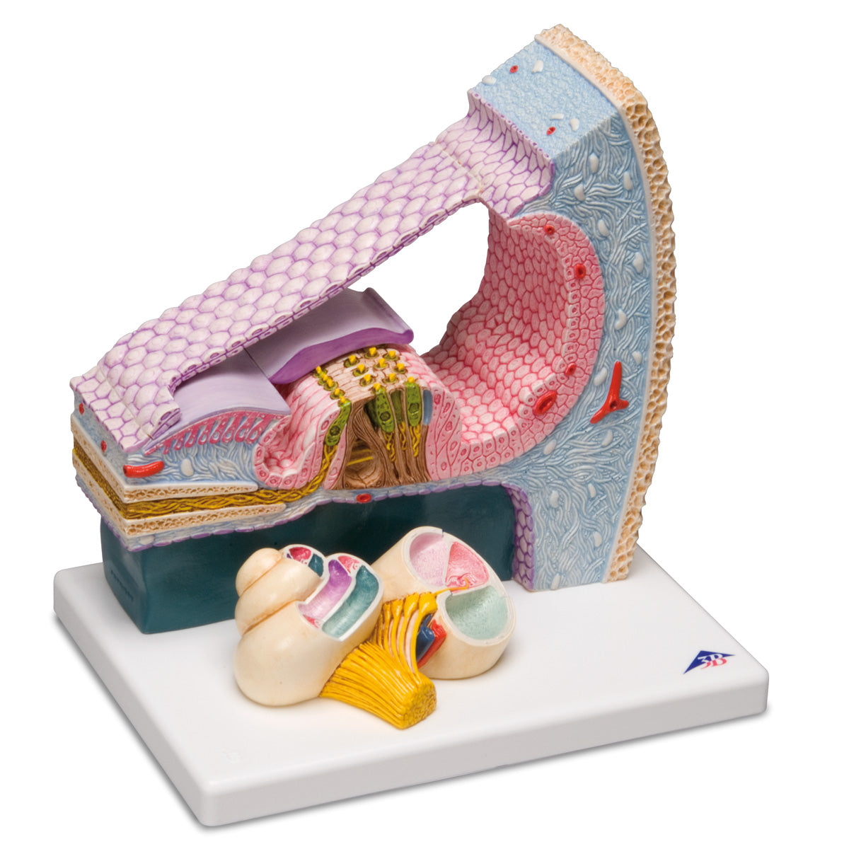
Anatomically speaking, the model shows 2 parts: A small part which shows almost the entire cochlea and a much larger part which shows a cross-section from the organ.


The small part gives an overall insight into the cochlea and its division into the 3 parallel systems/runs, which are educationally shown in different colours. In the middle of this system is the ductus cochlearis with the endolymph (also called scala media). The scala vestibuli and scala tympani, on the other hand, contain the perilymph, and they proceed respectively above and below the cochlear duct.


The large part of the model shows a cross-section through the ductus cochlearis with the scala vestibuli and scala tympani respectively. over and under. Centrally, the membrana basilaris is seen, which is set into resonant oscillations in connection with hearing. These oscillations are registered by the complex system of sensory cells (the cortical organ) which is attached to the said membrane.


The model thus provides the opportunity to study the anatomical structures that are crucial for the formation of the sensory impression (via the sensory cells), which is sent to the brain with the auditory nerve (sound perception).


You can see i.a. the following structures:


Membrana tectoria and membrana vestibularis (Reissner's membrane)
Many cell types such as Hensen cells, Claudius cells and Böttcher cells
Cuniculus internus (cortical tunnel)
Ligament spiral
N. cochlearis and spiral ganglion

Product flexibility

Clinical features

Clinically, the ear model can be used to understand disease, although most people will probably use it to study the cochlea and its internal structures.


The model can, for example, be used to understand disease, injury or congenital deformity in the hair cells of the cochlea as well as cochlear implantation.

View full details

A safe deal

For 19 years I have been at the head of eAnatomi and sold anatomical models and posters to 'almost' everyone who has anything to do with anatomy in Denmark and abroad. When you shop at eAnatomi, you shop with me and I personally guarantee a safe deal.

Christian Birksø
Owner and founder of eAnatomi ApS

Read more about eAnatomi