Skip to product information
1 of 4

SKU:EA1-1000320

Educational model showing ovulation, fertilization and implantation in the uterus

Educational model showing ovulation, fertilization and implantation in the uterus

Regular price 4 248 SEK
Regular price Sale price 4 248 SEK
Sale Sold out
Not including taxes if ordering from outside EU. Shipping calculated at checkout.

Out of stock

this product is made to order. To place an order please call or write us.

If you are looking for an educational model to demonstrate and understand the stages from the egg cell's maturation, release and fertilization to implantation in the lining of the uterine cavity, this model is ideal.


The model is highly illustrative, and compared to the organs of the woman, they are 2 times enlarged. Read more about the anatomy below. The dimensions of the model are 35 x 21 x 20 cm and it weighs approximately 1.1 kg. It is delivered on a stand with educational illustrations of the development of the fertilized egg.


An overview has been made in Latin, English and several other languages ​​(but not Danish), where the model's anatomical structures and numerous details regarding the development of the fertilized egg is named via pictures and lines.

The overview also includes a short description of the stages, etc. The latter description is, among other things, made in English and German (but again not Danish). The overview is not included with the purchase, but you can instead download and print it.

Anatomical features

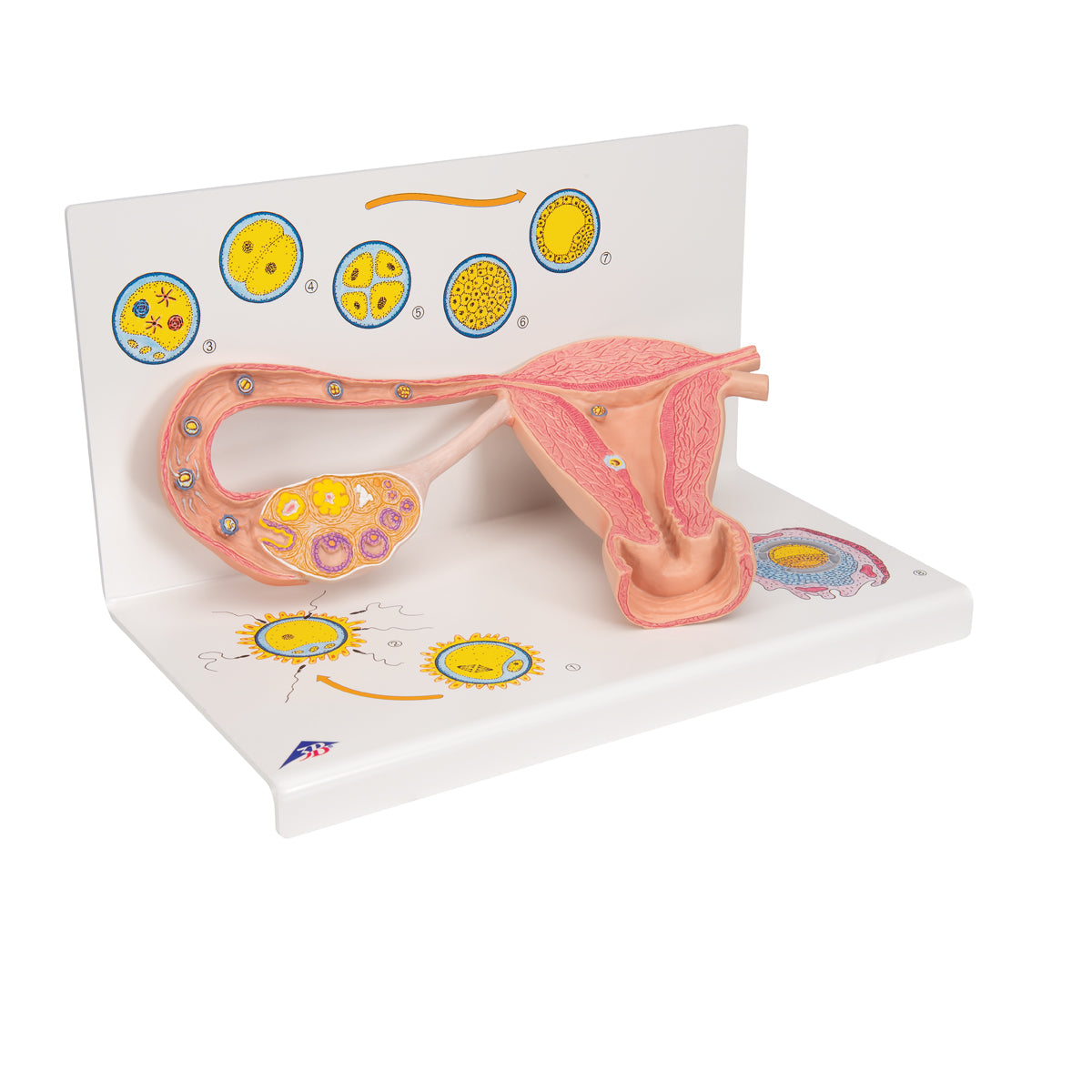
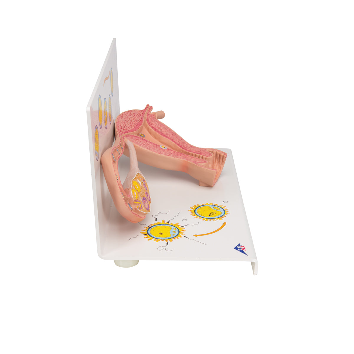
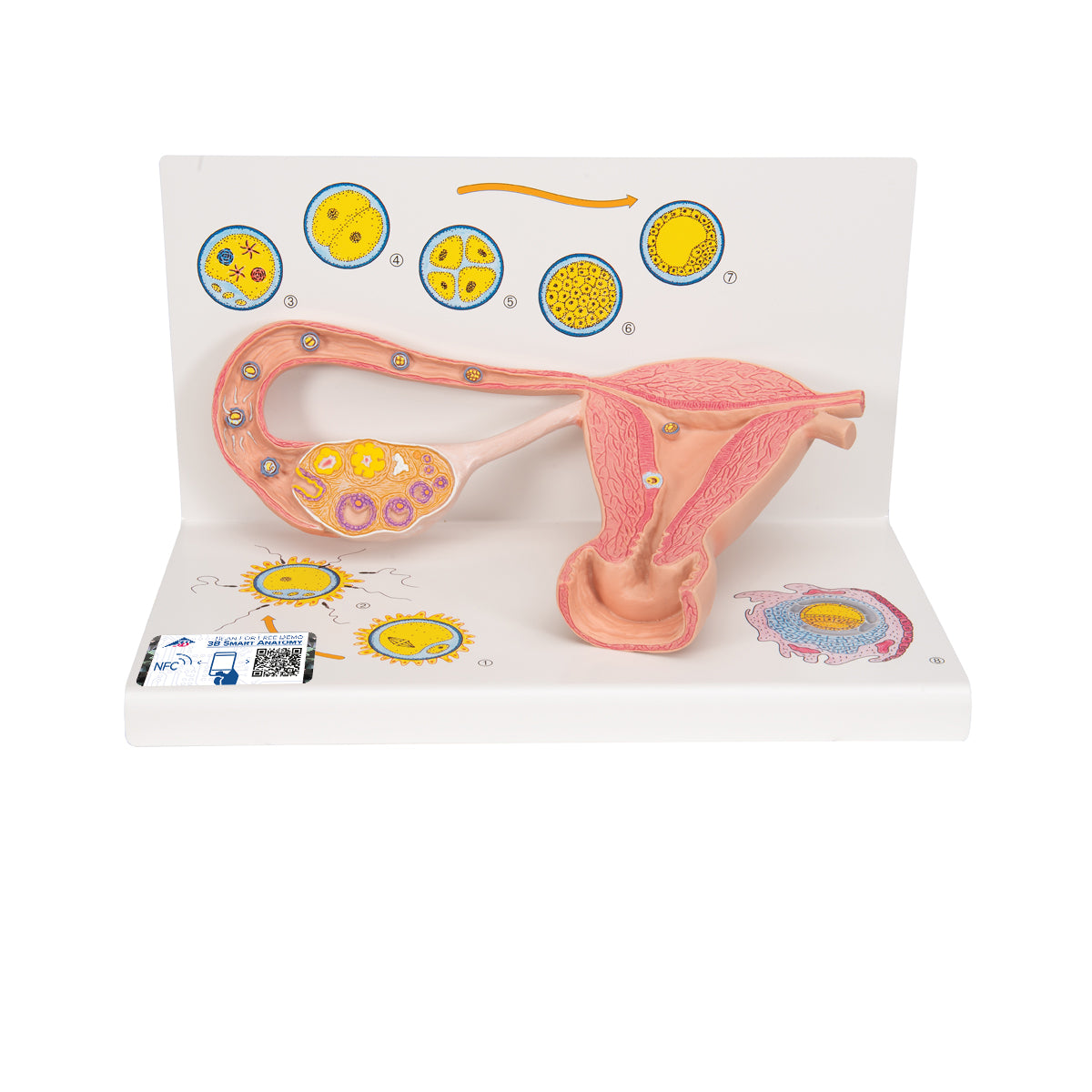
Anatomically and embryologically, the model shows in a particularly illustrative and educational manner the various stages from the egg's maturation, release and fertilization to implantation (insertion) in the uterine cavity. In combination with indicative illustrations on the stand, the most important details are shown, which include:


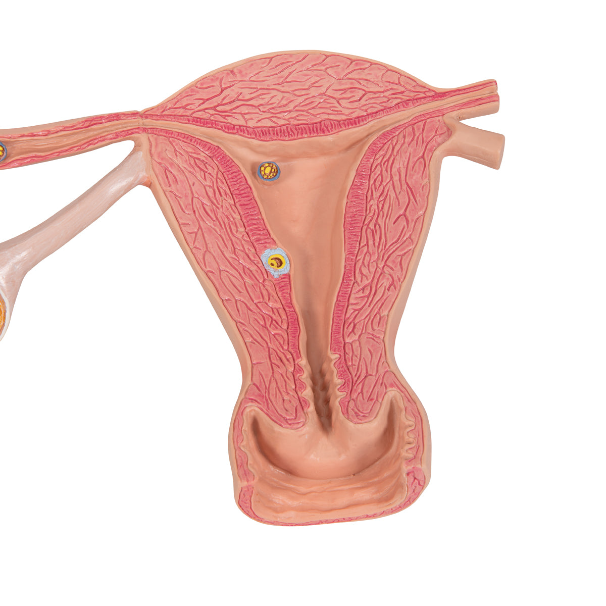
Right ovary (ovary), right tuba uterina (fallopian tube) and uterus (womb)
The maturation process of the ovum in the ovary
The ovulation
Fertilization with a spermatozoon in the ampulla (widest part) of the tuba uterina
The subsequent mitotic divisions with i.a. two-celled stage and Morula
The implantation of the blastocyst in the endometrium (the lining of the uterus)

Product flexibility

Clinical features

Clinically speaking, the model can be used, for example, in connection with sex education, pregnancy preparation and fertility treatment (artificial insemination).

View full details

A safe deal

For 19 years I have been at the head of eAnatomi and sold anatomical models and posters to 'almost' everyone who has anything to do with anatomy in Denmark and abroad. When you shop at eAnatomi, you shop with me and I personally guarantee a safe deal.

Christian Birksø
Owner and founder of eAnatomi ApS

Read more about eAnatomi