SKU:EA1-A614
Enlarged brain model with many areas in educational colors. Can be separated into 4 parts
Enlarged brain model with many areas in educational colors. Can be separated into 4 parts
Out of stock
this product is made to order. To place an order please call or write us.Couldn't load pickup availability
If you are looking for an enlarged, inspiring and highly educational brain model that makes it as easy as possible to identify important areas in the brain, we highly recommend this one.
The model is cast in very hard and robust plastic. Unlike many of our other brain models, which are molded in hollow and flexible plastic, the material of this model can neither be pinched nor moved. Some would argue that this makes it less pleasant to touch and work with when it needs to be taken apart and studied. Others believe it is beneficial.
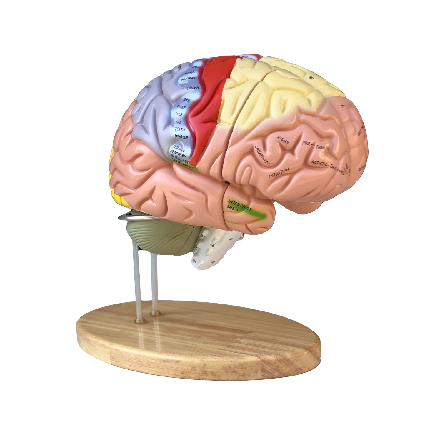
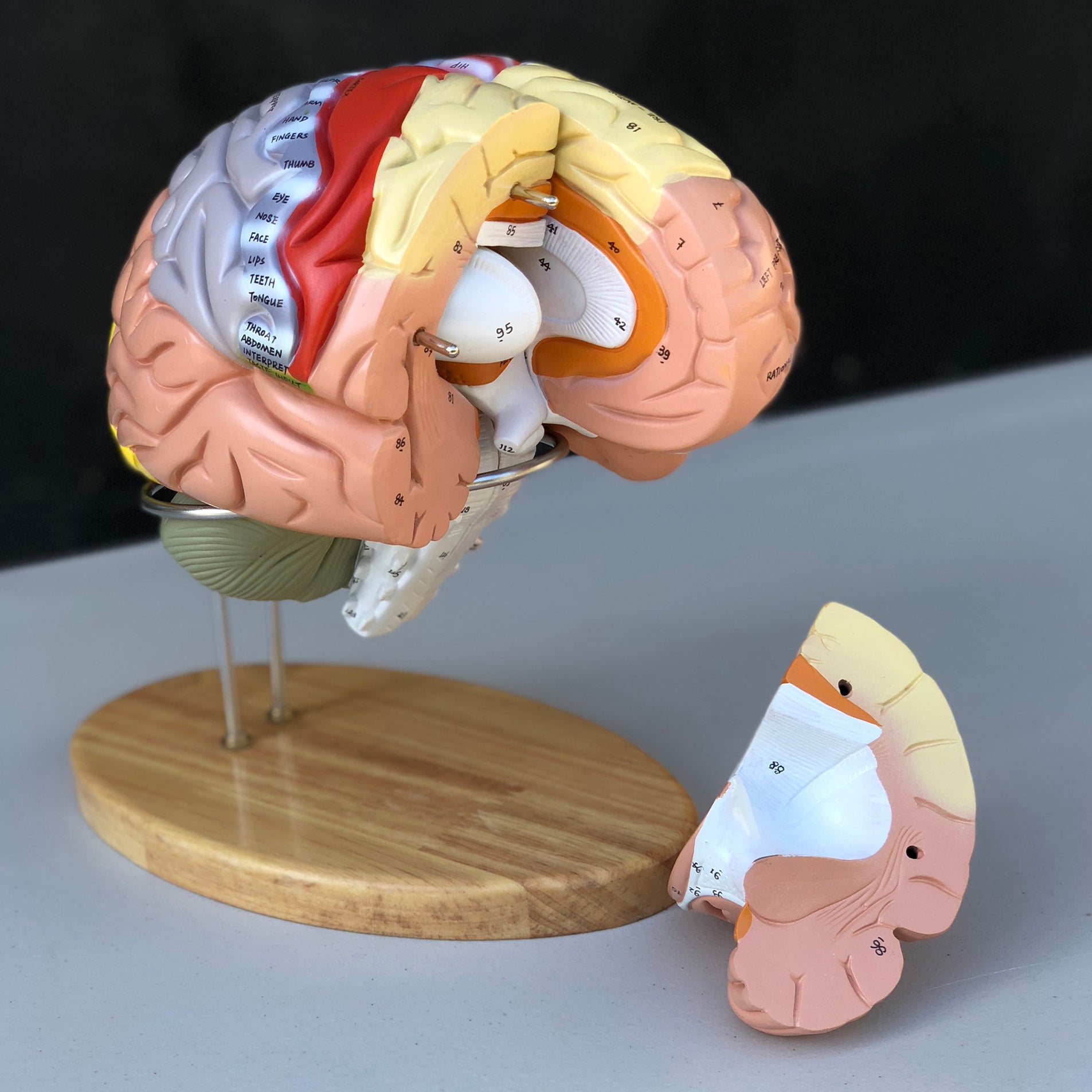
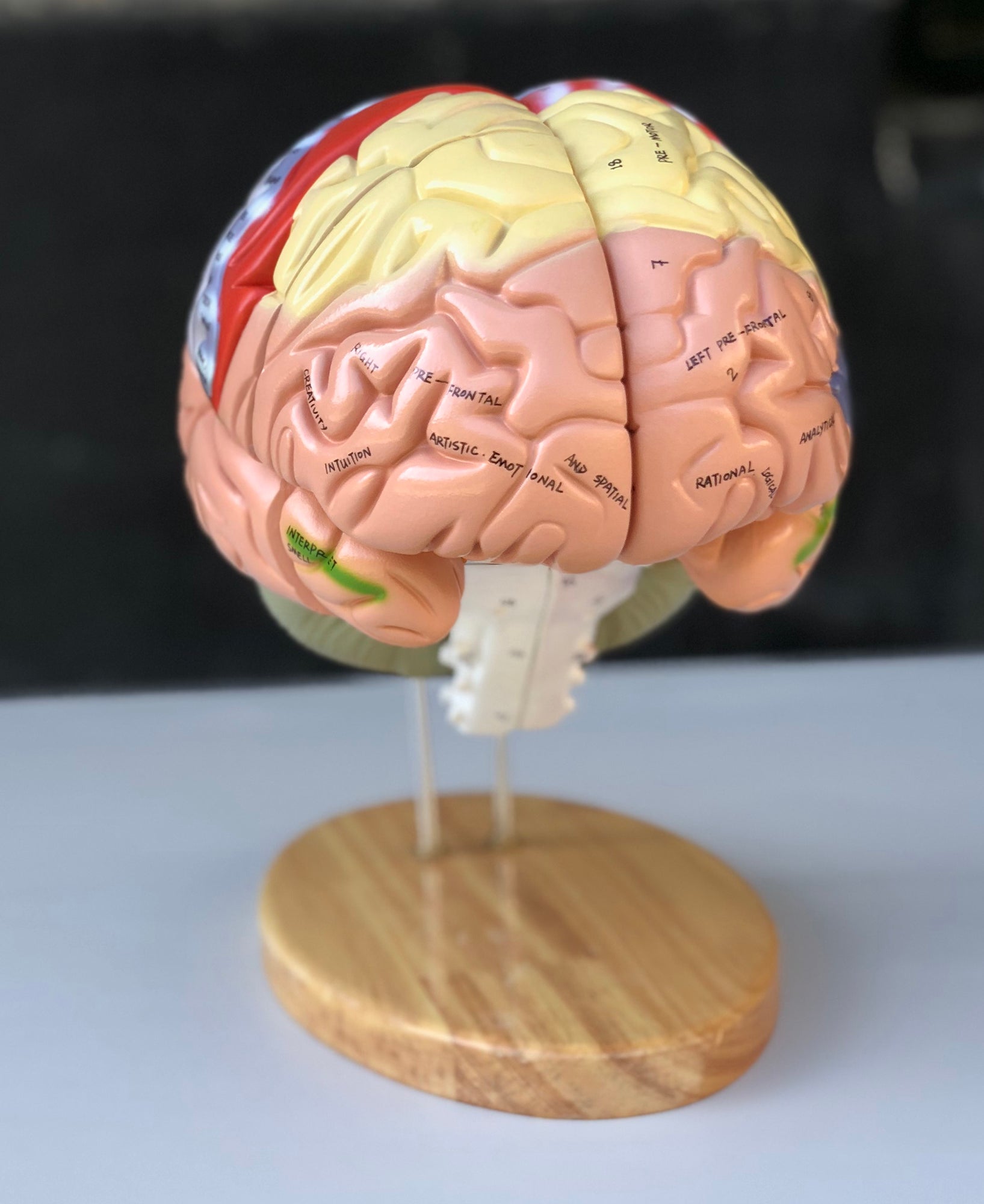
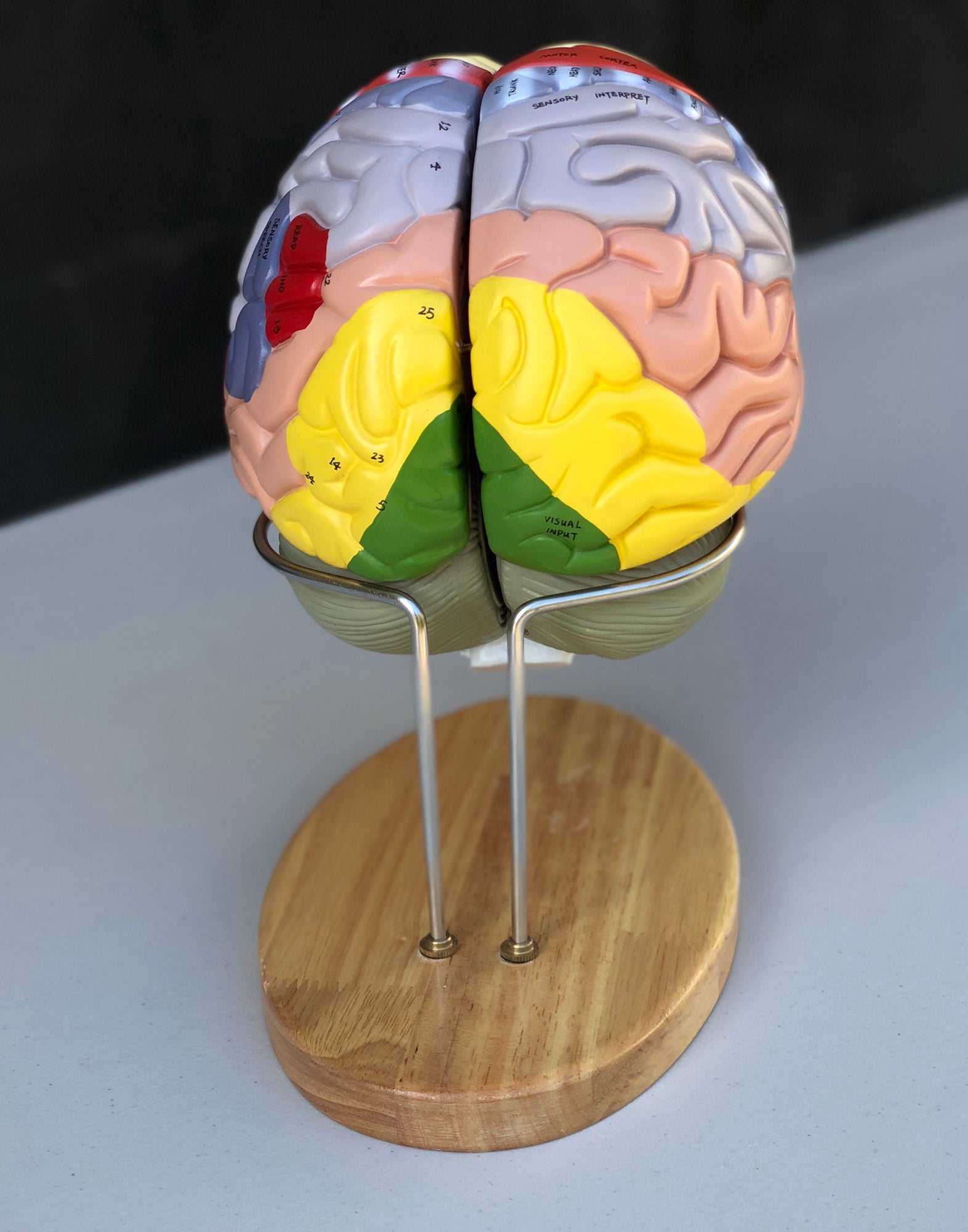
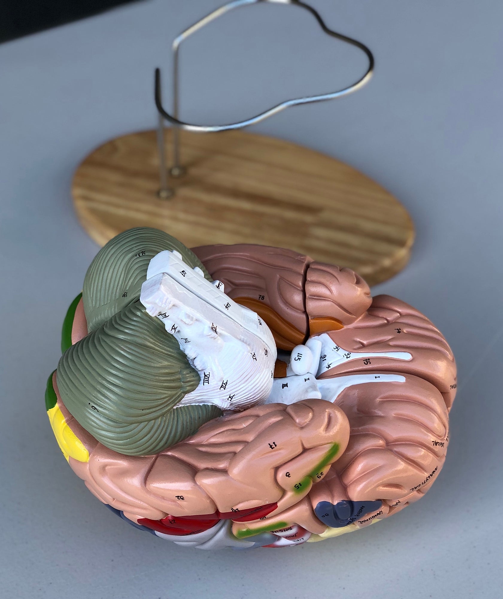
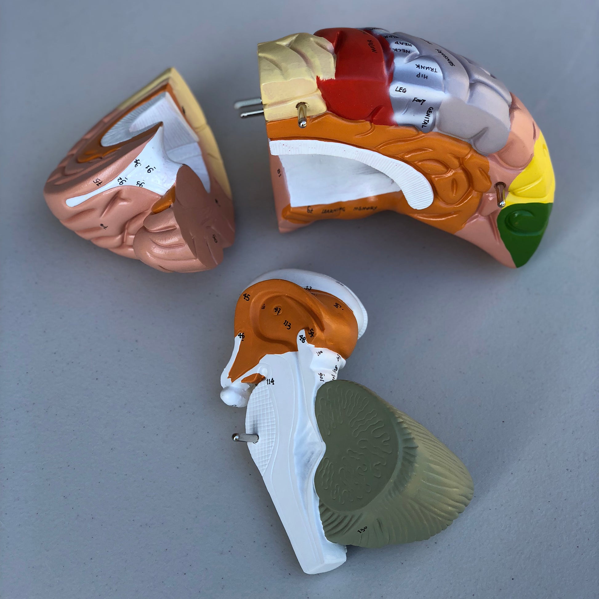
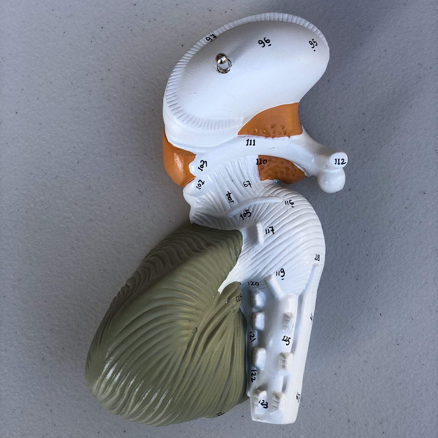
The model can be separated into 4 parts, which are held together via metal pins. Below you can read more about the anatomical details such as the colored areas and the limbic system. Compared to an adult, the model is approximately 1.5 times normal size. The dimensions are 22 x 18.5 x 19 cm and the weight is 1.9 kg. The model rests on a metal stand, which is mounted on a beautiful lacquered oak base with rounded edges (see pictures on the left). The model can be lifted off and separated very easily.
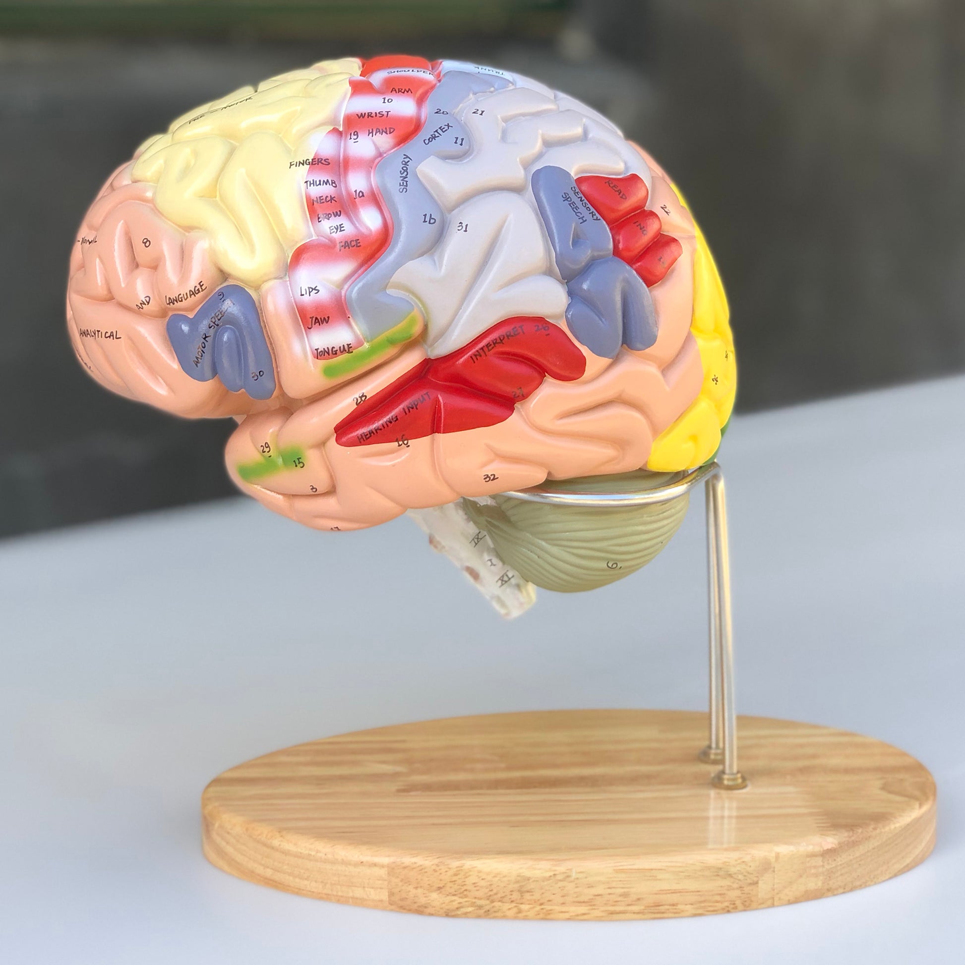
It should be emphasized that this brain model is the only one in our range that comes with both numbering of important anatomical structures as well as words written directly on special areas such as the primary motor area (see the images on the left and read more below).
NB: The numbering and naming are indicative. Therefore, be critical in your use, as figures for an anatomical structure may, for example, be located on the border of another structure.
Anatomical features
Anatomical features
Anatomically, the model shows the human brain, which can generally be divided into the cerebrum (cerebrum), the cerebellum (cerebellum) and the brain stem (truncus encephali).
These 3 structures are clearly separated via different colors, but the difference between gray and white matter cannot be seen on this model.
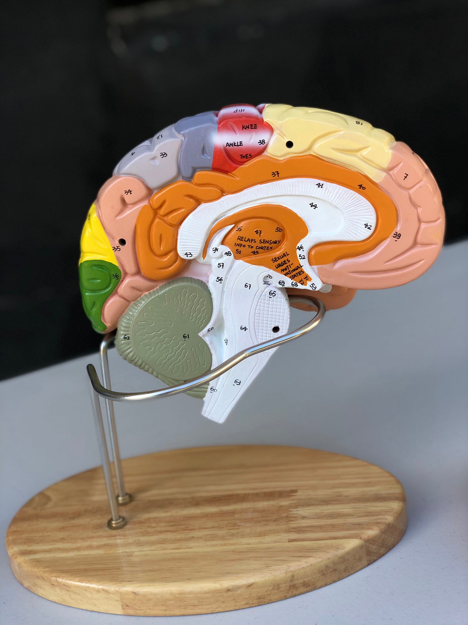
In the cerebrum (telencephalon and diencephalon), the lobes of the brain, as well as the thalamus and hypothalamus (and the pituitary gland) are primarily seen
In the cerebellum, the vermis cerebelli and the cerebellar hemispheres (hemisperium cerebelli) are seen
The brainstem shows its 3 parts (the midbrain, the pons and the medulla oblongata) as well as the apparent origins of the cranial nerves using Roman numerals (also called the cranial nerves)
Other structures such as the brain stem, fornix, ventricular system and the first 2 cranial nerves (the olfactory and optic nerves) are also seen, which do not originate from the brainstem
The pictures on the left show how the model can be separated into 4 parts. This makes it possible to study the internal structures of the brain, and some of these are seen in 3 dimensions.
Colored areas
The colors on this model are muted and very different. As seen in the images on the left, it is not only the lobes of the brain that are colored. Colors are also seen in special areas such as the pre- and postcentral gyrus, Broca's area, Wernicke's area and deeper structures.
As the model is both educationally colored and comes with English words written directly on the important areas of the model, you can very easily and quickly identify areas of interest. Examples are the primary motor area (gyrus precentralis) with indication of body regions ("neck, hip, shoulder, fingers" etc.) and areas such as "language" and speech".
The limbic system
Many of our customers ask about the limbic system in connection with the purchase of brain models. Hence this description.
The limbic system includes various anatomical structures in the central nervous system (CNS), and is primarily responsible for emotional functions such as anxiety, aggressiveness, mood, memory and social adaptability. Clinically, it is therefore often related to psychiatric disorders.
The limbic system includes, among other things amygdala, hippocampus, gyrus parahippocampalis, hypothalamus, fornix, corpus mammillare, the prefrontal cerebral cortex and the monoaminergic systems of the brainstem. The list is quite a bit longer - especially because numerous fiber connections connect the limbic structures. Many customers ask in particular about the amygdala and hippocampus (which is why they are mentioned first in this section).
NB: In this brain model, the hippocampus can be seen as well as some of the other limbic structures such as the fornix - but not the amygdala.
The amygdala is involved in anxiety and emotional coloring of sensory impressions. It lies as an almond-shaped nucleus IN FRONT of the hippocampus in the anterior pole of the temporal lobe (amygdala and hippocampus are therefore separate).
The hippocampus is involved in memory. It lies as an irregular twisted structure in the medial part of the temporal lobe.
If one of these 2 structures is to be seen clearly on a brain model, the brain tissue must be shown in a so-called frontal/coronal section through the temporal lobe. The frontal incision roughly corresponds to the incision direction "from ear to ear".
Because the amygdala is IN FRONT of the hippocampus (roughly speaking further forward "toward the forehead"), both of these structures can only be seen on a brain model if the model includes at least 2 frontal sections through the temporal lobe - or if the brain model is partially transparent.
We have not yet seen a brain model that shows 2 cuts through the temporal lobe, so that both the amygdala and the hippocampus are seen. In our range, on the other hand, we have a partially see-through brain model in the highest price range, which shows both structures.
All brain models in our range can be separated into different parts. All models (both with and without educational colors) that can be separated into 4 or more parts show the hippocampus. On almost all of these models, the hippocampus is also numbered and named on an overview that can be downloaded from the product descriptions of the brain models.
Product flexibility
Product flexibility
Clinical features
Clinical features
Clinically speaking, this model is our most educational when a brain model is to be used to understand problems (symptoms) and damage caused by lesions in areas of the lobes of the cerebrum. These inconveniences and injuries can, for example, be disturbances in movements, sense of touch, language and vision.
The model can also be used to understand many other lesions and disorders such as epilepsy, brain tumours, hydrocephalus and lesions involving cranial nerves.
Although the brain's blood supply is not visible on the model, it can also be used to understand apoplexy (stroke).
Share a link to this product

A safe deal
For 19 years I have been at the head of eAnatomi and sold anatomical models and posters to 'almost' everyone who has anything to do with anatomy in Denmark and abroad. When you shop at eAnatomi, you shop with me and I personally guarantee a safe deal.
Christian Birksø
Owner and founder of eAnatomi ApS