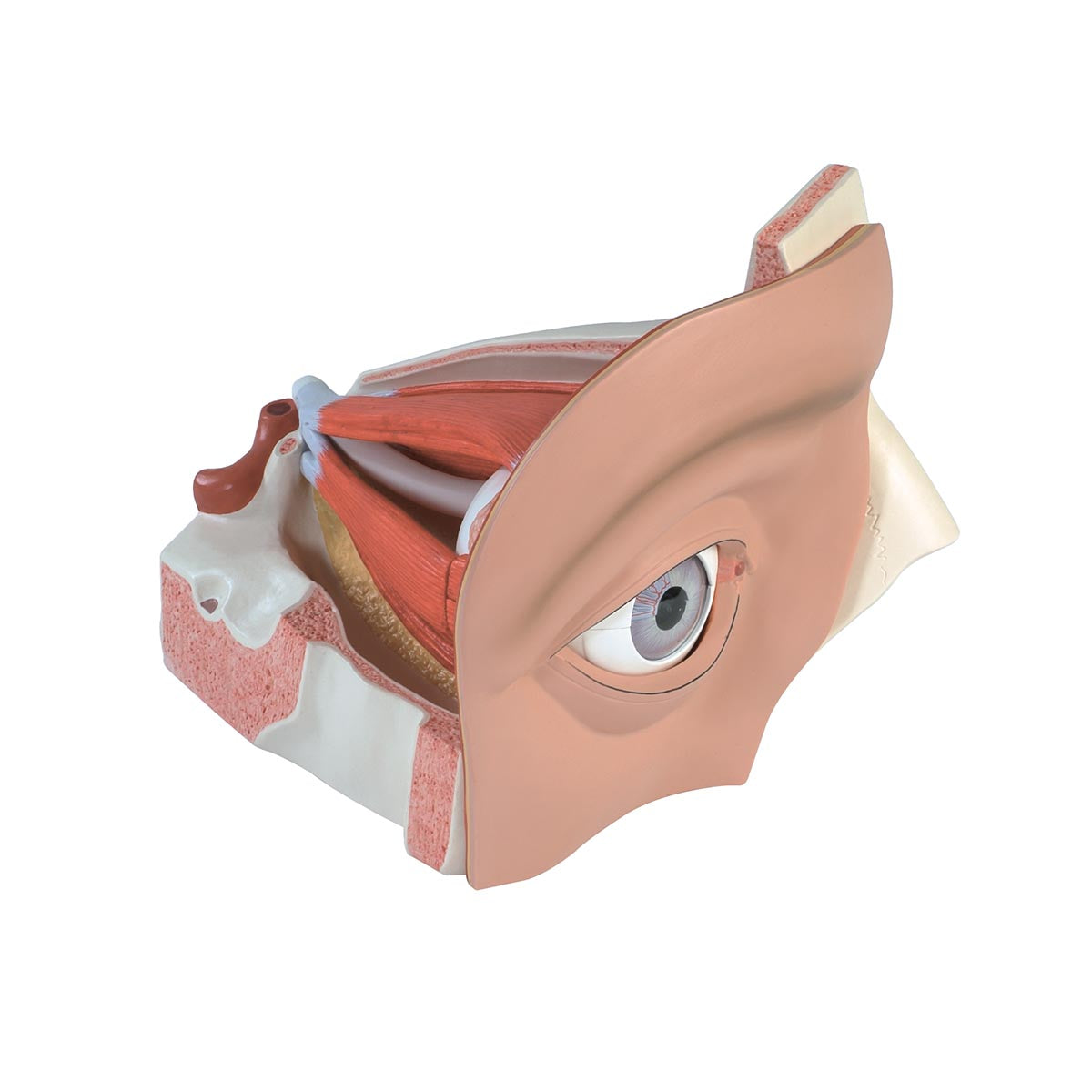
Anatomically speaking, the model gives an insight into the many structures found in the orbit. First of all, the model can be used to understand the individual layers of the eyeball:
The outermost layer is made up of the transparent cornea, which at the limbus is connected to the sclera (the white tendon that makes up the back 5/6).
The middle layer (called the uvea) which consists of the iris (rainbow) incl. the pupil, the corpus ciliare (the ray body) of which the ciliary muscle is primarily seen and the choroid (the choroid).
The innermost layer is called the retina.
The outermost layer of the eyeball can be halved and removed, so that the vitreous body (corpus vitreum) and the lens become visible. These can also be taken out and studied.
Furthermore, it can be seen how the optic nerve (N. opticus) leaves the eyeball at the back, accompanied by the vessels that supply the nerve. The optic nerve can be removed on this model.
In addition, the model shows the 3 muscles of the eyeball, which respectively control the diameter of the pupil and the deflection of the lens:
M. dilator pupillae
M. constrictor pupillae
M. ciliaris
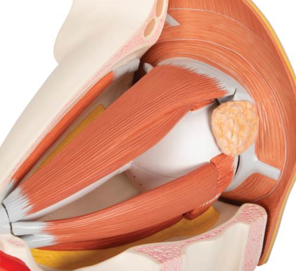
On the outside of the white tendon membrane (sclera) you can see the 6 striated muscles that control eyeball movements. These are divided into the 4 straight muscles (musculi recti) and the 2 oblique muscles (musculi obliqui). 2 of the rectus muscles are removable.
In addition, the model shows the lacrimal apparatus, which consists of the lacrimal gland (glandula lacrimalis) and the tear ducts (consisting of the lacrimal ducts, the lacrimal sac and the lacrimal duct). Only the tear duct is not visible on the model.
As something special on this model, the surface anatomy of the eye is also shown, just as the model shows the structure of the eyelids. For example, the free bone skeleton of the eyelids (tarsus superior and inferior) and the muscles found in the eyelids (M. orbicularis oculi and M. levator palpebralis superior) can be seen.