Skip to product information
1 of 7

SKU:EA1-1000216

Model of the head with exposed muscles, vessels and nerves

Model of the head with exposed muscles, vessels and nerves

Regular price 16 883 SEK
Regular price Sale price 16 883 SEK
Sale Sold out
Not including taxes if ordering from outside EU. Shipping calculated at checkout.

Out of stock

this product is made to order. To place an order please call or write us.

If you are looking for a model that provides a complete overview of the anatomy of the head and neck, we recommend this one.


The model can be separated into 4 separate parts, as the head is divided in the saggital plane, and therefore can be divided into two halves. When this is done, a section of the internal structures of the head is seen, including the oral cavity, nasal cavity and brain. The left hemisphere of the brain is removable, and when this is removed the inside of the skull can be seen.


Finally, the head can be separated from the neck so that the structures passing through the neck can be seen in the horizontal plane. The surface of the model is also divided into two halves, the right half is covered with skin, while the left half shows the superficial musculature as well as vessels and nerves.


The model is 26 cm tall, weighs 1.85 kg and is delivered on a white stand.

Anatomical features

Anatomically, the model can be used to gain an understanding of many different structures.


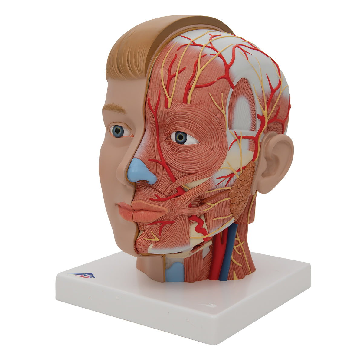
The left half of the face shows how the head's largest vessels and nerves run in relation to the surrounding musculature.

In this connection, muscles from many of the head's muscle groups are shown, including:


The mimic facial muscles
The muscles of mastication
The superficial neck muscles


In relation to these, the largest arteries that supply the superficial facial muscles can be seen:


Branches from A. carotis communis: A. facialis with side branches, A. auricularis posterior, A. occipitalis
A. temporalis superficialis with A. transversae faciei, A. zygomaticofacialis and the terminal branch ramus frontalis.


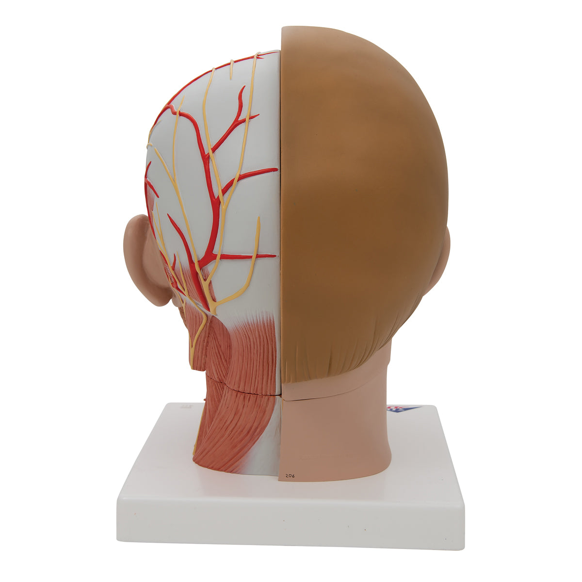
In addition, the largest nerves that supply the area can be seen, including branches from N. ophthalmicus and N. Mandibularis, individual branches from the cervical plexus, N. facialis with its terminal branches and finally sensory nerves to the neck and back of the head.


Of other structures, the parotid gland (one of the three large paired salivary glands) is seen with its duct running over the M. masseter and through the M. buccinator.


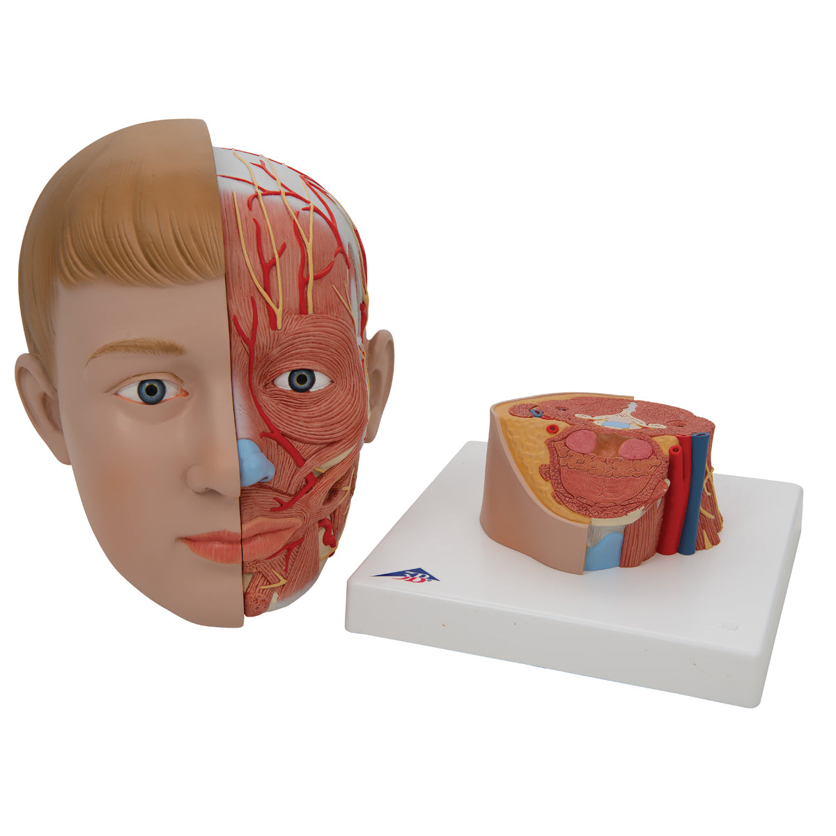
When the head is separated from the neck, the structures that pass through the neck are seen in a horizontal section, including:


Internal and external carotid arteries and internal jugular vein
No. cervicales, plexus brachialis and N. accessorius
Suprahyoid and infrahyoid muscles, the scalene muscles, individual pharyngeal muscles and the back muscles that attach around the neck.
The thyroid cartilage (cartilago thyroidea) and parts of the larynx
Palatine and lingual tonsils


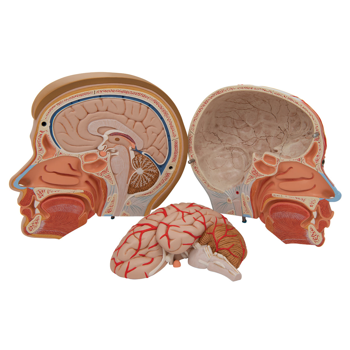
When the two halves of the head are separated from each other, a saggital section is seen through the oral cavity, nasal cavity and brain. This section also shows:


The sinuses (sinus sphenoidalis, ethmoidalis and frontalis)
The hard and soft palate (palatum durum and soft palate)
The turbinates (concha basalis superior, media and inferior)
In relation to the pharynx, the following are seen: recessus pharyngeus, ostium pharyngeum tuba auditory and tonsilla pharyngea


As mentioned, the brain is shown both in a cross-section, but can also be taken out from the skull. Structures can be seen:


telencephalon
The midbrain (diencephalon)
cerebellum
The brainstem
Some of the venous sinuses

Product flexibility

Clinical features

Clinically speaking, the model can be used to understand disease problems related to the muscles, vessels, nerves and surrounding structures of the head.


For example, facial paresis, formation of salivary stones in the salivary glands (carotid stenosis), and disorders related to the jaw muscles and jaw joint such as temporomandibular dysfunction.


Furthermore, the model can be used to understand lesions and disorders in specific areas of the brain. Examples are epilepsy, brain tumors, hydrocephalus, lesions involving cranial nerves and sclerosis (multiple sclerosis).

View full details

A safe deal

For 19 years I have been at the head of eAnatomi and sold anatomical models and posters to 'almost' everyone who has anything to do with anatomy in Denmark and abroad. When you shop at eAnatomi, you shop with me and I personally guarantee a safe deal.

Christian Birksø
Owner and founder of eAnatomi ApS

Read more about eAnatomi