Skip to product information
1 of 4

SKU:EA-1000158

Easy-to-understand model of lumbar vertebrae with pathological conditions

Easy-to-understand model of lumbar vertebrae with pathological conditions

Regular price 2 624 SEK
Regular price Sale price 2 624 SEK
Sale Sold out
Not including taxes if ordering from outside EU. Shipping calculated at checkout.

Out of stock

this product is made to order. To place an order please call or write us.

This easy-to-understand model of the lumbar vertebrae shows the nerves, degenerative changes and disc herniation.


The model measures 22 cm in height and weighs approximately 0.7 kg. The model can be completely disassembled into all its components. It is supplied on a removable stand.

Anatomical features

Anatomically, the model shows the 5 lumbar vertebrae in the lower back as well as spinal nerves. Therefore, the model shows the two most important joint types in the spinal column, which are the symphyses (disci/band discs) between the vertebral bodies and the facet joints, which are sliding joints between the vertebrae's pivots. In addition, cartilage-covered joint surfaces are colored in a light blue color (at the facet joints).


Overall, the bone quality is very good, and from top to bottom (L1 "down to" L5), the model illustrates more and more severe degenerative changes in the vertebral bodies and discs:


Disc on top of L1 (upper lumbar vertebra): Nothing abnormal
L1: Nothing abnormal
Discus on top of L2: Protrusion is seen
L2: Incipient degenerative changes
Disc on top of L3: A median prolapse is seen
L3: Advanced degenerative changes
Disc on top of L4: A lateral prolapse to the right is seen
L4: Severe degenerative changes and narrowing of the left intervertebral foramen
Disc above L5: Extreme disc degeneration is seen
L5: Very severe degenerative changes and pressure on the spinal nerve on the left side

Product flexibility

In terms of movement, this model is not flexible. It cannot be moved at all and therefore cannot be used to demonstrate the movements of the spine.


The model, on the other hand, can be completely disassembled into all its components (lumbar vertebrae, discs and nerves). In this way, all degenerative changes can be studied easily and conveniently.

Clinical features

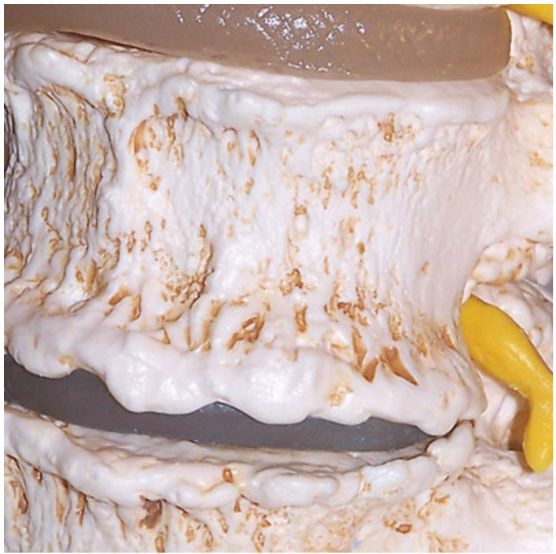
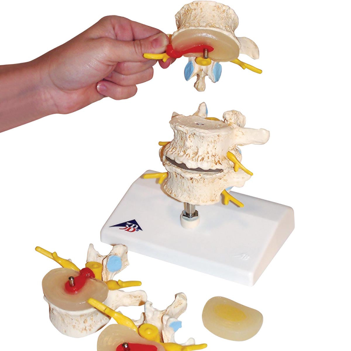
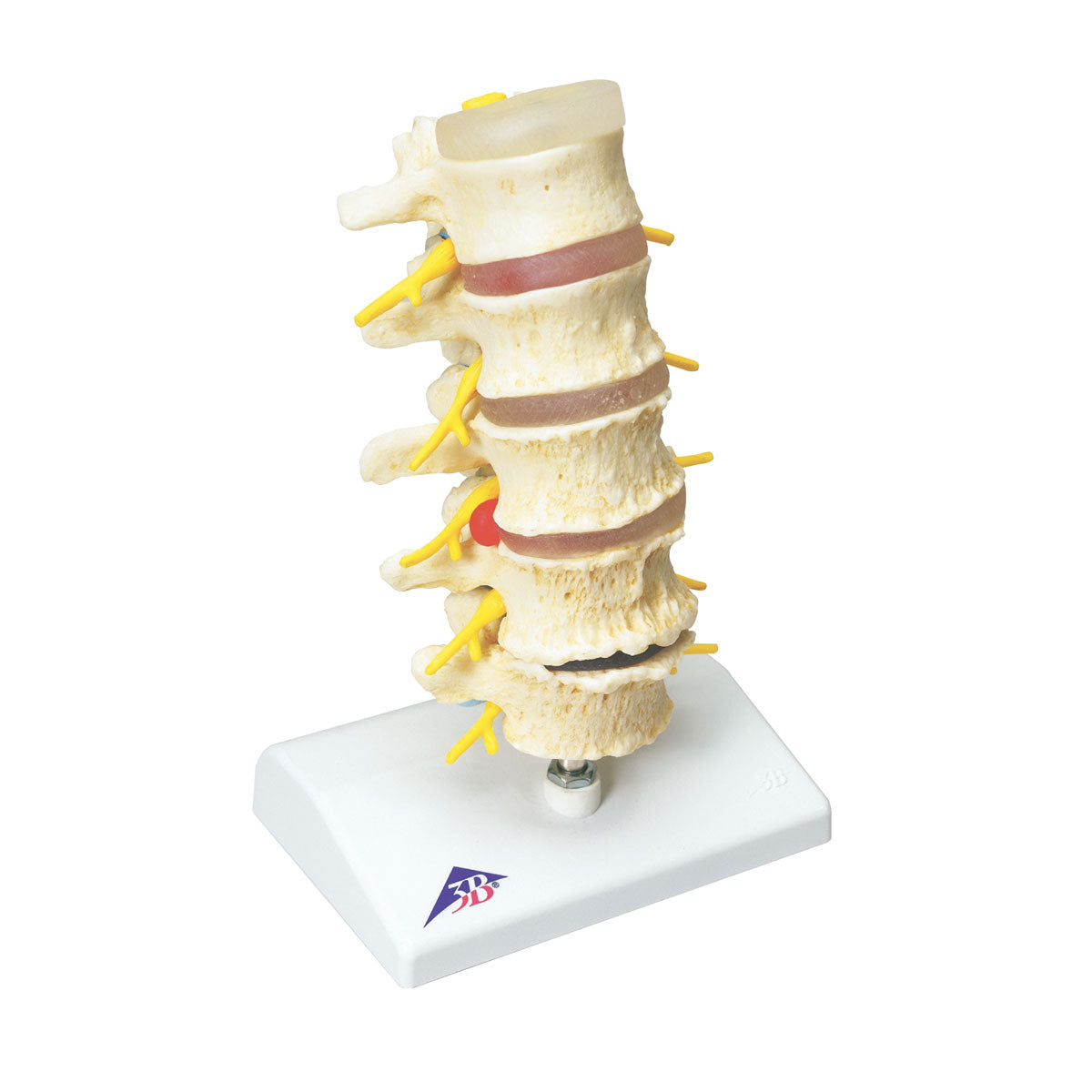
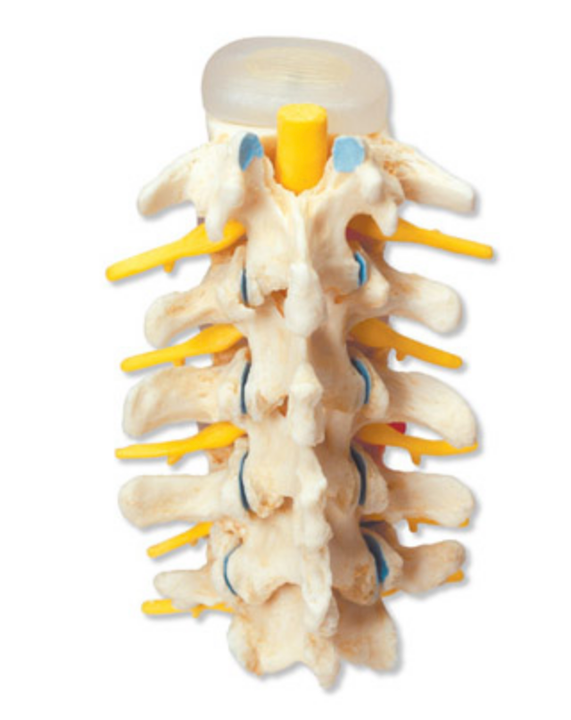
Clinically speaking, the model has been developed to understand degenerative changes in vertebrae and discs. Furthermore, the model can be used to understand treatment options such as spondylosis and treatment of disc herniation with percutaneous endoscopic technique.


The model can also be used to understand many other disorders such as fractures, scoliosis, back pain, etc


NB: The model shows the degenerative changes in the lower back. In Denmark, acute back strain is the cause of most cases of lumbar disc prolapse (in the lower back), and therefore this type of prolapse occurs most frequently in younger people. Degenerative changes, on the other hand, are the cause of the vast majority of cases of cervical disc herniation (in the neck), and these occur most frequently in the elderly.

View full details

A safe deal

For 19 years I have been at the head of eAnatomi and sold anatomical models and posters to 'almost' everyone who has anything to do with anatomy in Denmark and abroad. When you shop at eAnatomi, you shop with me and I personally guarantee a safe deal.

Christian Birksø
Owner and founder of eAnatomi ApS

Read more about eAnatomi