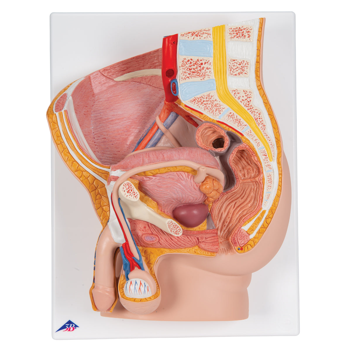
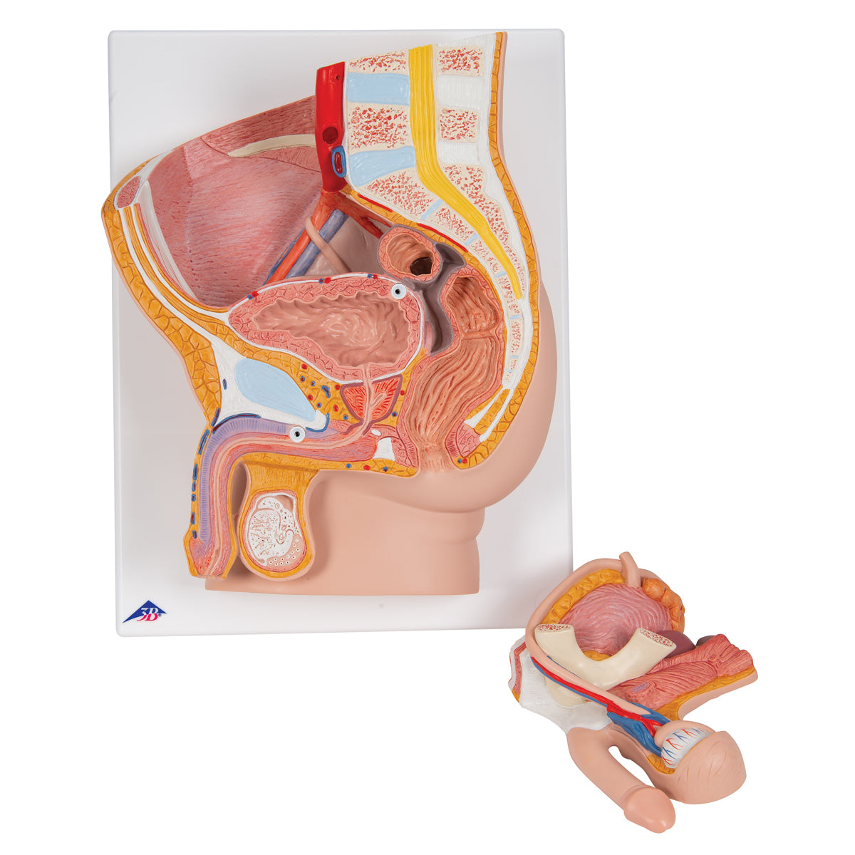
Anatomically, the model shows many different tissues and organs, some of which can be seen in 3 dimensions.
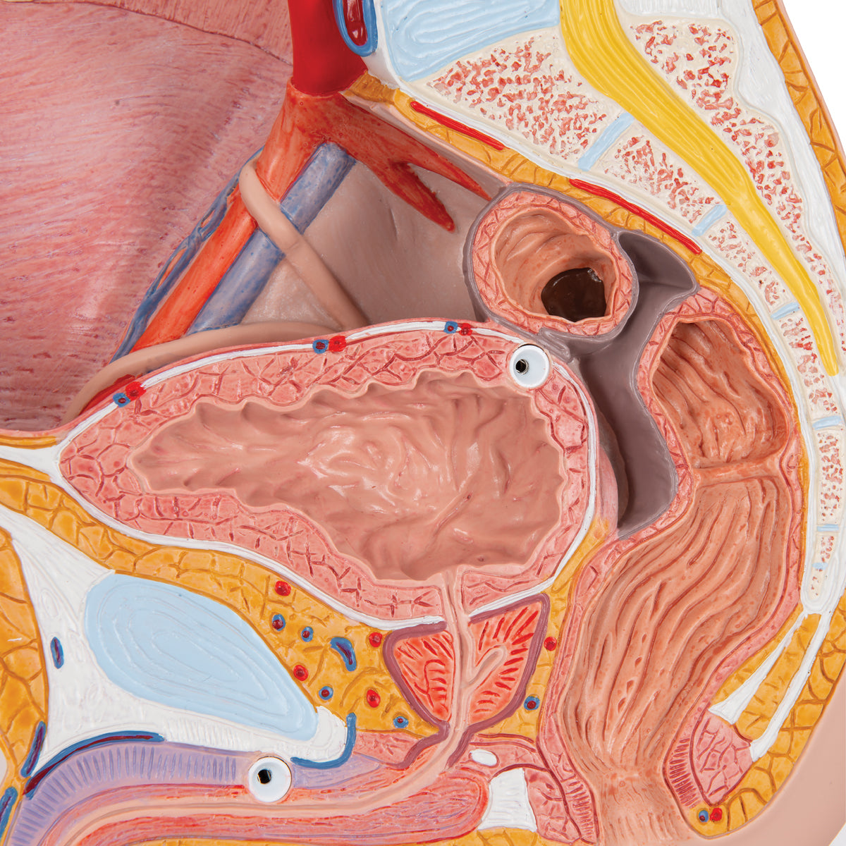
The pelvis (pelvic ring), which consists of bones and the pelvic floor, can only be seen on the model. For example, a cross-section of the symphysis (the front joint) is seen in the pelvic ring. On the other side, part of the large intestine (colon sigmoideum), the rectum (rectum), one ureter (ureter), the urinary bladder (vesica urinaria), the large artery and vein of the pelvis and parts of their branches can be seen. Ligaments are largely invisible, while nerves are not visible at all (however, the lower part of the spinal cord is visible).
Some tissues/organs are also seen in the horizontal plane (also called the transverse plane) when viewing the model "from above". Here you can see i.a. back and abdominal muscles as well as the kidney tissue in the right kidney. NB: All this cannot be seen in the images on the left but in the PDF file.
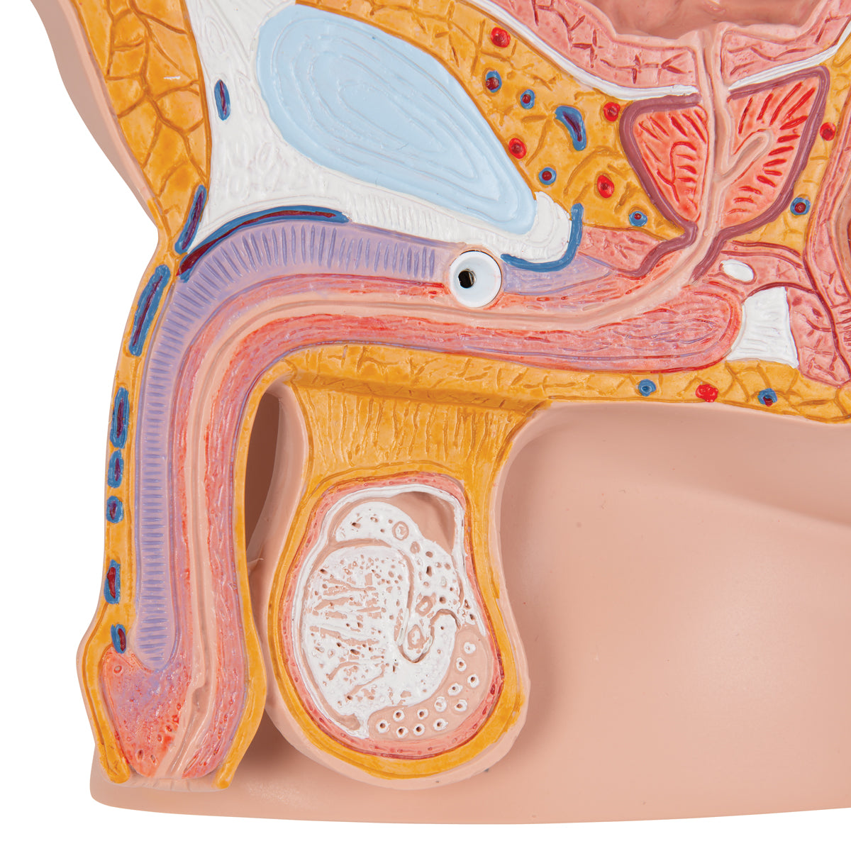
The internal genitalia include the testicles (testes) and the epididymides, which are seen in relation to the scrotum and the spermatic cord (funicle). The contents of the spermatic cord are clearly seen, while this is not the case for its stipules/leaves. In addition, the internal structures of one testicle can be seen - although not with small details.
The seminal ducts include the ductus deferens (the vas deferens), the ductus ejaculatorius and the urethra masculinum (the urethra), all of which are visible (the ductus ejaculatorius can only be felt).
In relation to the accessory gonads, the prostate (bladder neck gland) and vesiculae seminales (seminal vesicles) are seen, but not gll. bulbourethrales and etc. urethral. The export corridors and their connection can only be guessed.
The external genitalia include the penis and the scrotum, which are also seen. The internal structure of the penis is also seen in small details.
Many of the pelvic organs function as a channel or a reservoir (e.g. the rectum and the urinary bladder), which is why they are seen in a pedagogical way with an air-filled cavity (cavity).