SKU:EA1-QS 1
Particularly lifelike skull model in adult size - can be separated into 2 parts
Particularly lifelike skull model in adult size - can be separated into 2 parts
Out of stock
this product is made to order. To place an order please call or write us.Couldn't load pickup availability
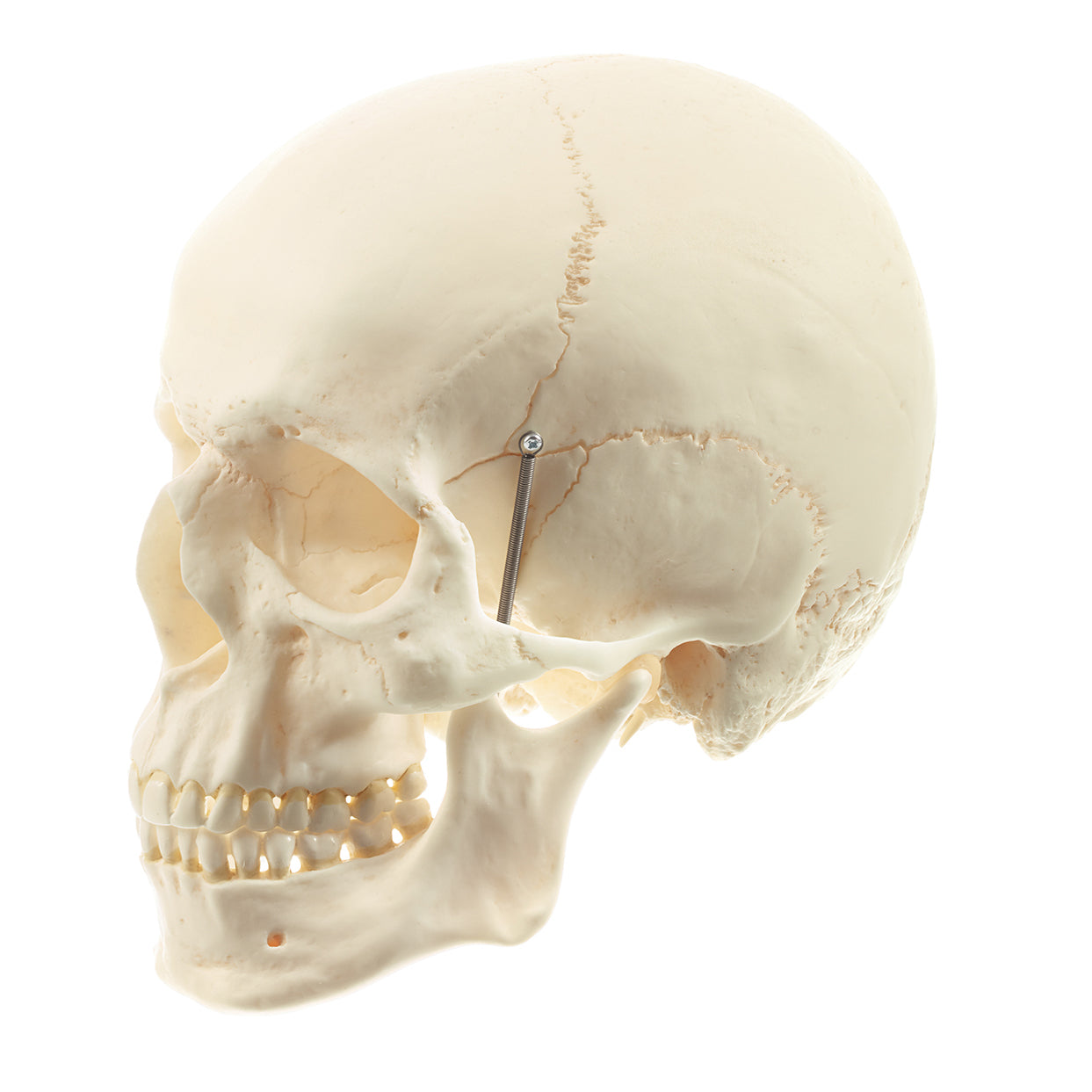
This skull model offers the most lifelike bone structure, the most accurate reproduction of the anatomical details and the feeling of touching real bones. Furthermore, the bone color is slightly yellowish, which makes the skull model appear more realistic. It is produced in SOMSO plastic by the manufacturer SOMSO, which is world-renowned for very high quality.
The model is suitable for basic anatomy teaching and close studies of the external anatomy of the skull. The skull is also used for drawing lessons, due to its beautiful lifelike and detailed surface.
The model is cast in a size that corresponds to an adult person. The length is 17.5 cm and the width is 14.1 cm. It weighs approximately 700 grams.
The mandible is attached with a spring, and can thus be easily and quickly removed. The skull model can thus be separated into 2 parts.
Anatomical features
Anatomical features
Anatomically, the human skull can be divided into 2 parts, and the skull model therefore shows the following:
1) The braincase (neurocranium), which is intended to enclose the brain and the organs of hearing and balance
2) The facial skeleton (viscerocranium) which surrounds the nasal cavity and forms the tooth-bearing framework around the oral cavity. The 32 teeth are also included
The braincase consists of 8 bones. There are 4 unpaired (the frontal, sphenoid, sphenoid and occipital bones) and 2 paired (the occipital and temporal bones). All these bones as well as sutures can be identified on the skull model.
The facial skeleton includes 6 paired bones (the maxilla, palatine, cheekbone, nasal bone, lacrimal bone, and lower conchbone) and 3 unpaired bones called the mandible, the ploughshare, and the zygomatic bone (some do not count the zygomatic bone as part of the facial skeleton). All these bones and sutures can also be identified on the skull model. NB: The hyoid bone is also included in the facial skeleton but cannot be seen on this skull model.
The human skull contains many holes and channels containing vessels and nerves. Overall, there are connections between the braincase and the neck, to and from the eye socket, to and from the pterygo-palatine fossa and to and from the nasal cavity.
On this skull model, all holes and canals are visible, but only from the outside of the skull, as the skull top is not removable. Overall, the bone quality is extremely good, which is why, for example, knots and depressions in the bone tissue can be seen very clearly. The scope of "osseous landmarks" also includes the smallest details.
Compared to the cheaper skull models in our range, this one also feels more real. You can feel the authenticity when you hold the bones or the whole skull at the same time and feel the weight.
Product flexibility
Product flexibility
In terms of movement, it is only relevant to mention the jaw joint. The lower jaw bone ("mandible") is held in place via a relatively long metal spring, which is attached via screws to the mandible and palatine bone. The metal spring can be easily and quickly removed from the screw in the mandible without the screw having to be removed. Therefore, the mandible can be completely removed in a few seconds.
There are also large pieces of rubber mounted in the joint bowls (fossa mandibularis) of the jaw joints, which hold the joint head (caput mandibulae) "like in a rubber bowl" (see the pictures on the left).
The model's jaw joints are not very flexible. Natural movements in the jaw joint require that, among other things, the articular head can slide forward and downward until the articular head is forward on the tuberculum articulare. The latter is the bony part with a weak guiding groove in front of the depression (fossa mandibularis). This can only be demonstrated on the model if the rubber in the joint cups is removed, which the model is not designed for.
If you take a firm hold of the mandible, you can however move it up and down and to the sides, although the natural sliding joint mechanism as described above cannot be demonstrated at all.
Clinical features
Clinical features
Clinically speaking, the skull model is ideal for understanding diseases, disorders and disorders in this part of the skeleton.
Share a link to this product

A safe deal
For 19 years I have been at the head of eAnatomi and sold anatomical models and posters to 'almost' everyone who has anything to do with anatomy in Denmark and abroad. When you shop at eAnatomi, you shop with me and I personally guarantee a safe deal.
Christian Birksø
Owner and founder of eAnatomi ApS
