Skip to product information
1 of 3

SKU:EA1-1000293

Model of mole cancer in different stages

Model of mole cancer in different stages

Regular price 1 265 SEK
Regular price Sale price 1 265 SEK
Sale Sold out
Not including taxes if ordering from outside EU. Shipping calculated at checkout.

Out of stock

this product is made to order. To place an order please call or write us.

If you are looking for an educational skin model for understanding mole cancer (malignant melanoma), we would highly recommend this one.


The skin model measures 14 x 10 x 11.5 cm and weighs 0.34 kg. Compared to the skin of a human, it is greatly enlarged and shows both skin without disease as well as 5 stages of mole cancer.

It comes on a stand where 5 original color images of different types of mole cancer are also displayed.


An overview has been made in Latin, English and several other languages ​​(but not Danish), where the anatomical structures of the model are named via pictures and lines. The overview includes a description of the disease (in, among other things, English but not Danish), the ABCDE criteria, determination of thickness (including Breslow level) etc.

Anatomical features

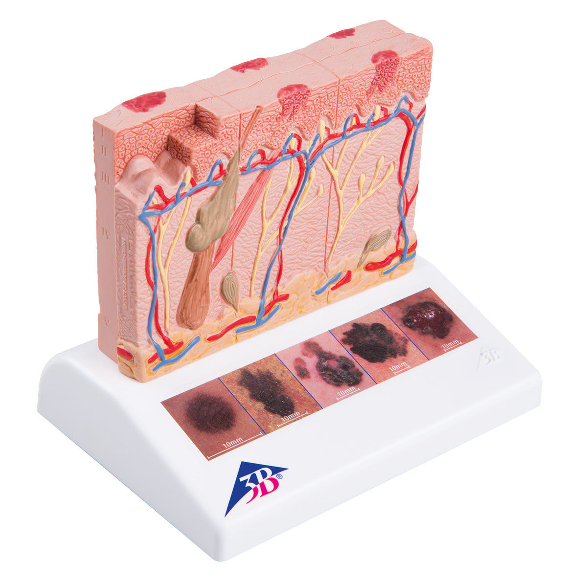
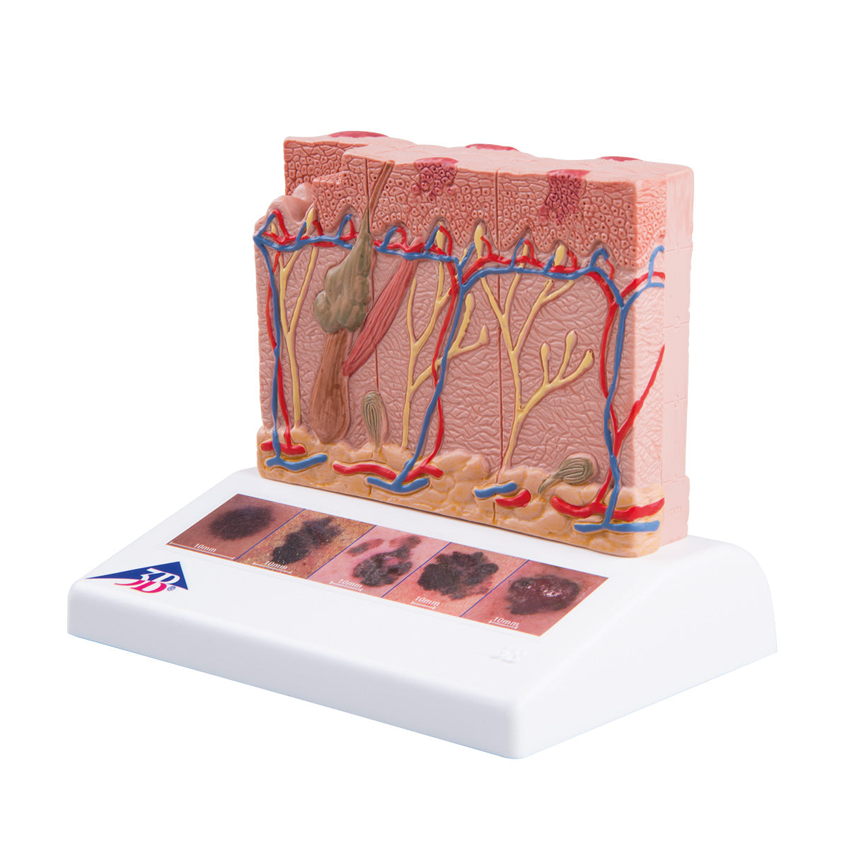
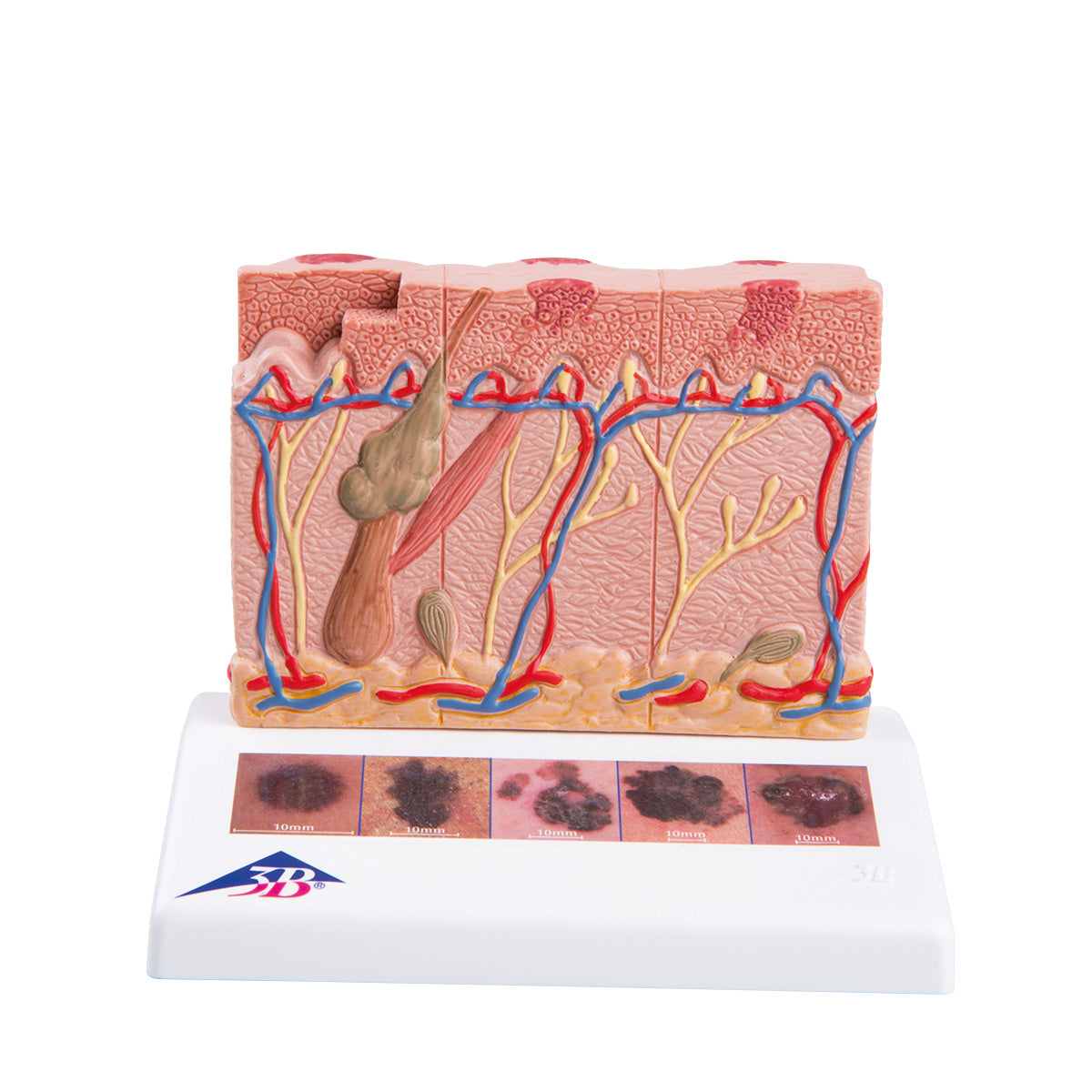
Anatomically, the model shows the skin's 2 layers, which are the epidermis and dermis. The skin (subcutis) is also seen.


The epidermis primarily consists of keratinocytes, and the model shows the layered structure which results from the displacement of these cells upwards towards the skin surface (see the different shapes of the cells). Furthermore, the basement membrane can be seen at the bottom.


The thick connective tissue layer with nerves, blood vessels and hair follicles with attached smooth muscle (m. arrector pili) can be seen in the dermis.


Some of the subcutaneous tissue is also shown in yellow at the bottom, which symbolizes fat cells (since the subcutaneous tissue mainly consists of fat cells).


The model's skin is divided into 6 "pillars" which cannot be separated. Each column shows both the epidermis, dermis and subcutaneous tissue. One column shows skin without disease, while the other 5 show mole cancer in different stages using darker cells (see images on the left).


All this means that the model can be used to understand the thickness of tumors via boats

Breslow level and Clark level.


Furthermore, there are 5 color images on the stand, which allow understanding of the English acronym ABCD (possibly + EFG), which is used in the clinical examination (where A means "Asymmetry", B means "Border", etc.).

Product flexibility

Clinical features

Clinically speaking, the skin model has been developed for understanding mole cancer.

View full details

A safe deal

For 19 years I have been at the head of eAnatomi and sold anatomical models and posters to 'almost' everyone who has anything to do with anatomy in Denmark and abroad. When you shop at eAnatomi, you shop with me and I personally guarantee a safe deal.

Christian Birksø
Owner and founder of eAnatomi ApS

Read more about eAnatomi