Skip to product information
1 of 5

SKU:EA1-4708

Skull model assembled with magnets in 22 parts - colored version

Skull model assembled with magnets in 22 parts - colored version

Regular price €307,95 EUR
Regular price Sale price €307,95 EUR
Sale Sold out
Not including taxes if ordering from outside EU. Shipping calculated at checkout.

Out of stock

this product is made to order. To place an order please call or write us.

Are you looking for a model of the adult skull that makes it easy to understand the complex relationships that the individual skull bones have to each other? This model is not only easy to take apart, but also provides an understanding of the design of the individual skull bones.

The skull is created as a natural cast of the skull of an adult European man and is therefore lifelike in size. The skull weighs 0.8 kg and can be separated into 22 separate parts. This makes it possible to study the individual skull bones very closely, while providing a good understanding of the relationships between the individual bones when the skull is reassembled.


As something special, this model is extremely easy to assemble and disassemble, as the individual bone parts are held together by magnets instead of nails.


The 22 bones are each marked with their own color to easily distinguish them from each other. In total there are 8 paired bones - two identical bones are marked with the same color. A total of 9 different colors are used.

Anatomical features

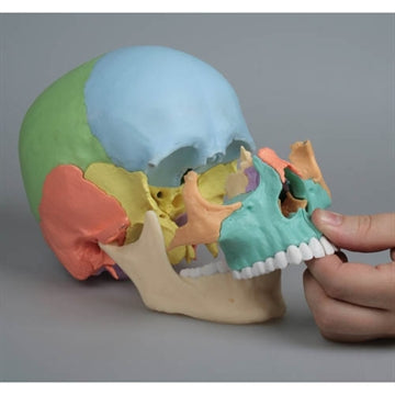
Anatomically speaking, the model shows the bones of the skull and their interrelationships. Overall, the skull is divided into 2 parts:


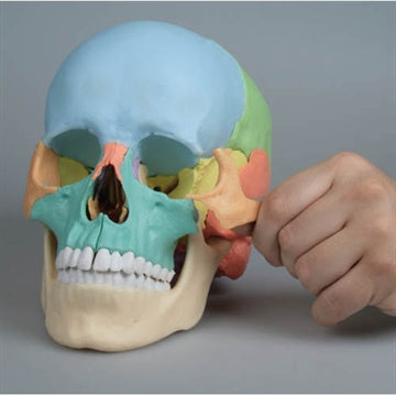
The neurocranium, which is the braincase that encloses and protects the brain.
The viscerocranium, or facial skull, which surrounds the eye sockets and nasal cavity and bears the 32 teeth.


The skull contains many holes and channels through which vessels and nerves run to and from the brain. Many of these, but not all, are shown on this model.


The model is very suitable for forming an overview of where these holes and channels begin and end, as they will typically have their beginning on the inside of the skull, and their end on the outside of the skull - or vice versa. Because the bones that make up the top of the skull can be removed, a good understanding of the structure of the interior of the skull can be obtained.


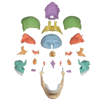
The following bones are shown on the model:


Os parietale (left and right)
Os occipital
Us frontal
Os temporale (left and right)
Us sphenoidal
Os ethmoidal
Stomach
Os zygomaticum (left and right)
Maxilla - upper jaw with teeth (left and right)
Us palatinum (left and right)
Concha nasalis inferior (left and right)
Os lacrimale (left and right)
Us nasal (left and right)
Mandible - lower jaw with teeth


It should be noted that the hyoid bone (os hyoideum) is also included in the skull bones, but this is not included in the model.


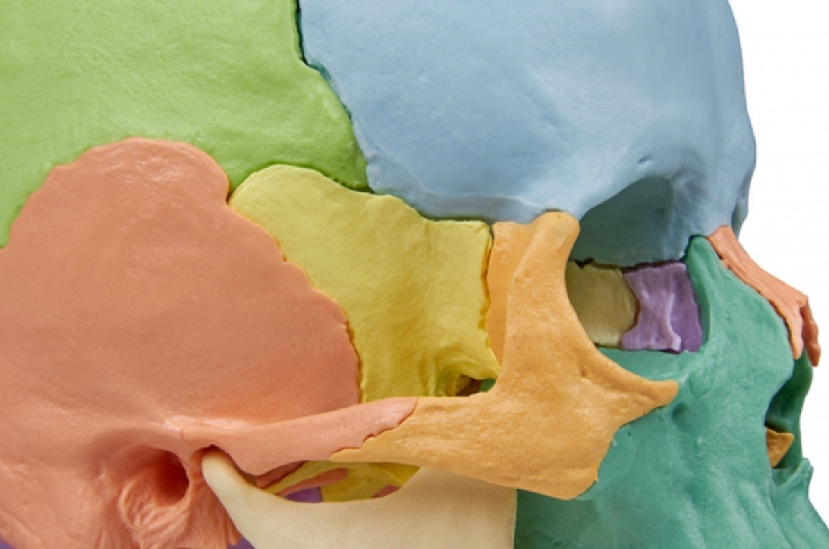
In addition, the model shows the various sutures found between the skull bones, which in the adult are fused together.

Product flexibility

In terms of movement, it is relevant to mention the jaw joint. The lower jaw is only held in place by magnets, so the model is not ideal for demonstrating the various movements that can be performed in the jaw joint.


If this is attempted, it requires holding a hand under the lower jaw (mandible) so that the joint head remains in the socket.

Clinical features

Clinically, the skull model can be used to understand specific skull deformities, such as craniosynostosis.


The model is also ideal for understanding the individual skull bones and their connections, as well as diseases, disorders and disorders in this part of the skeleton.

View full details

A safe deal

For 19 years I have been at the head of eAnatomi and sold anatomical models and posters to 'almost' everyone who has anything to do with anatomy in Denmark and abroad. When you shop at eAnatomi, you shop with me and I personally guarantee a safe deal.

Christian Birksø
Owner and founder of eAnatomi ApS

Read more about eAnatomi