Skip to product information
1 of 4

SKU:EA1-BS 23/4

Advanced brain model with arteries, color markings, meninges etc.

Advanced brain model with arteries, color markings, meninges etc.

Regular price €1.516,95 EUR
Regular price Sale price €1.516,95 EUR
Sale Sold out
Not including taxes if ordering from outside EU. Shipping calculated at checkout.

Out of stock

this product is made to order. To place an order please call or write us.

Anatomical model of the brain from SOMSO Modelle. One in highest educational value, guarantee of accuracy and highest material quality. The SOMSO Model is recognized worldwide for producing the best anatomical models on the market.


The model here is divisible into 10 parts and includes e.g. the arteries of the brain with the circle of Willis, part of the skull and colored areas of special interest, e.g. in relation to localization of motor and cognitive functions.


The model weighs approximately 1.3 kg. and has the dimensions: 22 x 20 x 15 cm


Supplied on a green base with explanatory booklet.

Anatomical features

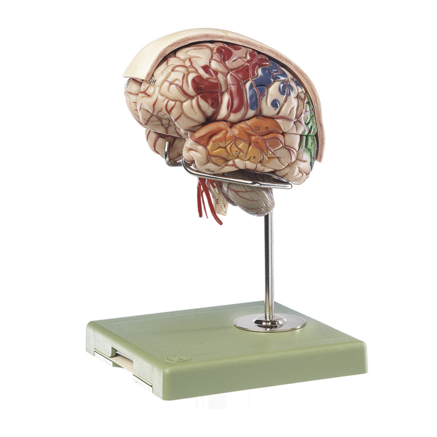
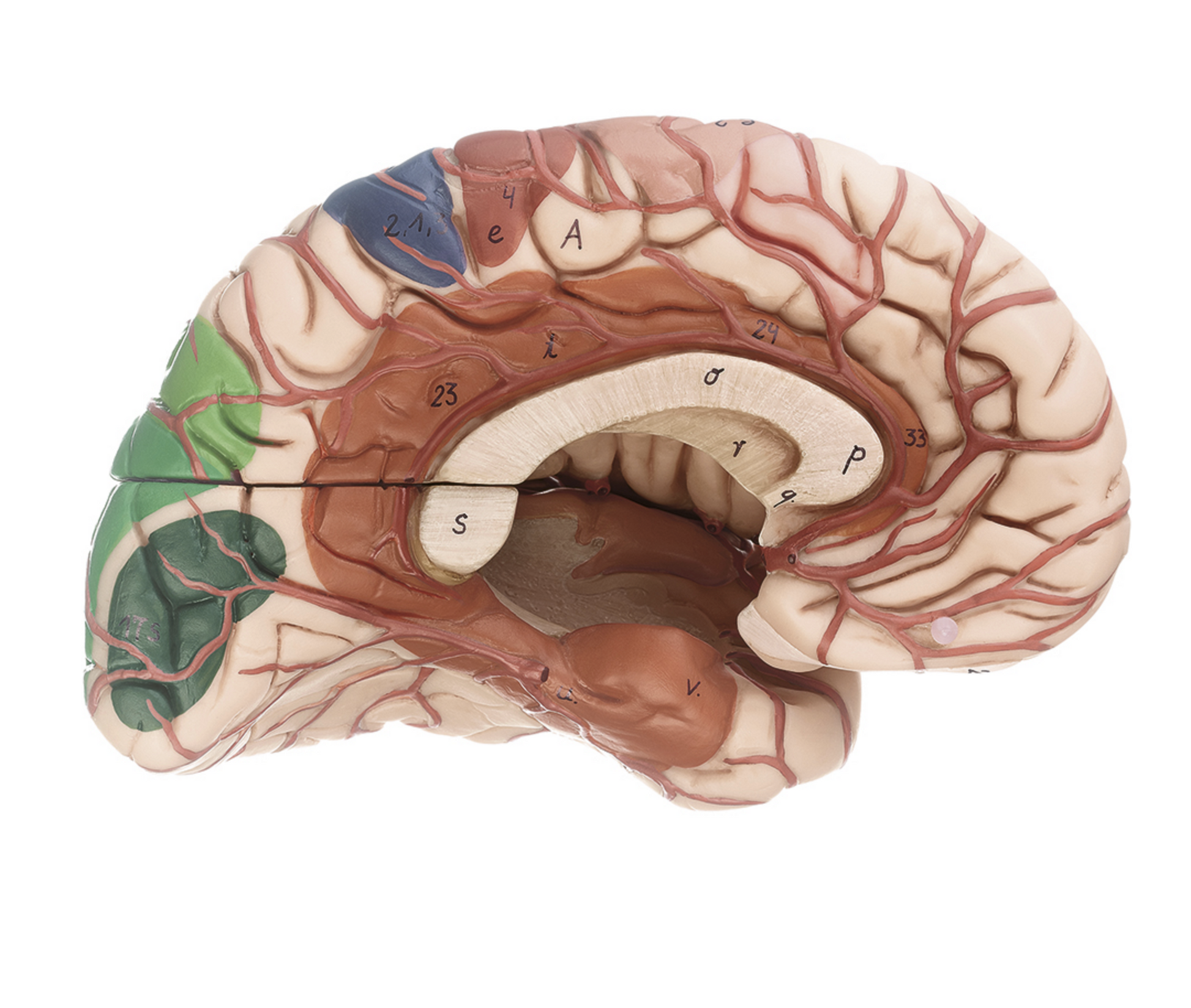
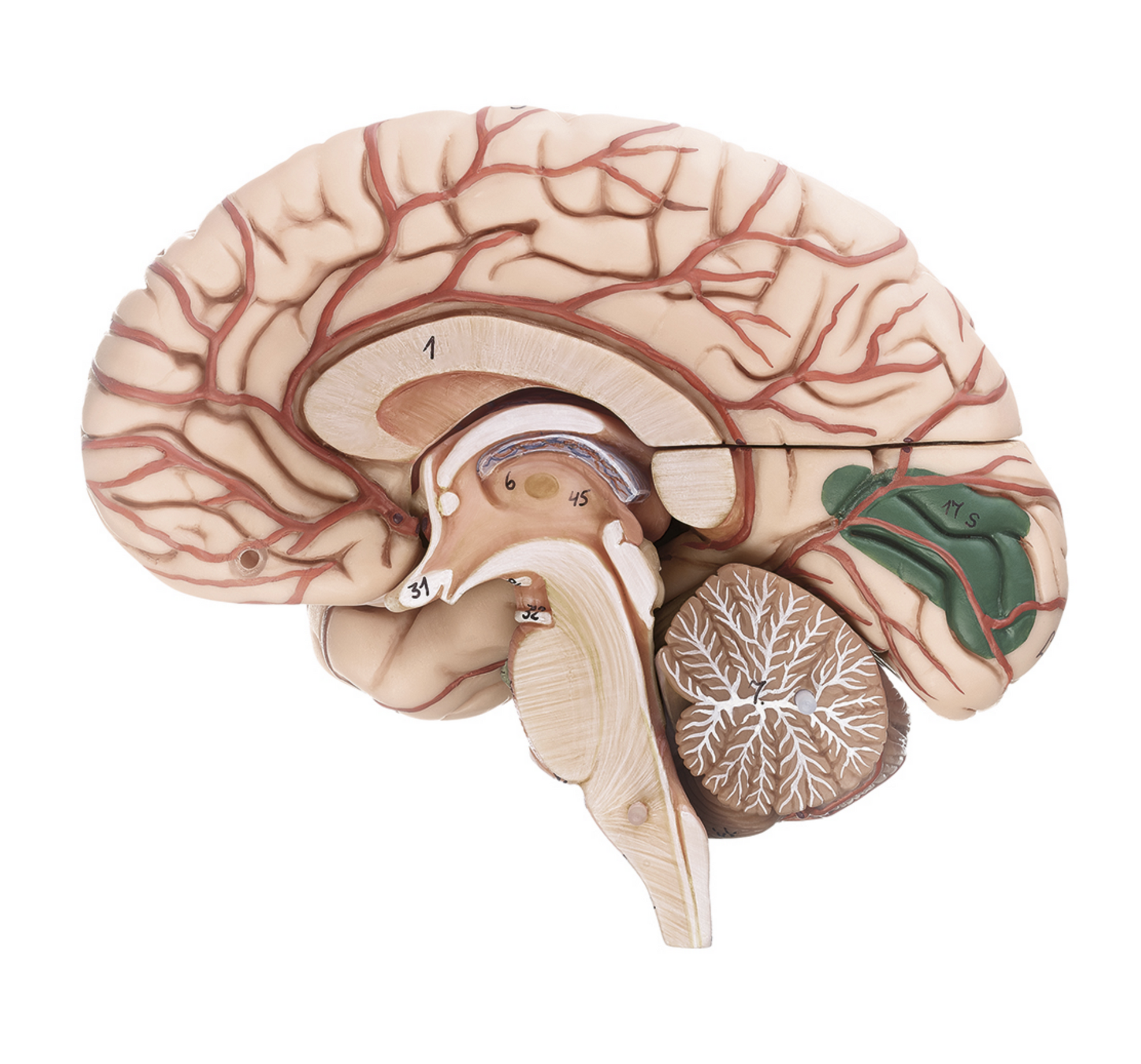
Anatomically, the model shows the human brain, which can generally be divided into the cerebrum (cerebrum), the cerebellum (cerebellum) and the brain stem (truncus encephali).


These 3 structures are clearly separated and the difference between gray and white matter can clearly be seen on this model.


In the cerebrum (telencephalon and diencephalon), the lobes of the brain, as well as the thalamus and hypothalamus (and the pituitary gland) are primarily seen


In the cerebellum, the vermis cerebelli and the cerebellar hemispheres (hemisperium cerebelli) are seen


The brainstem shows its 3 parts (the midbrain, the pons and the medulla oblongata) as well as the apparent origins of the cranial nerves using Roman numerals (also called the cranial nerves)


Other structures such as the brain stem, fornix, ventricular system and the first 2 cranial nerves (the olfactory and optic nerves) are also seen, which do not originate from the brainstem

Product flexibility

Clinical features

Clinically, the model can be used to understand lesions and disorders in specific areas of the brain. Examples are epilepsy, brain tumors, hydrocephalus, inflammation of the meninges, and sclerosis (multiple sclerosis). The model can also be used to understand apoplexy (stroke).

View full details

A safe deal

For 19 years I have been at the head of eAnatomi and sold anatomical models and posters to 'almost' everyone who has anything to do with anatomy in Denmark and abroad. When you shop at eAnatomi, you shop with me and I personally guarantee a safe deal.

Christian Birksø
Owner and founder of eAnatomi ApS

Read more about eAnatomi