Skip to product information
1 of 4

SKU:EA1-1008493

Model of alveoli with surrounding structures

Model of alveoli with surrounding structures

Regular price €350,95 EUR
Regular price Sale price €350,95 EUR
Sale Sold out
Not including taxes if ordering from outside EU. Shipping calculated at checkout.

Out of stock

this product is made to order. To place an order please call or write us.

Are you looking for a model that shows the structure of the functional lung tissue in very fine detail? This model shows how the conductive airways transition to the respiratory ones and how these end up in the alveoli, where gas exchange takes place.


The model is produced in 130 times natural size, has the dimensions 26 x 33 x 19 cm (width x height x length) and weighs 1.35 kg. The model is delivered on a white stand.

On the back of the model, a 1000 times magnification of a single alveolus with surrounding capillary network is seen. In a pedagogical way, this gives an understanding of the barrier that the air must move over when it has to go from the lung tissue into the blood.

In addition, an illustration of the branching of the bronchial tree from segmental bronchus to alveolus can be seen on the stand.

The individual structures of the model are not numbered, but when purchasing the model, a manual is included that shows the names of the model's structures in a high level of detail.

Anatomical features

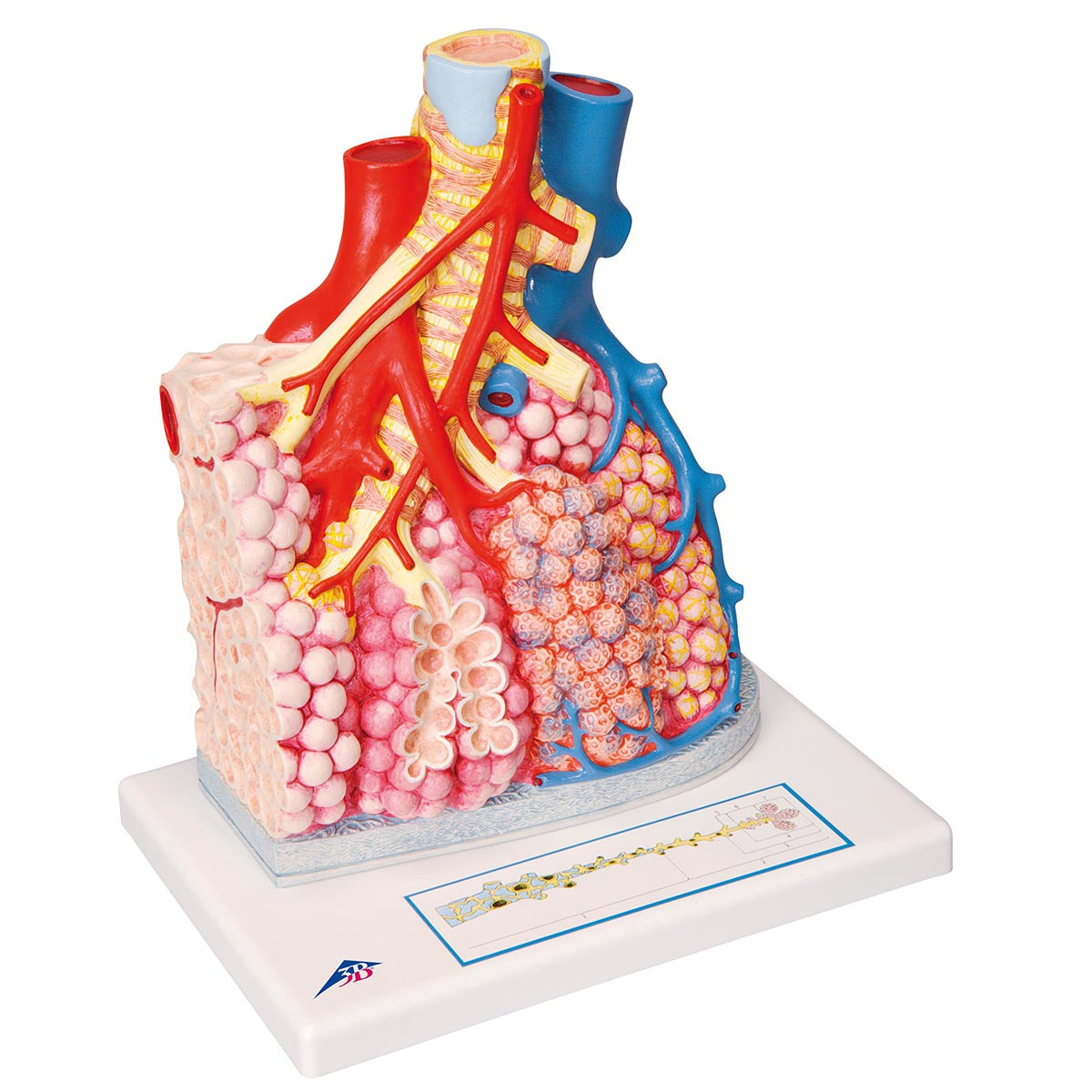
Anatomically speaking, the model shows, in great detail, which changes take place when you move down the airways.

The model shows the following divisions of the airways:

Segmental bronchus
Bronchioles
Terminal bronchiole (without alveoli)
Respiratory bronchiole (with alveoli)
Alveolar duct
Alveolar sac with alveoli


In addition, the model illustrates different divisions of the lung tissue:

Respiratory acinus: are all the alveoli that receive air from the same terminal bronchiole

Lungelobulus: an area of ​​the lung tissue corresponding to 3-5 respiratory acini which is surrounded by connective tissue.

The entire model thus illustrates a lung lobule.

The model also shows how the transition between bronchi and bronchioles is seen where the cartilage in the wall is replaced by an increasing amount of smooth muscle.

Furthermore, the model demonstrates how the various vessels found in relation to the lung tissue run in relation to the bronchi and bronchioles. The following vessel can be seen on the model:

- Branch from A. pulmonalis: leads the oxygen-poor blood from the heart to the alveoli, where it is oxygenated.
- Branch from A. bronchialis: supplies the lung tissue itself with oxygen and nutrients.
- Branch from V. pulmonalis: leads the now oxygenated blood back to the heart.

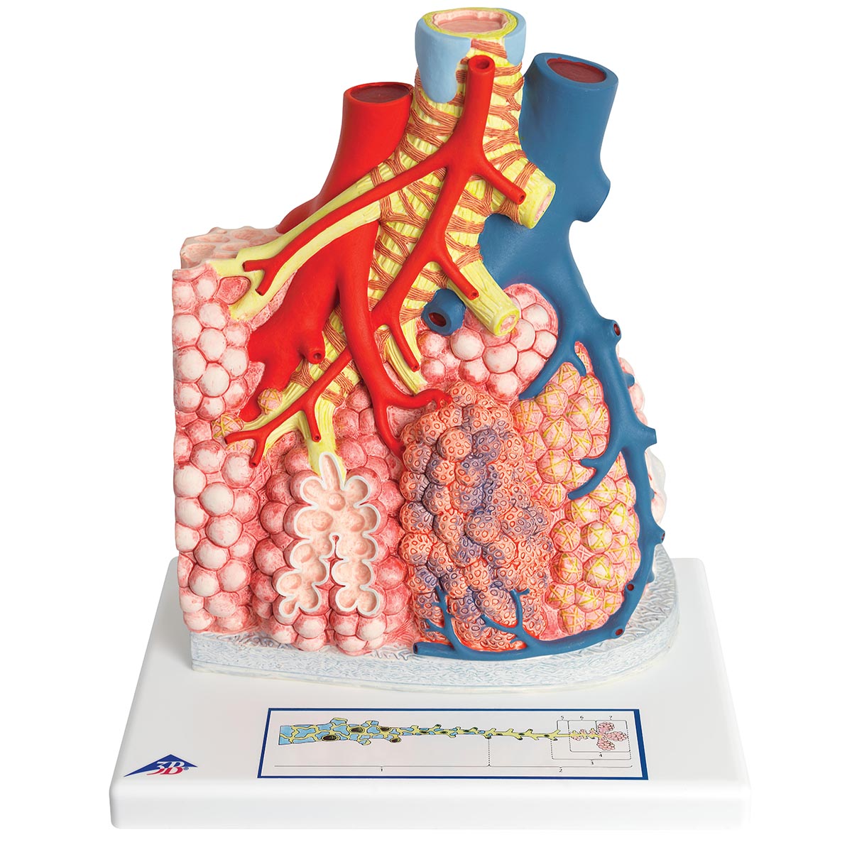
On the back of the model, the enlarged alveolus shows the difference between the different cells that make up the wall of the alveolus. These are:


- Type-I pneumocytes
- Type-II pneumocytes
- Alveolar macrophages - located on the inside of the alveoli

Product flexibility

Clinical features

Clinically speaking, the model can be used to understand diseases in the lungs. Examples are blood clots in the lungs (pulmonary embolism), obstructive lung diseases, emphysema, atelectasis, pulmonary fibrosis and cancer of the lungs.

View full details

A safe deal

For 19 years I have been at the head of eAnatomi and sold anatomical models and posters to 'almost' everyone who has anything to do with anatomy in Denmark and abroad. When you shop at eAnatomi, you shop with me and I personally guarantee a safe deal.

Christian Birksø
Owner and founder of eAnatomi ApS

Read more about eAnatomi