Skip to product information
1 of 7

SKU:EA1-1000257

Complete eye model which is enlarged and can be separated into 8 parts

Complete eye model which is enlarged and can be separated into 8 parts

Regular price €443,95 EUR
Regular price Sale price €443,95 EUR
Sale Sold out
Not including taxes if ordering from outside EU. Shipping calculated at checkout.

Low stock: 1 left

This complete, educational and enlarged model shows the most important anatomical structures both outside and inside the eye.


The eye model weighs approximately 1.2 kg and the dimensions are 20 x 18 x 21 cm. It shows the eye in 5 times normal size and can be separated into 8 parts. It is delivered on a stand (white plastic sheet).

Anatomical features

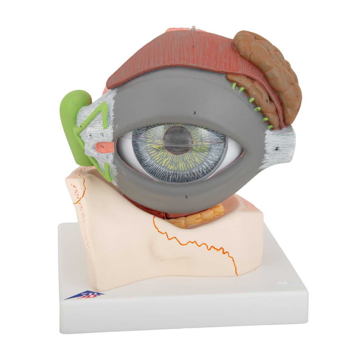
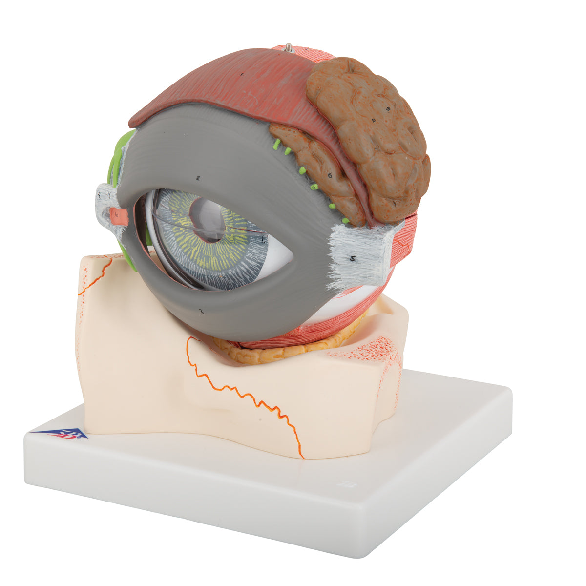
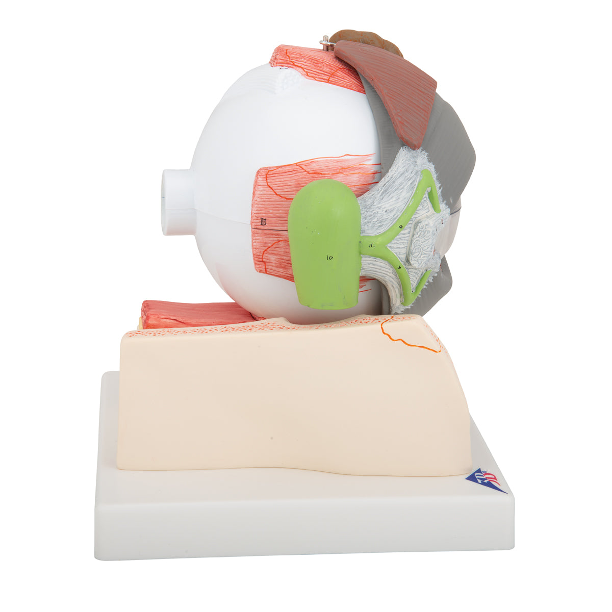
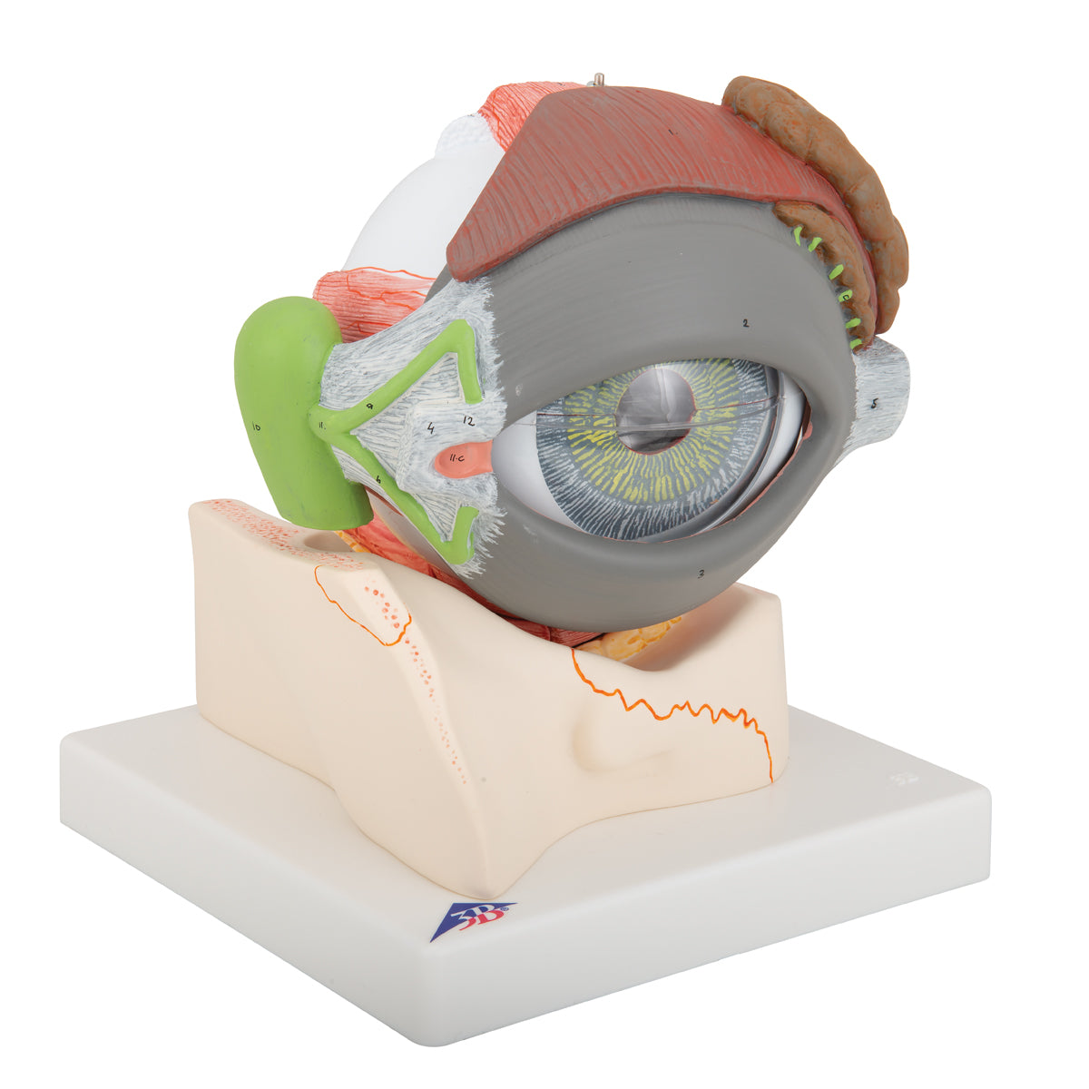
Anatomically, the model shows the layers of the eye, which consist of the following:


The outermost layer, made up of the transparent cornea, which at the limbus is connected to the sclera (the white tendon that makes up the back 5/6)


The middle layer (called the uvea) which consists of the iris (rainbow) incl. the pupil, the corpus ciliare (the ciliary body) which can only be seen on the model and the choroid (choroid)


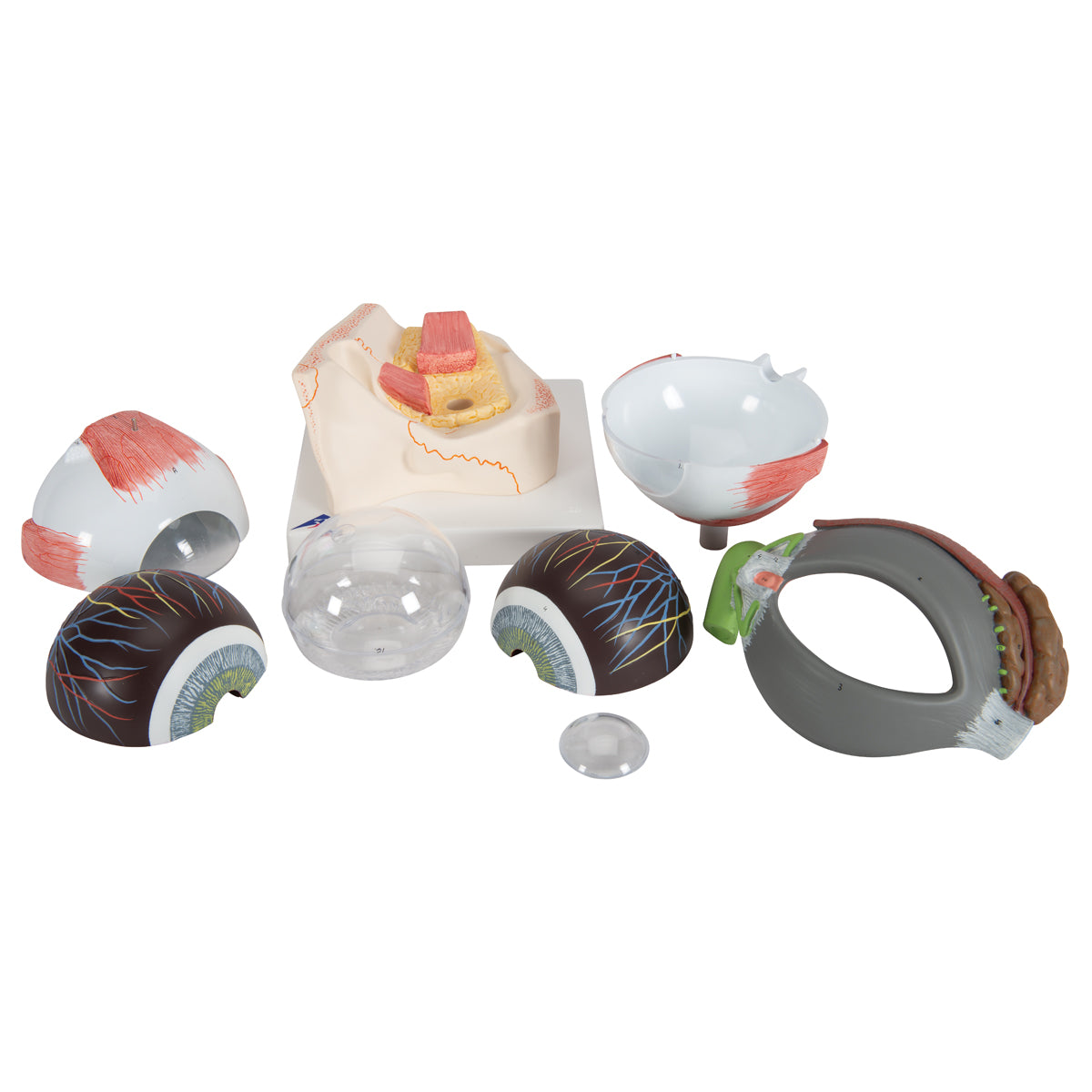
The inner layer called the retina


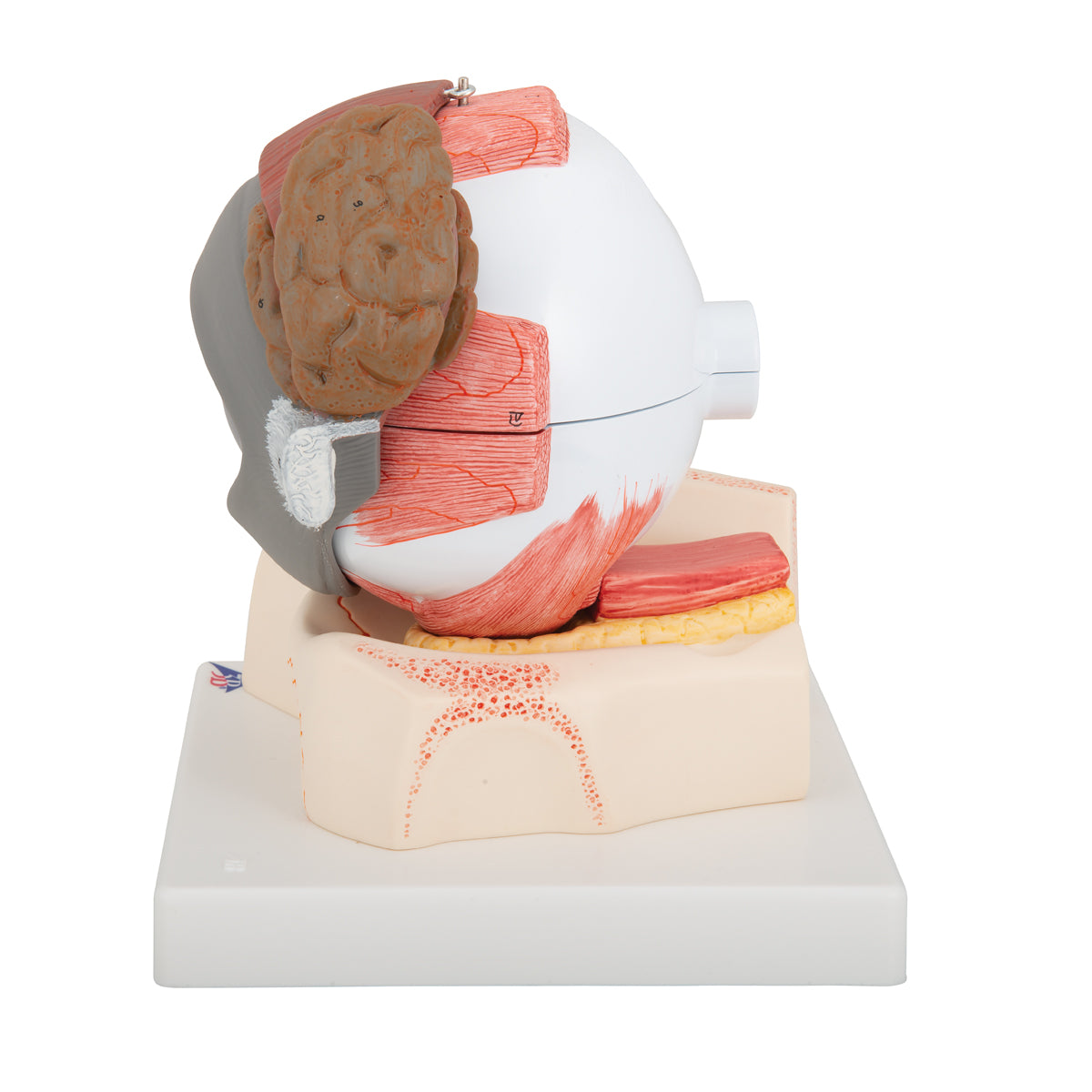
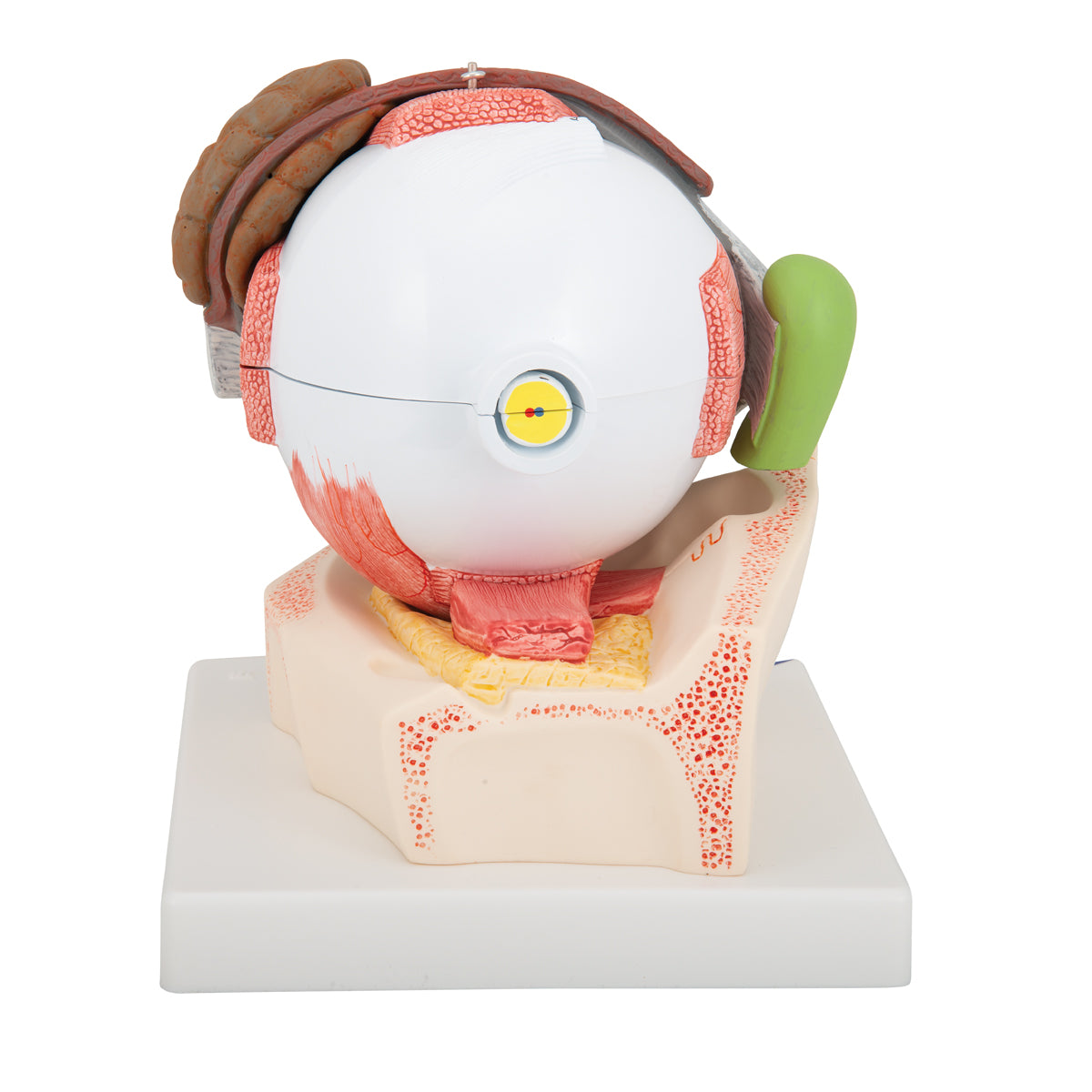
Inside the eye model is the vitreous body (corpus vitreum), and at the back is the optic nerve (nervus opticus) in relation to blood vessels.


The eye model can be split so that the layers can be studied (see images to the left). Furthermore, the lens and the entire vitreous body can be removed.


Outside the white tendon, the eye's movement apparatus is seen, which consists of 6 striated muscles - the 4 straight muscles called musculi recti and the 2 oblique muscles called musculi obliqui.


In addition, a bit of bone tissue, other soft tissues such as ligaments and fatty tissue and the lacrimal apparatus, which consists of the lacrimal gland and lacrimal ducts (the lacrimal canals, the lacrimal sac and the lacrimal duct, of which only the latter is not visible), are seen.

Product flexibility

Clinical features

Clinically, the model can be used to understand diseases of the eye. It can be, for example, "red eye", retinal detachment, vitreous collapse, diabetic eye disease, uveitis, scleritis as well as metastases and primary tumors such as malignant melanomas (birthmark cancer) in the uvea.


Furthermore, the model can be used to understand diseases of the lacrimal gland and tear ducts, such as lacrimal duct stenosis.

View full details

A safe deal

For 19 years I have been at the head of eAnatomi and sold anatomical models and posters to 'almost' everyone who has anything to do with anatomy in Denmark and abroad. When you shop at eAnatomi, you shop with me and I personally guarantee a safe deal.

Christian Birksø
Owner and founder of eAnatomi ApS

Read more about eAnatomi