Skip to product information
1 of 6

SKU:EA1-1000358

Complete hand model with ligaments, muscles, tendons, vessels and nerves - can be separated into 4 parts

Complete hand model with ligaments, muscles, tendons, vessels and nerves - can be separated into 4 parts

Regular price €384,95 EUR
Regular price Sale price €384,95 EUR
Sale Sold out
Not including taxes if ordering from outside EU. Shipping calculated at checkout.

Low stock: 1 left

If you want to enlarge a model of the hand, which gives a detailed insight into the various anatomical layers, and at the same time shows both bones, muscles, tendons and ligaments in a high degree of detail, we recommend this one.


The model is produced in life size, weighs 0.561 kg and has the dimensions 33 x 18 x 18 cm (height x length x width). 3 of the model's superficial structures are removable, whereby the deeper structures are exposed. The model is delivered on a white stand.

Anatomical features

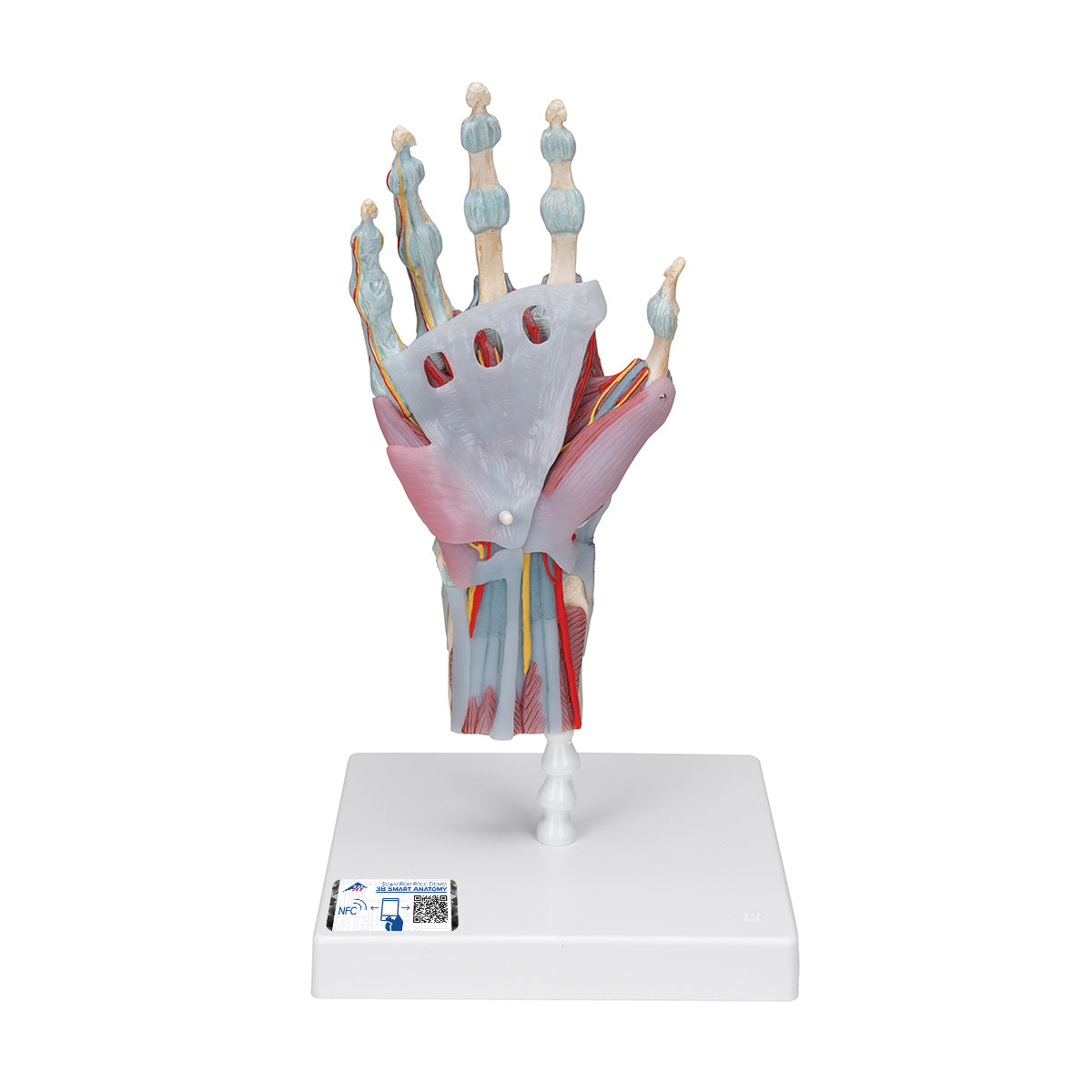
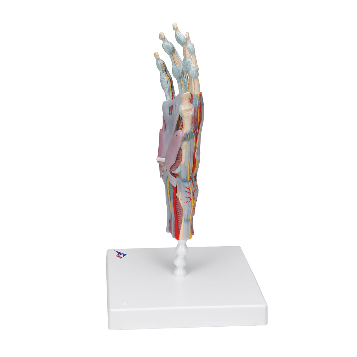
Anatomically, the model shows the majority of the structures found in the hand. On the model, all the skin has been removed, and you can therefore clearly see the muscles, tendons and ligaments.


On the dorsal side of the hand (the side where the back of the hand is located) the extensor retina is shown, where the tendons from the extensor muscles of the forearm pass through before they attach to the bottom of the hand.


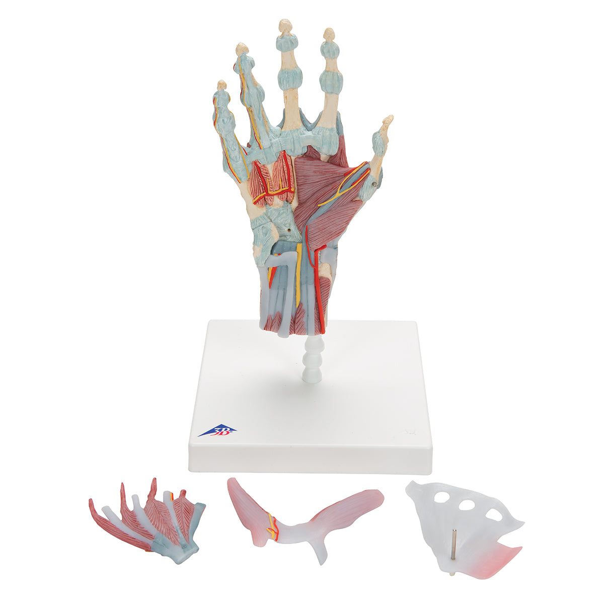
On the palmar side of the hand (the side where the palm is located) the model is divided into 3 layers, of which the outermost 2 can be removed. The following parts are removable:


Aponeurosis palmaris with M. palmaris brevis (outermost removable layer)
M. abductor pollicis brevis, M. flexor pollicis brevis, M. abductor digiti minimi, M. flexor digiti minimi brevis, flexor retinaculum (inner removable layer)


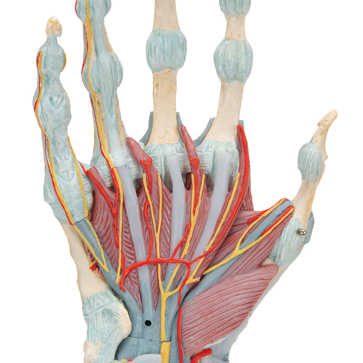
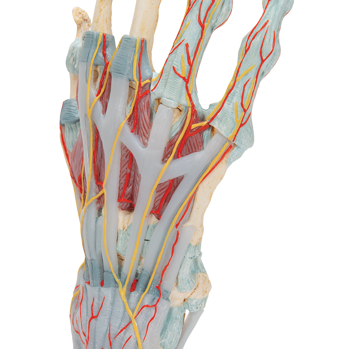
When these are removed, the deep-lying structures in the hand are seen, just as the vessels and nerves that pass out to the fingers become visible.

Among other things, the canalis carpi with the contents of the median nerve, tendo M. flexor pollicis longus, tendo M. flexor digitorum superficialis and tendo M. flexor digitorum profondus are seen.


In addition, the model shows the 3 joints of the fingers with associated ligaments, as well as how the superficial and deep flexor tendons pass in relation to each other on their way out to the finger bones (chiasma tendinum).


Vessels show the large arteries that pass from the forearm down to the hand, as well as how the fingers receive their blood supply from the arcus palmaris superficialis. Veins are not visible on this model.

Product flexibility

In terms of movement, the model's joints are not movable, and the model cannot therefore be used to demonstrate the movements of the wrist and finger joints.

Clinical features

Clinically, this model is ideal for understanding disorders such as carpal tunnel syndrome, wrist tendinitis, trigger finger (trigger finger/digitus saltans) and Dupuytren's contracture (driver's fingers).


Furthermore, it can also be used to understand luxation (joint slip), ligament injury, bone fracture and more.

View full details

A safe deal

For 19 years I have been at the head of eAnatomi and sold anatomical models and posters to 'almost' everyone who has anything to do with anatomy in Denmark and abroad. When you shop at eAnatomi, you shop with me and I personally guarantee a safe deal.

Christian Birksø
Owner and founder of eAnatomi ApS

Read more about eAnatomi