SKU:EA1-1000260
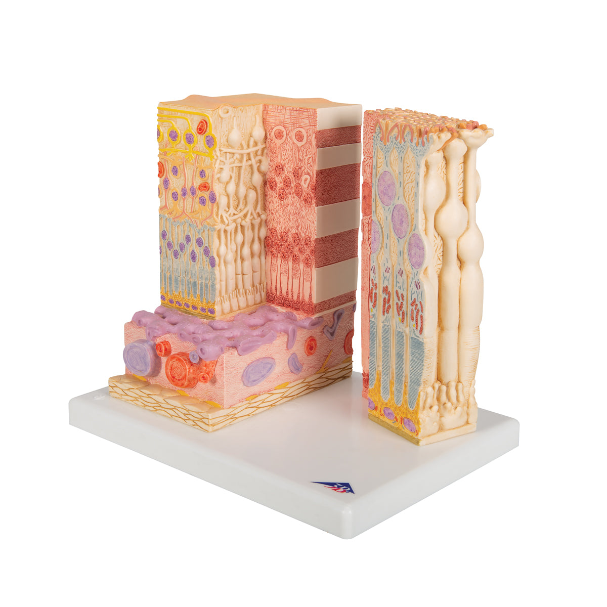
Detailed model of the cells in the eye's retina, choroid and sclera in a microscopic perspective
Detailed model of the cells in the eye's retina, choroid and sclera in a microscopic perspective
ATTENTION! This item ships separately. The delivery time may vary.
Couldn't load pickup availability
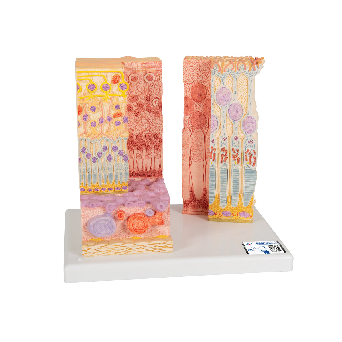
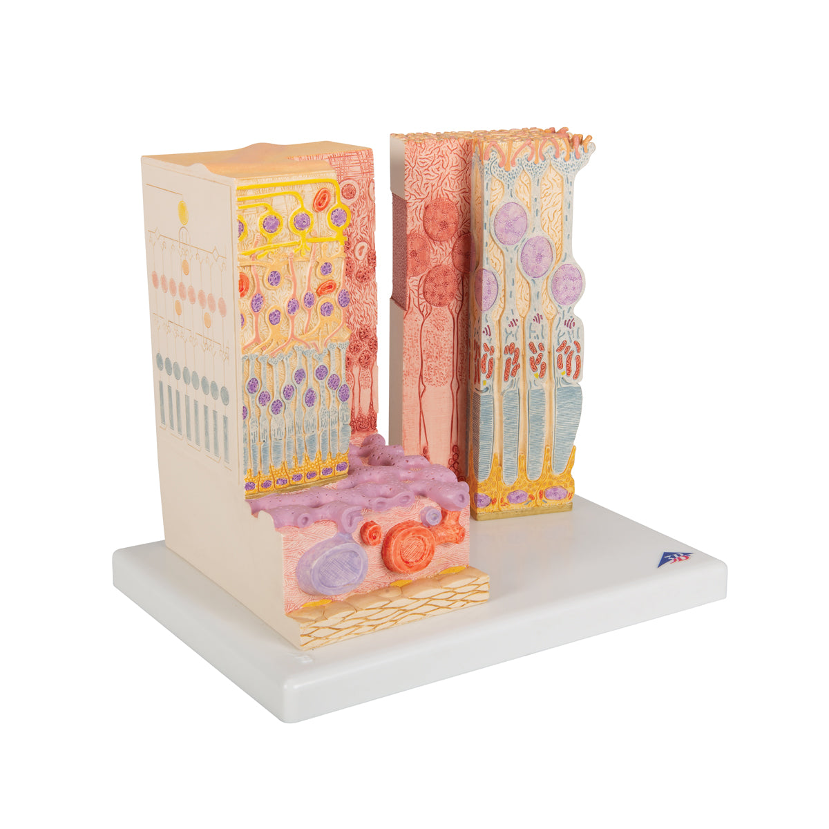
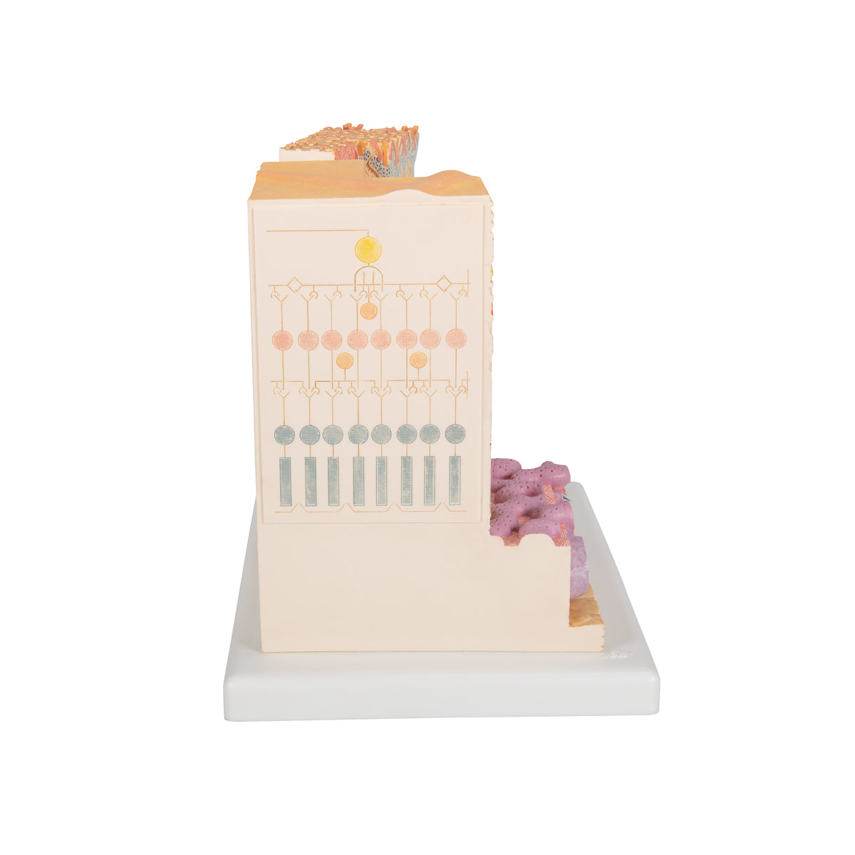
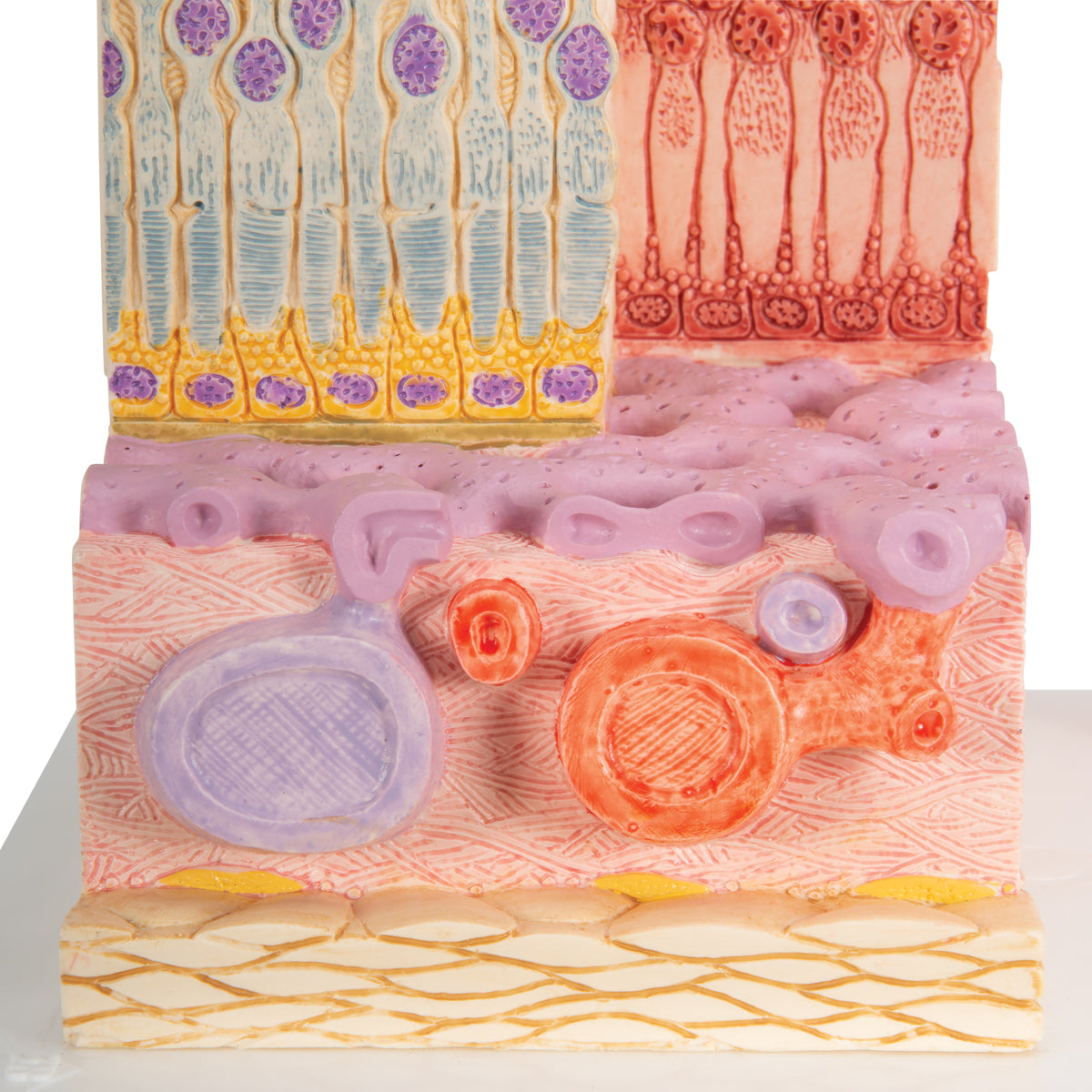
This model shows the cells in the eye's retina (retina), choroid (choroid) and sclera (tendon) as well as the branches of related nerve cells.
The entire model weighs 1.1 kg and measures 25 x 23 x 18.5 cm. The left part of the model is magnified 850 times and the right part 3800 times compared to the tissue of the eye in an adult person. It is all delivered together on a white stand (plastic sheet).
Anatomically speaking
Anatomically speaking
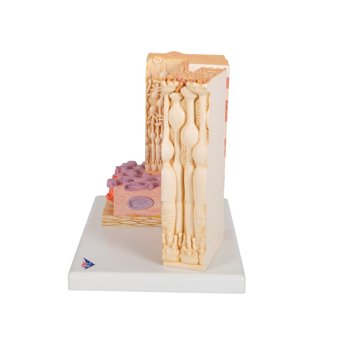
Anatomically, the model shows the cells in the eye's retina (retina), choroid (choroid) and sclera (tendon) via 2 different parts:
In the left part of the model, the tissue is magnified 850 times. Here you can see the complete structure of the retina's many layers as well as the choroid and part of the sclera
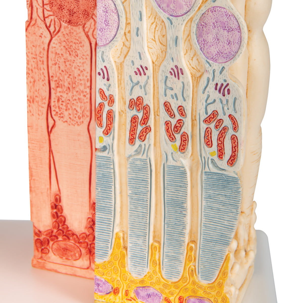
In the right part of the model, the tissue is magnified 3800 times. Here you can see the structure of photoreceptors (rod and cone layer/stratum photosensorium) and the pigment epithelium (pars pigmentosa)
Flexibility
Flexibility
Clinically speaking
Clinically speaking
Clinically, the model can be used to understand diseases of the eye. It can be, for example, retinal detachment, AMD, uveitis, scleritis as well as metastases and primary tumors.
Share a link to this product

A safe transaction
For 19 years I have been managing eAnatomi and sold anatomical models and posters to 'almost everyone' who has anything to do with anatomi in Scandinavia and abroad. When you place your order with eAnatomi, you place your order with me and I personally guarantee a safe transaction.
Christian Birksø
Owner and founder of eAnatomi and Anatomic Aesthetics

