Skip to product information
1 of 7

SKU:EA1-1000269

Enlarged and hand-painted heart model with trachea and esophagus

Enlarged and hand-painted heart model with trachea and esophagus

Regular price €536,95 EUR
Regular price Sale price €536,95 EUR
Sale Sold out
Not including taxes if ordering from outside EU. Shipping calculated at checkout.

Out of stock

this product is made to order. To place an order please call or write us.

This heart model is hand painted and twice the size of a natural heart. Both externally and internally, it appears far more "alive", inspiring and better looking compared to cheaper models.


The model is ideal for understanding the heart's relationship to the trachea and esophagus. It can be divided into 5 pieces. Supplied on a removable stand and measures 32 x 18 x 18 cm.

Anatomical features

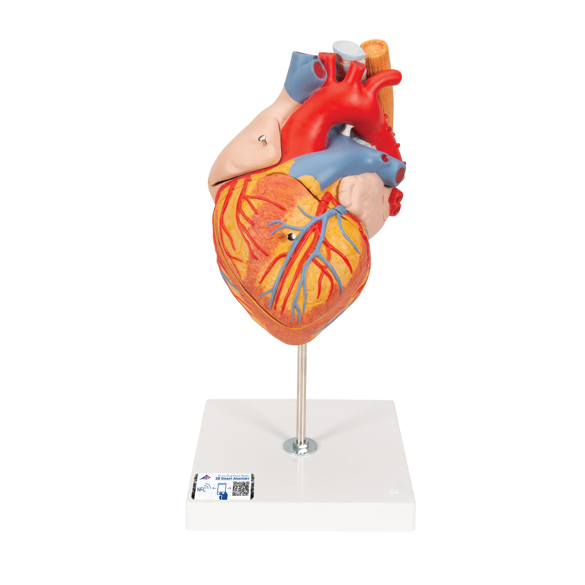
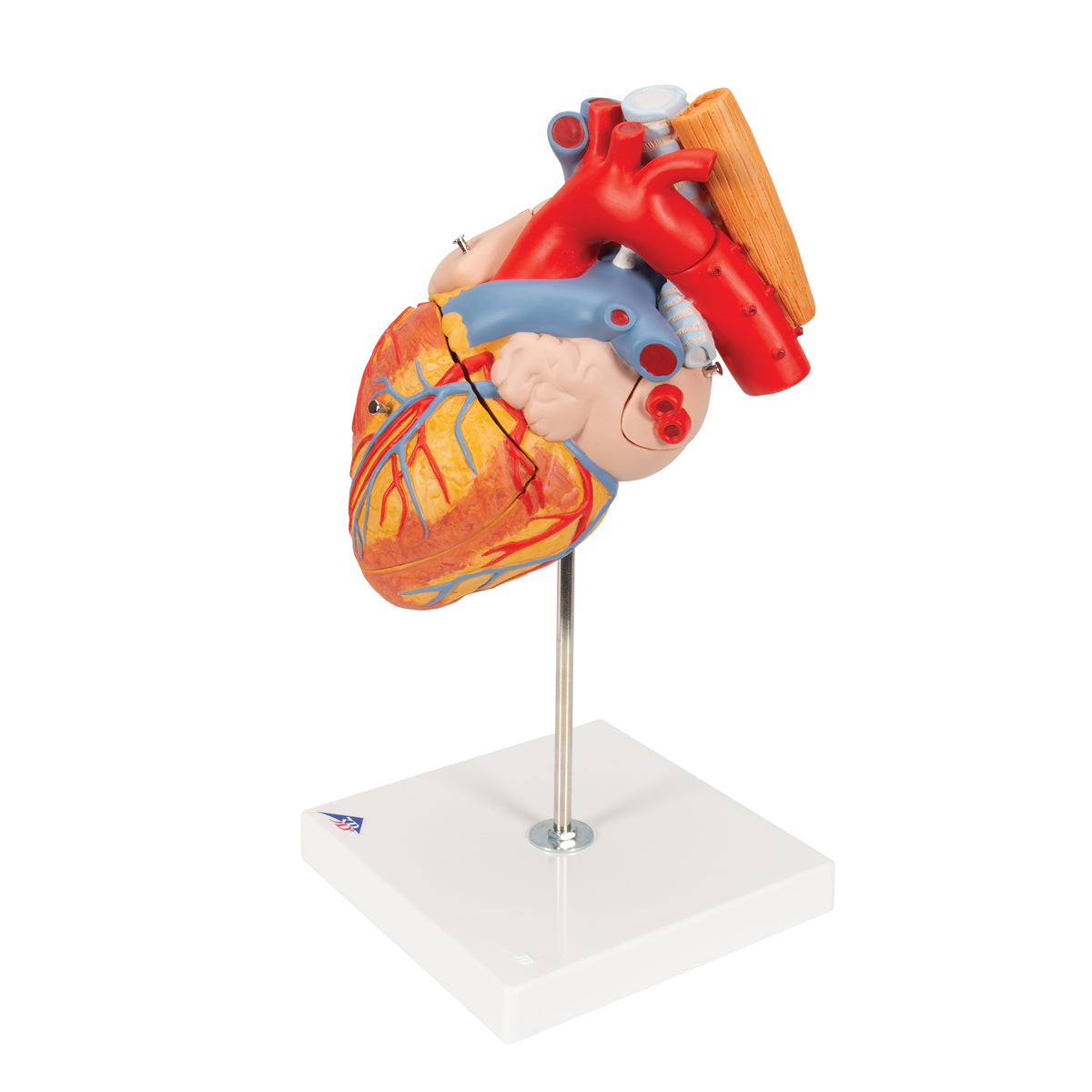
Anatomically speaking, the heart model primarily shows the 4 chambers, the auricles, the heart's blood supply (arteries and veins), the 4 valves based on the sac valve and lobe valve system, neat details of the heart's structure (i.e. endo-, myo- and pericardium) as well as the beginning of the blood vessels that carry blood to and from the heart. However, the aorta is shown with both the ascending aorta, the aortic arch and the beginning of the descending aorta. The heart's impulse conduction system is not seen.


Generally speaking, the structure of the heart consists of 3 layers: Endocardium (a thin endothelial-covered connective tissue), myocardium (the thick muscle) and epicardium at the outer end, which is a serous membrane. In the ventricles, the structure is extremely complex, because the myocardium is made up of muscular muscle bundles, which run downward (and back again) in spiral-like shapes. This results in the ventricles shortening during a contraction (during the pumping action).


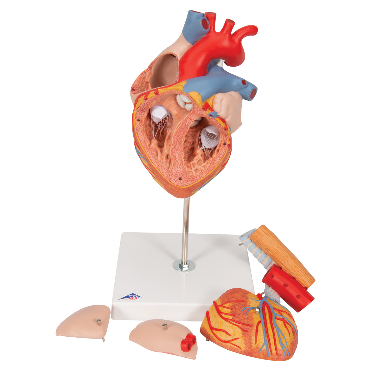
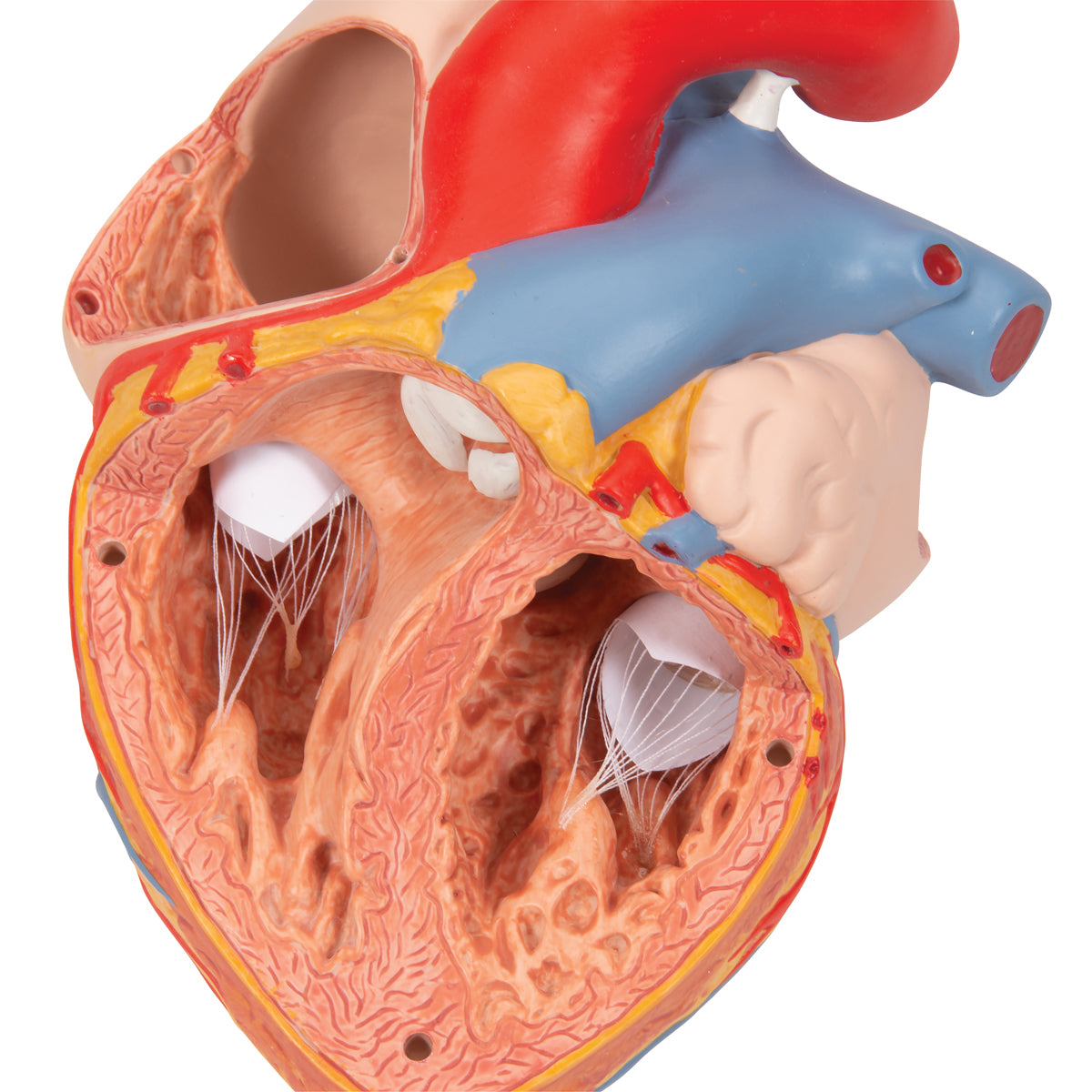
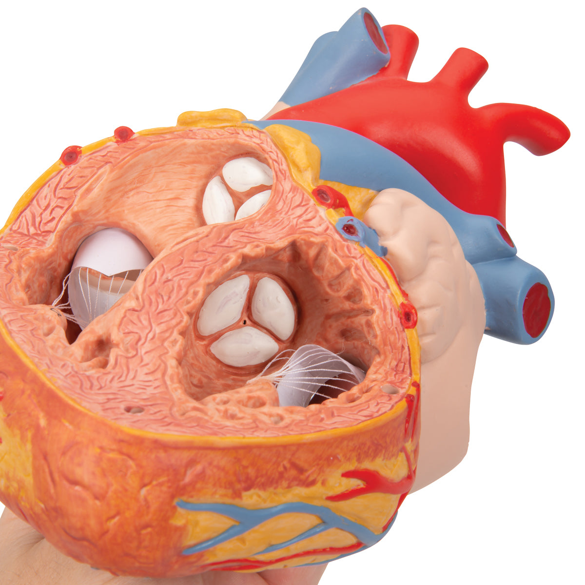
If the front of the heart model is removed, the complex muscular structure of the ventricles is clearly seen. The inner surfaces of the ventricles are made up of protruding muscle ridges (trabeculae carneae), which cross each other at different angles. This can be seen particularly clearly on the heart model.


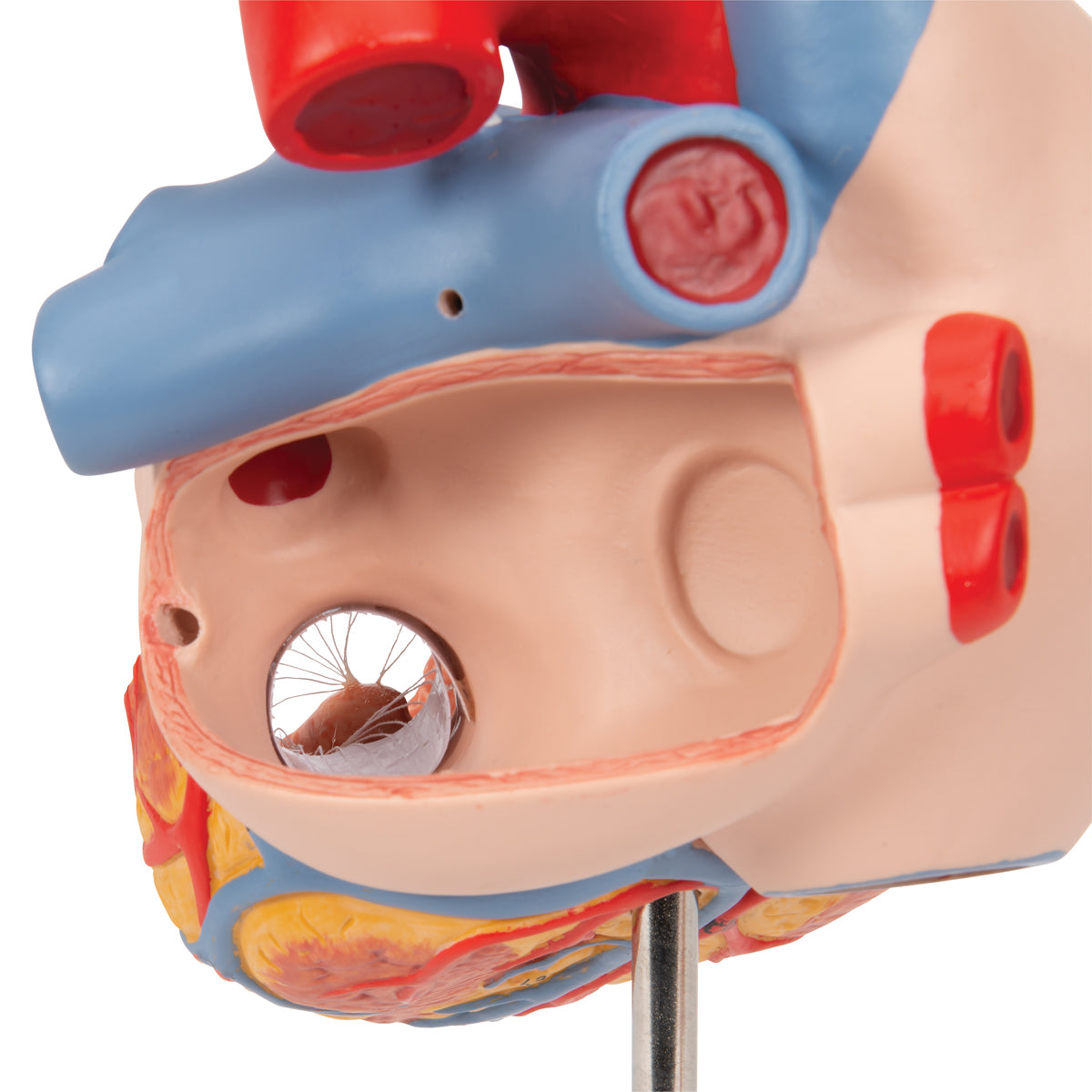
The 2 AV valves (between atria and ventricles) are tied to papillary muscles in the ventricles, which is also very clear on the model.


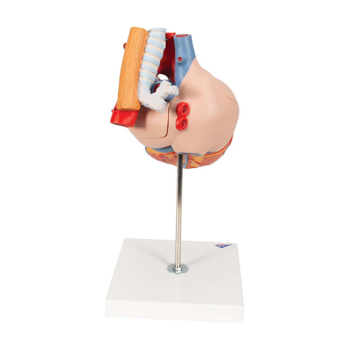
Furthermore, a part of the oesophagus, a large part of the trachea with the characteristic horseshoe-shaped rings of cartilage and the beginning of the main bronchi can be seen.

Product flexibility

Clinical features

Clinically speaking, the model does not show anything pathological (disease-wise), but it can be used to shed light on many heart diseases such as valvular diseases, inflammation, atherosclerosis and blood clots.


The model is also ideal for elucidating diseases in the anatomical region between the heart, trachea and esophagus. Finally, the model can also be used to explain ultrasound examination of the heart via the esophagus (called TEE).

NB: Read any also about our similar heart model which is also enlarged and hand painted. It costs a little less because it does not include the trachea and esophagus

View full details

A safe deal

For 19 years I have been at the head of eAnatomi and sold anatomical models and posters to 'almost' everyone who has anything to do with anatomy in Denmark and abroad. When you shop at eAnatomi, you shop with me and I personally guarantee a safe deal.

Christian Birksø
Owner and founder of eAnatomi ApS

Read more about eAnatomi