SKU:EA1-A574
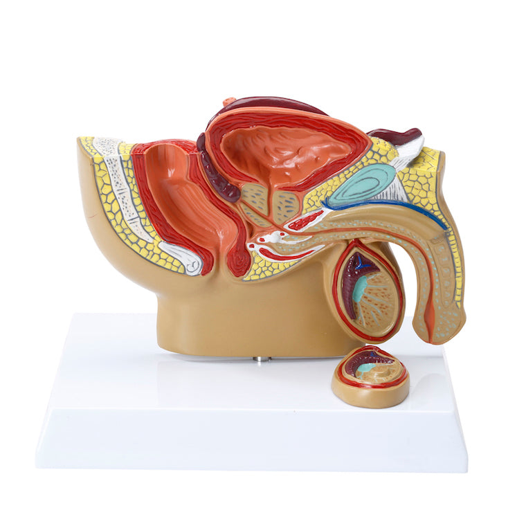
Miniature model of the male internal and external genitalia seen in a median section
Miniature model of the male internal and external genitalia seen in a median section
25 in stock
Couldn't load pickup availability
If you are looking for a model of the male genitalia in a particularly practical size, which is at once small, elegant, suitably detailed with the internal structure of the testicles and sold at an attractive price, we would highly recommend this model. It is also popular with our customers.
The model shows the man's genitals and the relationships to other organs with a total reduction of just over 50% compared to an adult man.
It measures 15 x 10 cm and therefore does not require much space on, for example, a desk or a bookcase. The model cannot be taken apart and is delivered on a stand.
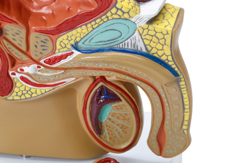
NB: The stand also shows a fixed part of a testicle with a tumor.
Anatomical features
Anatomical features
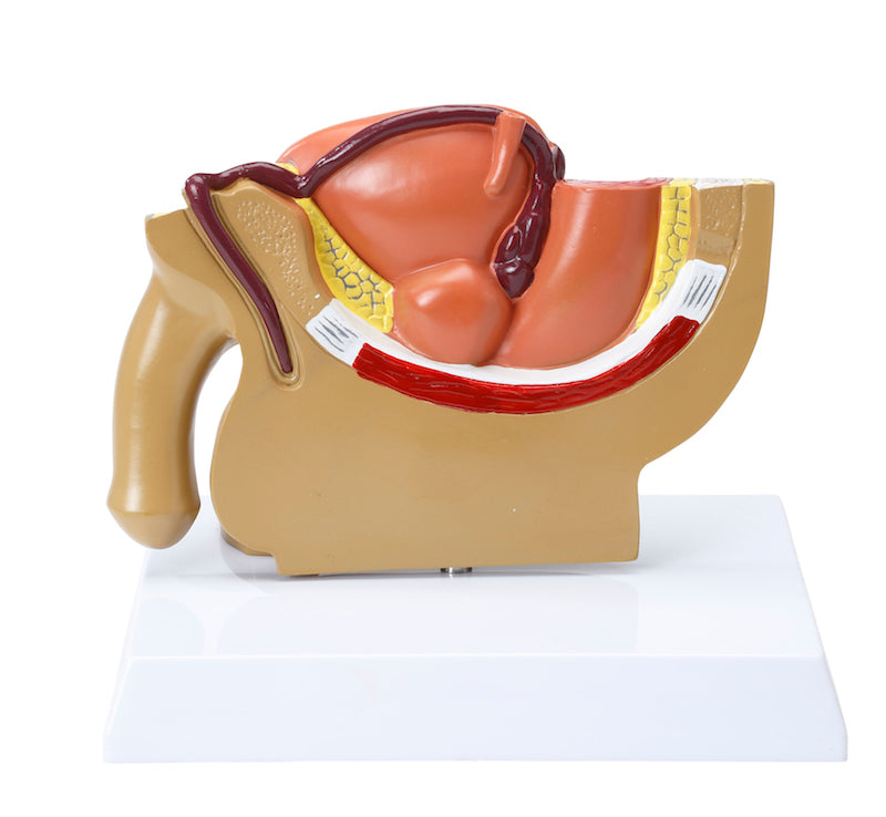
Anatomically, the model shows several different tissues and organs in the left side of the pelvis via a median section. Furthermore, the surface of some of the organs can be seen on the back of the model (see the pictures on the left).
The pelvis (pelvic ring), which consists of bones and the pelvic floor, can only be seen on the model. For example, a cross-section of the symphysis (the front joint) is seen in the pelvic ring. On the other side, some of the rectum (rectum) and urinary bladder (vesica urinaria) can be seen in detail.
The internal genitalia include the testicles (testes) and epididymides, which are seen in relation to the scrotum. The spermatic cord (funicle) can only be sensed with a really good will. In contrast, the internal structures of the testicles are seen, where the seminiferous tubules contorti and the septula testis can be seen.
The stand also shows a fixed part of a testicle with a clear tumor.
The seminal ducts include the ductus deferens (the spermatic cord), the ductus ejaculatorius and the urethra masculinum (the urethra), all of which are visible.
In relation to the accessory gonads, the prostate (bladder neck gland), vesiculae seminales (seminal vesicles) and etc. are seen. bulbourethrales. Gll. urethrales, on the other hand, are not seen. The export aisles and their connection are only partially seen in detail.
The external genitalia include the penis and the scrotum, which are also seen. The internal structure of the penis is also seen in small details.
Many of the pelvic organs function as a channel or a reservoir (e.g. the rectum and the urinary bladder), which is why they are seen in a pedagogical way with an air-filled cavity (cavity).
Product flexibility
Product flexibility
In terms of movement, the pelvis is not flexible. The few joints that are visible are not movable.
Clinical features
Clinical features
Clinically, the model is ideal for understanding testicular cancer. The model can also be used to understand diseases and disorders in the man's pelvis - for example in the genitals, the urinary bladder or the rectum. Examples are torsio testis, urethral stricture, anal fistula, anal fissure, BPH (benign prostatic hyperplasia) and prostate cancer.
Share a link to this product


A safe deal
For 19 years I have been at the head of eAnatomi and sold anatomical models and posters to 'almost' everyone who has anything to do with anatomy in Denmark and abroad. When you shop at eAnatomi, you shop with me and I personally guarantee a safe deal.
Christian Birksø
Owner and founder of eAnatomi ApS