Skip to product information
1 of 7

SKU:EA-1000214

Model of head and neck muscles in 5 parts

Model of head and neck muscles in 5 parts

Regular price €1.207,95 EUR
Regular price Sale price €1.207,95 EUR
Sale Sold out
Not including taxes if ordering from outside EU. Shipping calculated at checkout.

Out of stock

this product is made to order. To place an order please call or write us.

With this model, you get a complete overview of the anatomy of the head and neck. The model illustrates both the superficial and deeper musculature, and also shows vessels and nerves in relation to these.


The model can be separated into 5 parts, as the top of the skull can be removed, and the brain can be removed and separated into 3 parts.


The model has the dimensions 36 x 18 x 18 cm (height x length x width), weighs 2 kg and is delivered on a white stand, which is also removable.

Anatomical features

Anatomically, the model can be used to gain an understanding of many different anatomical structures.


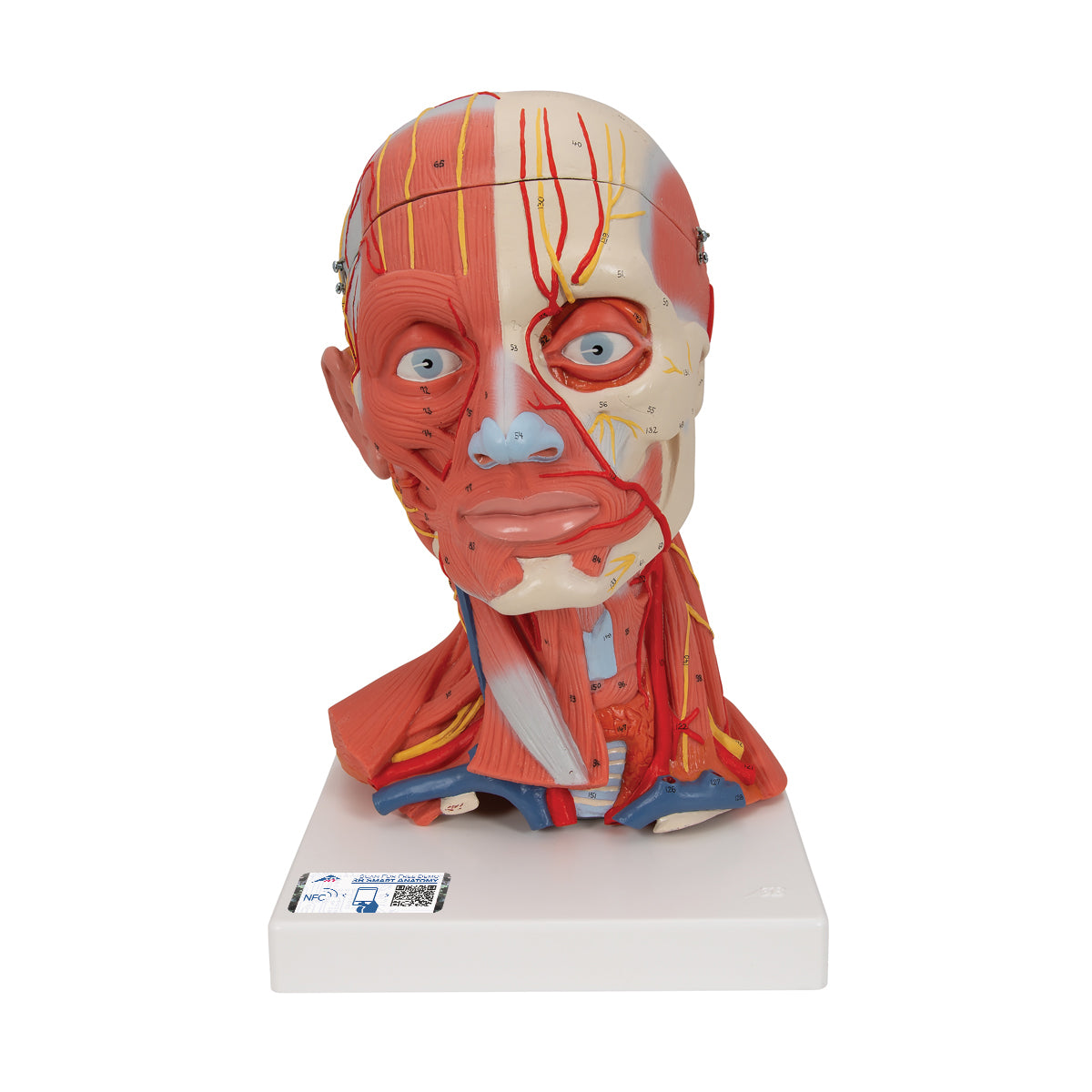
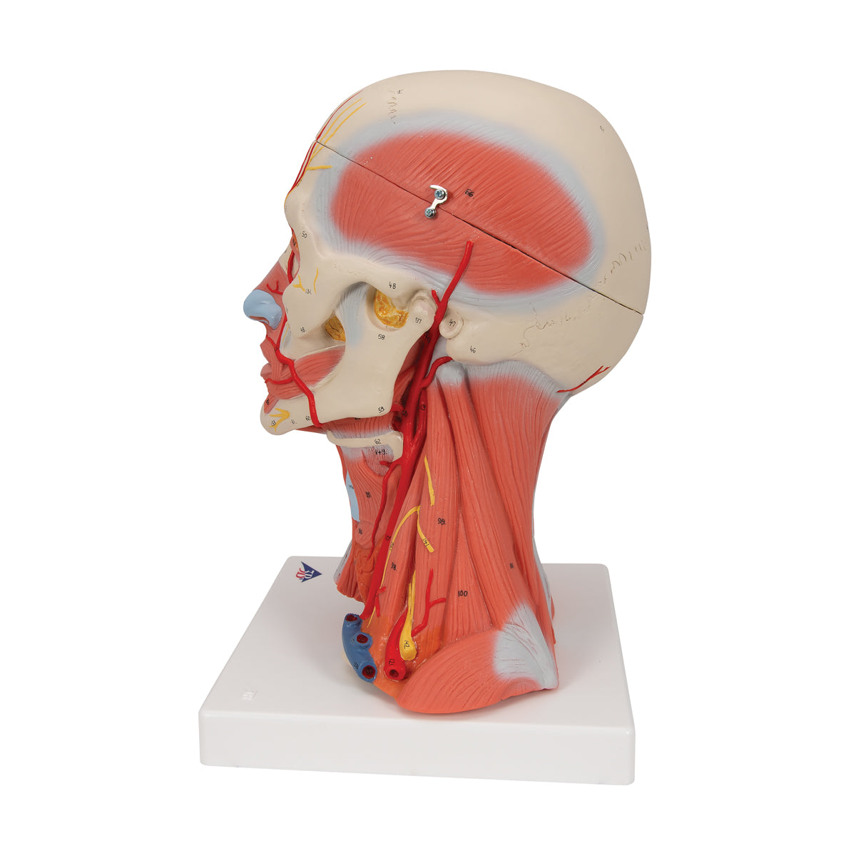
First of all, the model illustrates a large number of muscles from the head and neck muscle groups:


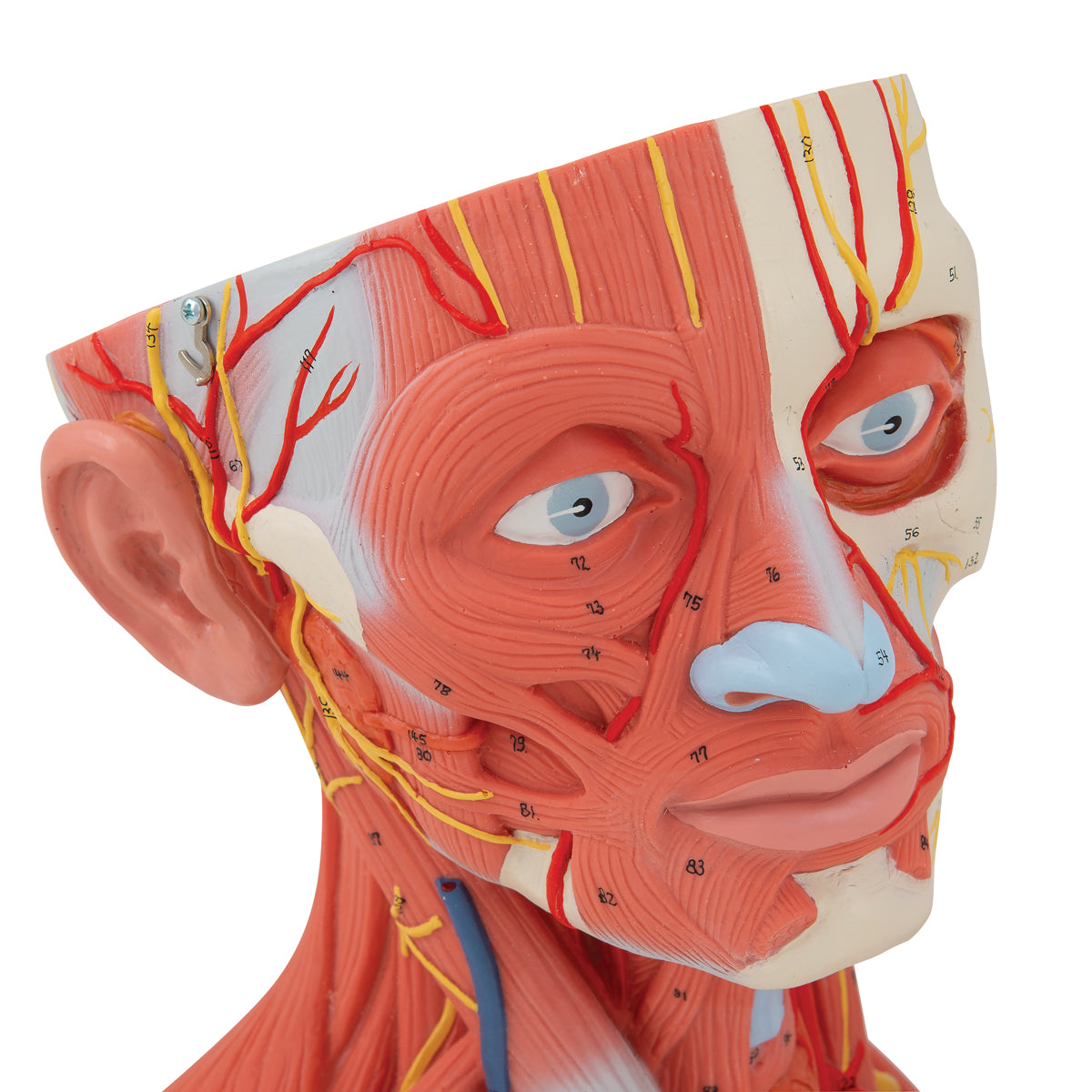
The mimic facial muscles
The muscles of mastication
The supra- and infrahyoid muscles
The superficial neck muscles
The scale group
Some back muscles
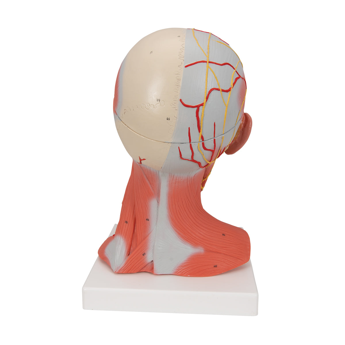
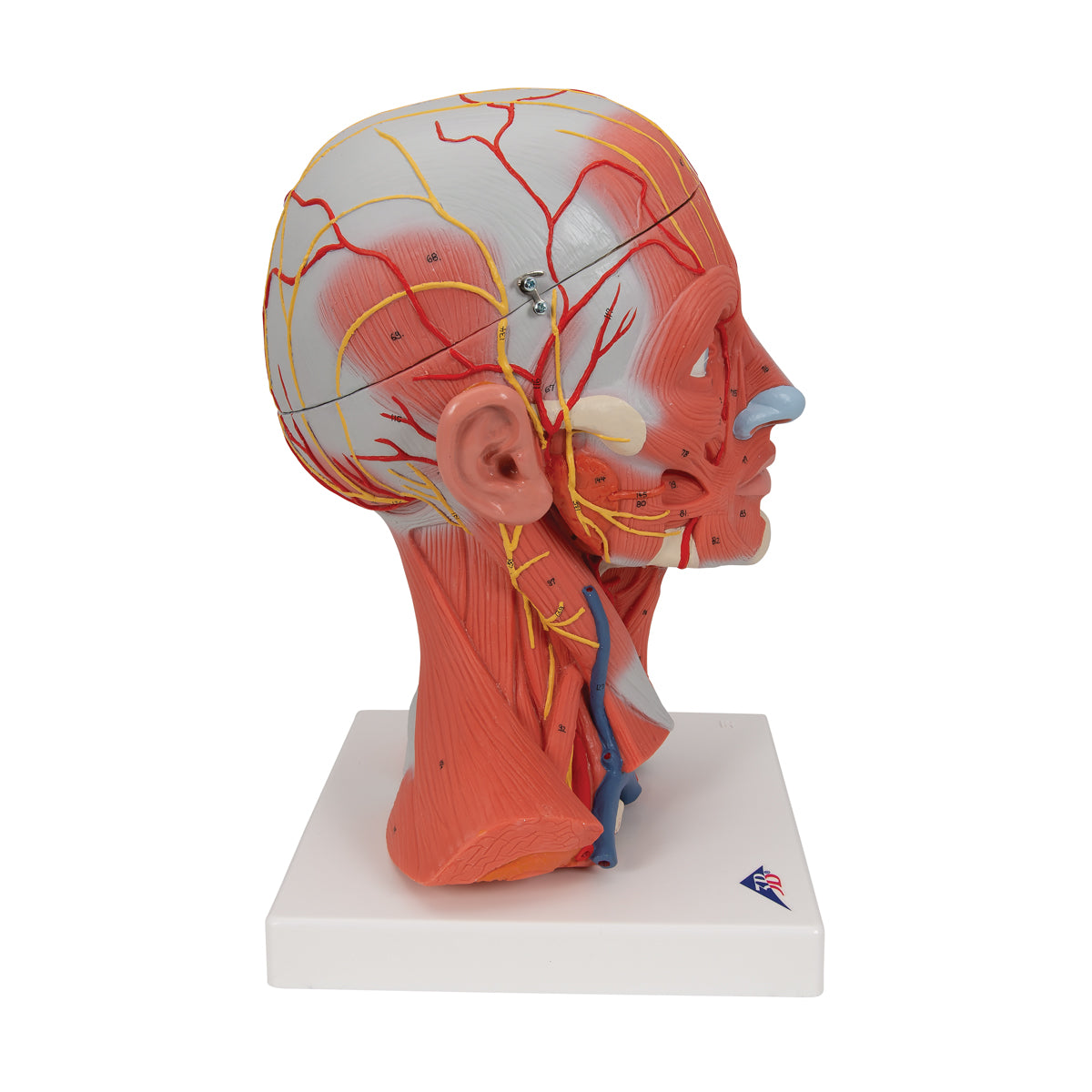
In relation to this, the largest vessels can be seen that run in relation to the muscles. In the face, only the arteries are shown, while the largest veins on the neck are included.


The model depicts all 12 cranial nerves and in addition the brachial plexus and nerves that depart from the cervical plexus can be seen.


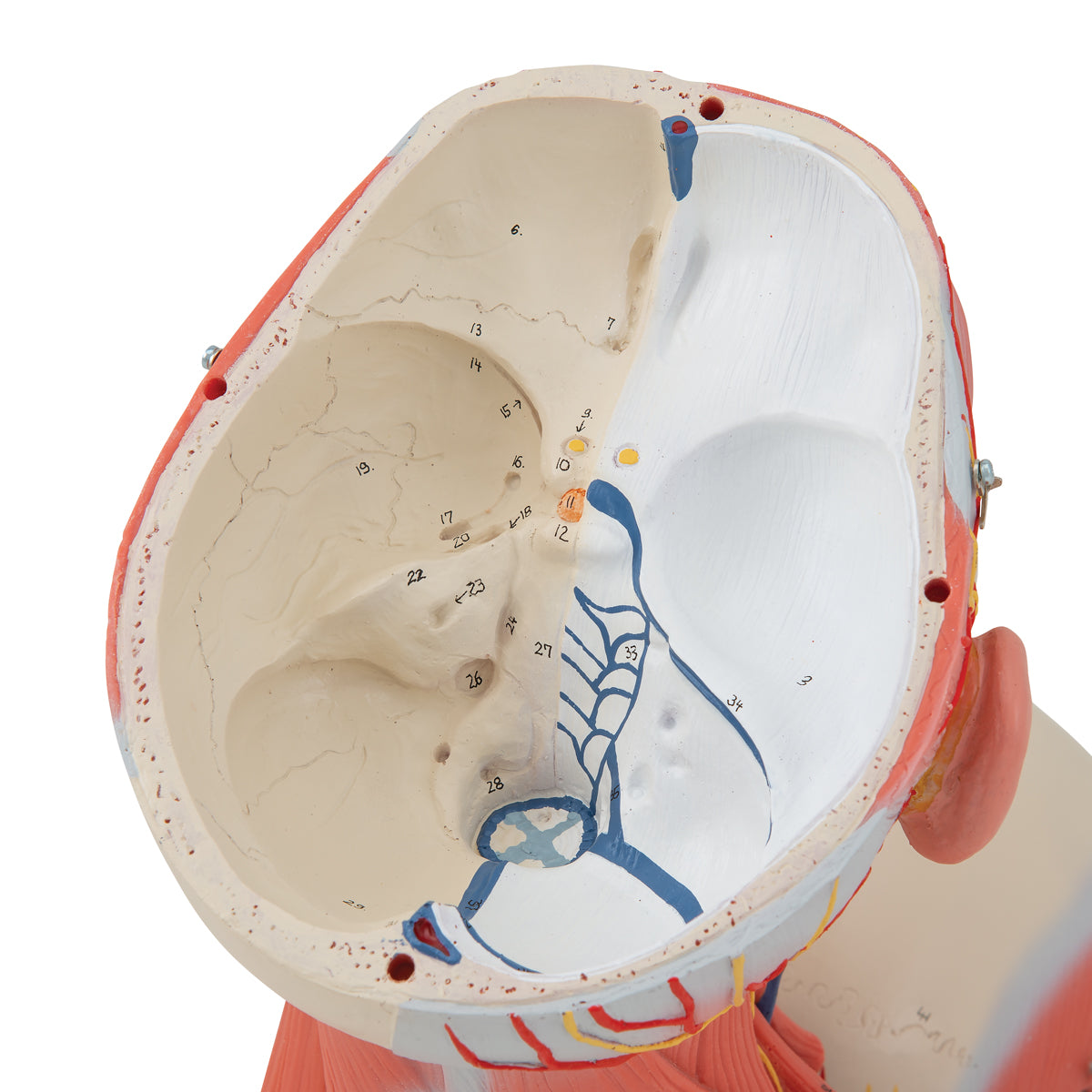
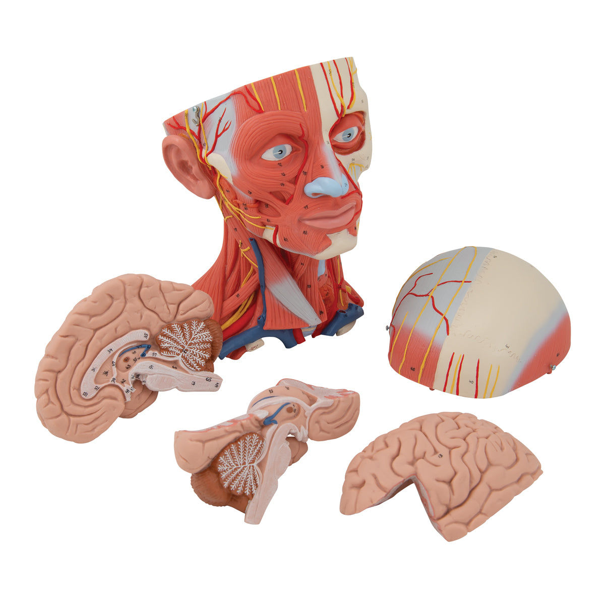
When the top of the skull is removed and the brain is removed, the inside of both the top and bottom of the skull is visible. When the brain is separated into 3 parts, the following structures can be seen, among other things:


telencephalon
cerebellum
The midbrain (diencephalon - with, among other things, the thalamus, hypothalamus, epithalamus and corpus mamillare)
The brainstem
The ventricular system

Product flexibility

Clinical features

Clinically speaking, the model can be used to understand disease problems related to the muscles, vessels, nerves and surrounding structures of the head.


For example, facial paresis, formation of salivary stones in the salivary glands (carotid stenosis), and disorders related to the jaw muscles and jaw joint such as temporomandibular dysfunction.


Furthermore, the model can be used to understand lesions and disorders in specific areas of the brain. Examples are epilepsy, brain tumors, hydrocephalus, lesions involving cranial nerves and sclerosis (multiple sclerosis).

View full details

A safe deal

For 19 years I have been at the head of eAnatomi and sold anatomical models and posters to 'almost' everyone who has anything to do with anatomy in Denmark and abroad. When you shop at eAnatomi, you shop with me and I personally guarantee a safe deal.

Christian Birksø
Owner and founder of eAnatomi ApS

Read more about eAnatomi