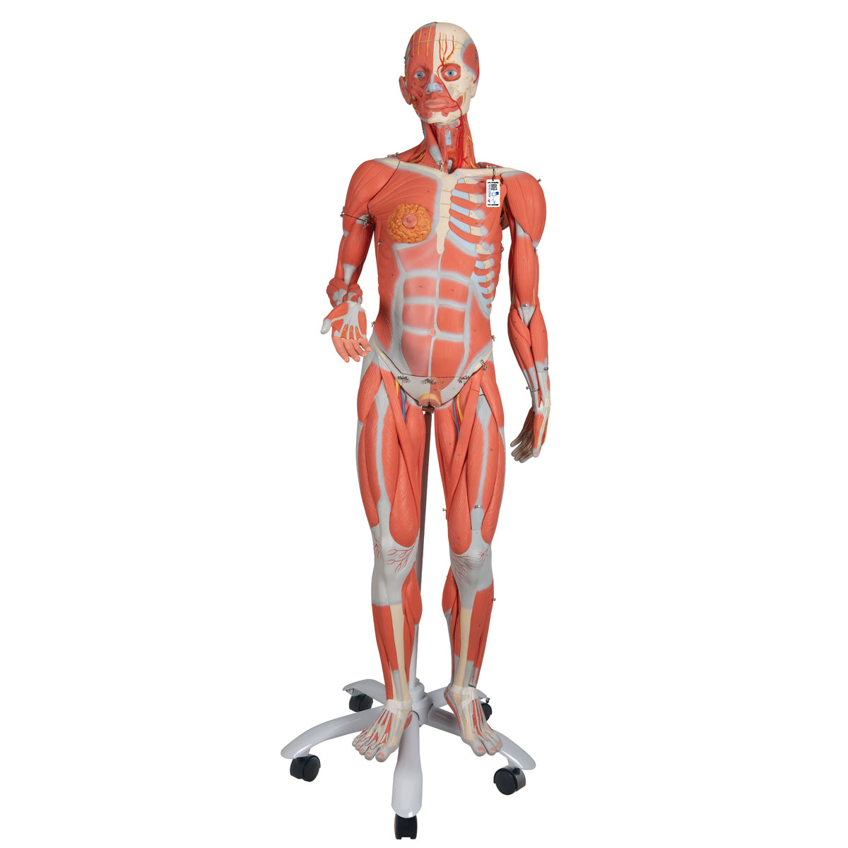
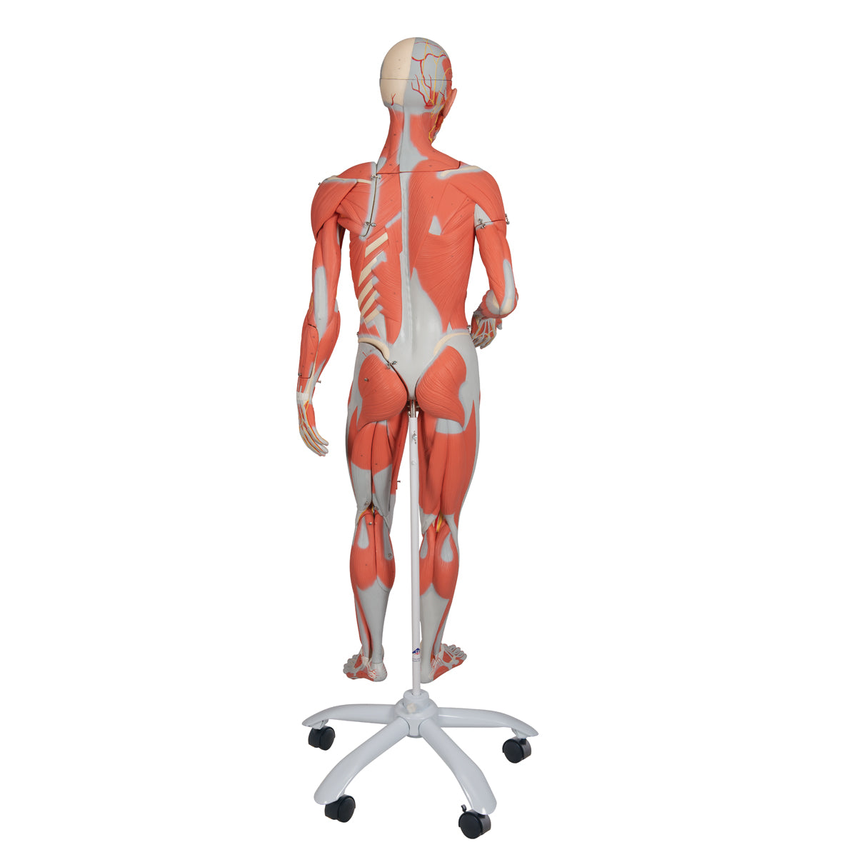
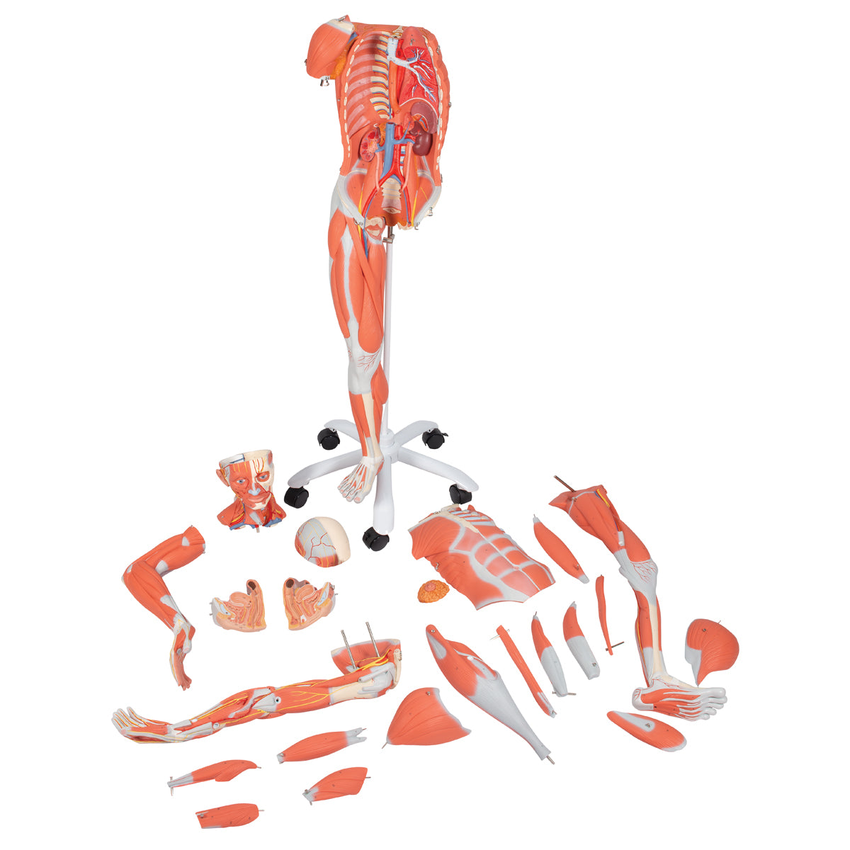
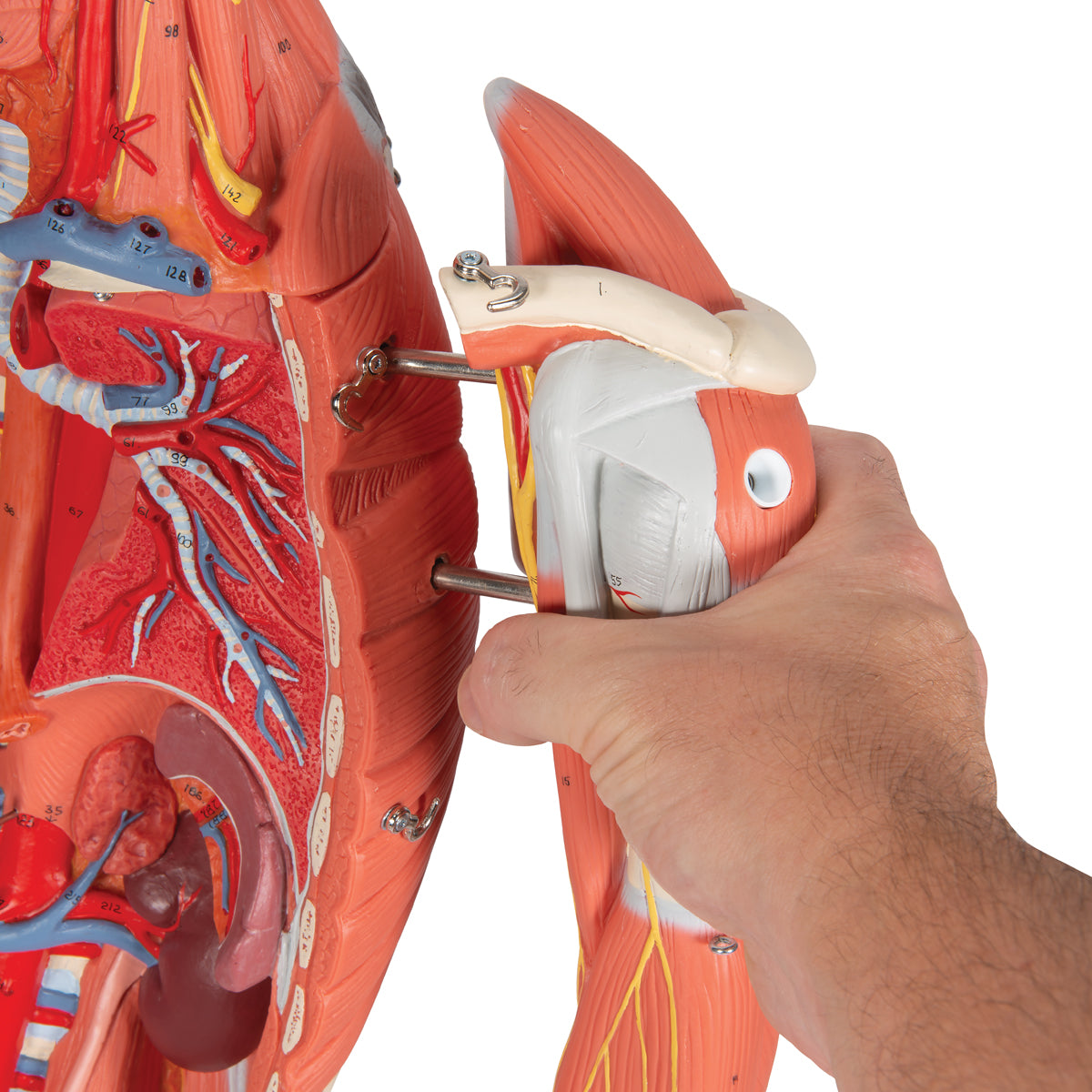
Anatomically speaking, the model focuses on illustrating the skeletal muscles. For this purpose, both arms as well as the left leg can be removed and studied in greater detail.
The following muscles can be removed from the right arm:
M. deltoideus (part of the shoulder muscles)
M. brachioradialis, M. extensor carpi radialis longus and brevis (all part of the radial extensor log)
M. biceps brachii
M. pronator teres, M. flexor carpi radialis, M. palmaris longus, M. flexor carpi ulnaris (the superficial muscles of the extensor tendon of the forearm)
M. extensor digitorum, M. extensor digiti minimi, M. extensor carpi ulnaris, M. anconeus (the superficial muscles of the extensor log of the forearm)
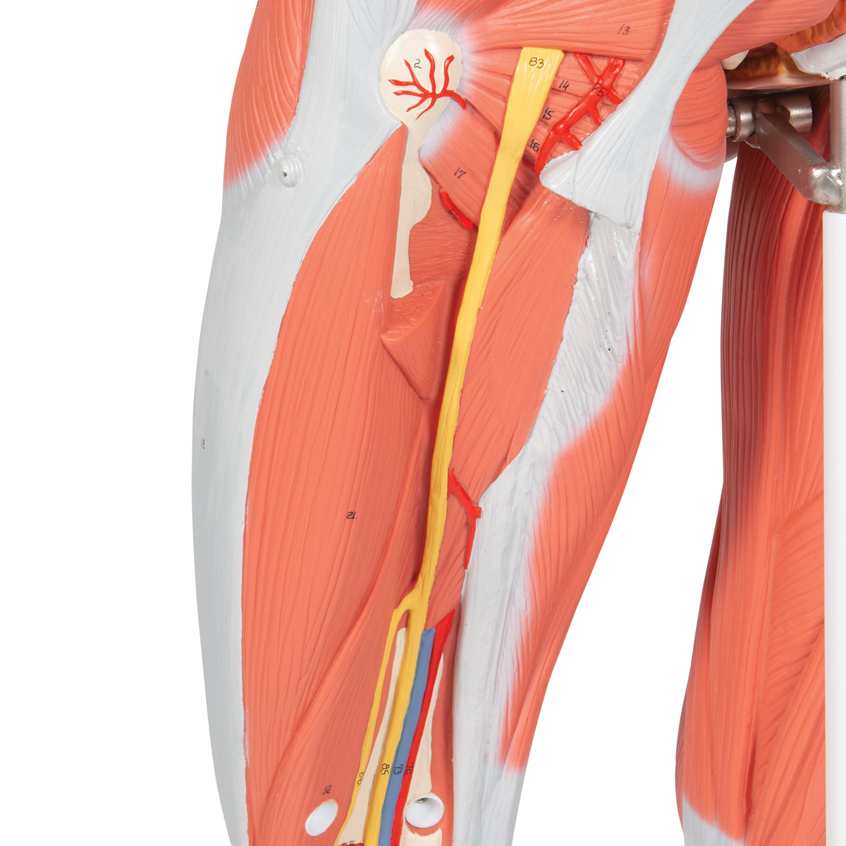
The following muscles can be removed from the right leg:
M. gluteus maximus (one of the gluteal muscles)
M. biceps femoris, M. semitendinosus and M. semimembranosus (back muscle group of the thigh)
M. soleus and M. gastrocnemius (the superficial muscles of the flexor joint of the lower leg)
M. sartorius and rectus femoris (part of the front muscle group of the thigh)
M. extensor digitorum longus, M. fibularis tertius and M. extensor hallucis longus (from the extensor log of the lower leg)
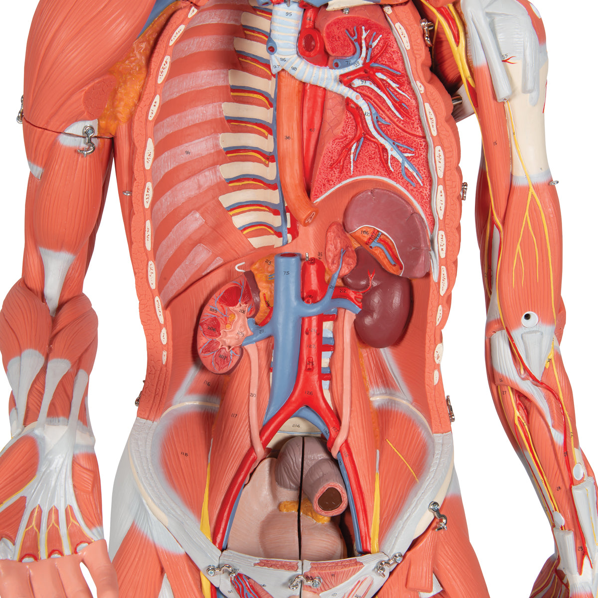
When the chest/abdominal wall is removed, individual organs are seen on the back of the chest/abdominal wall:
Left lung - seen in a frontal section, so that the branches of the bronchial tree and the accompanying A. and V. pulmonalis can be followed into the lung tissue
The esophagus - can be followed down into the abdominal cavity
Both kidneys as well as adrenal glands and ureters - the right kidney is seen in a frontal section, so that the structure of the organ is illustrated
The spleen
Aorta and inferior vena cava
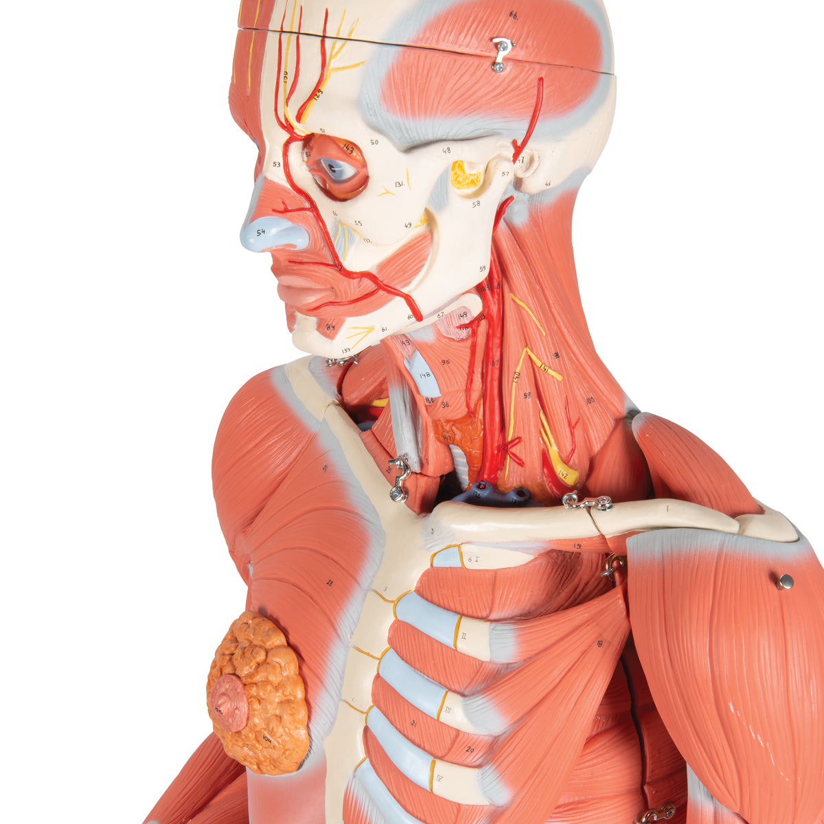
In addition, the model shows the largest vessels and nerves that ran in relation to the remaining structures of the model. The mammary gland on the left side is also depicted.