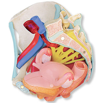
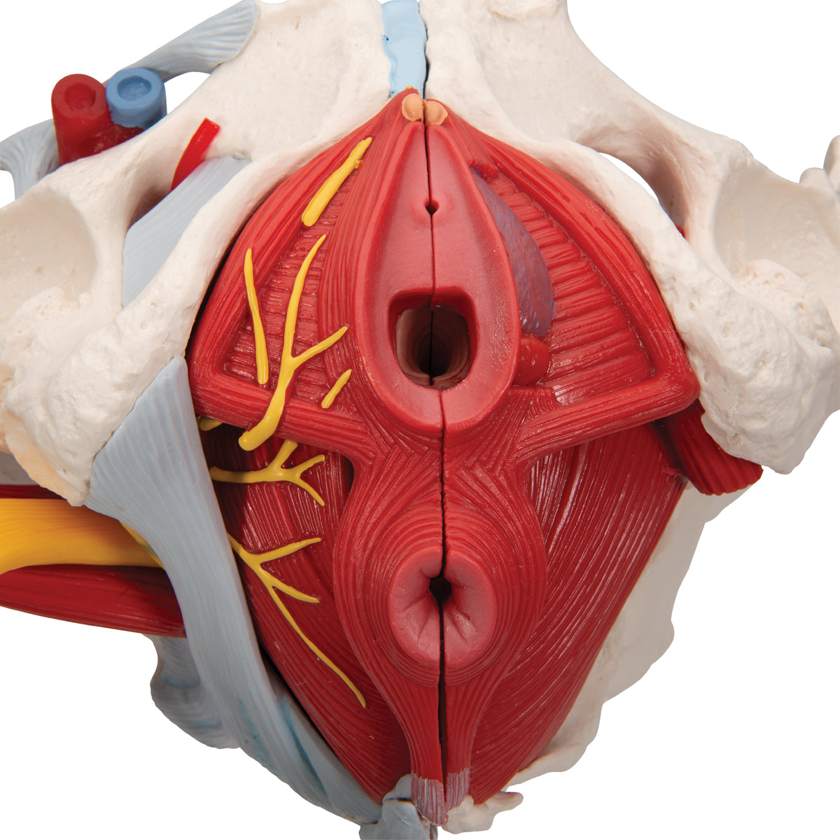
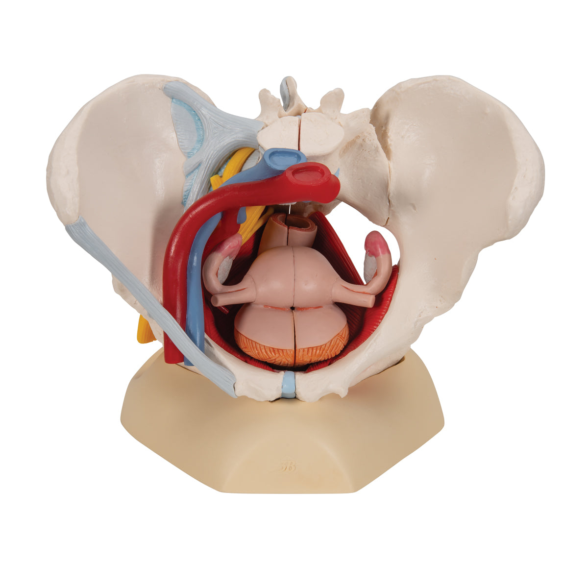
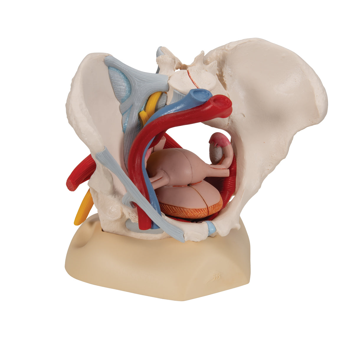
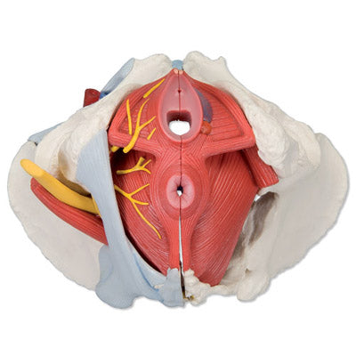
Anatomically, the model shows many different tissues. It shows both the woman's pelvis, the muscles in the pelvic floor with the relationships to the urinary bladder with the urethra and the rectum, the internal genitalia, some of the external genitalia as well as ligaments, nerves and blood vessels on its right side.
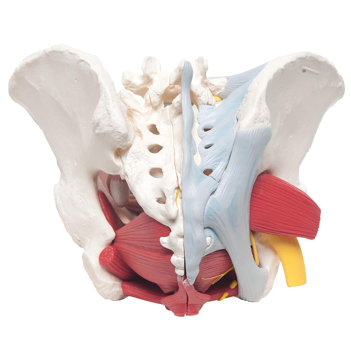
The pelvis consists of the 3 building blocks, which are the 2 hip bones and the sacrum. The model also shows the lower lumbar vertebra. In the bone tissue, the most important details can be seen, such as large nodules. Smaller details such as the linea glutaea are omitted. Overall, the model shows the following bones:
Os coxae (the 2 hip bones) of which both are composed
- Os ilium (iliac bone/hip bone)
- Os ischii (seat bone)
- Os pubis (pubic bone)
Us sacrum (sacrum) incl. os coccygis (coccyx)
5th lumbar vertebra (lumbar vertebra)
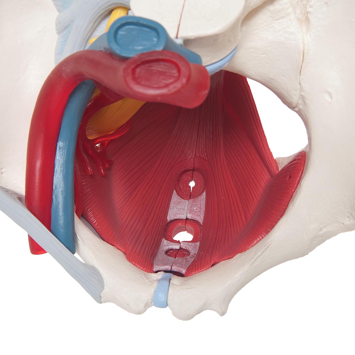
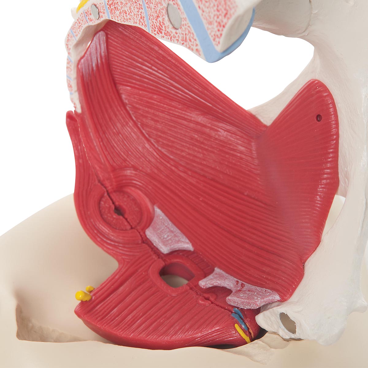
The pelvic floor (also called the pelvic floor or diaphragma pelvis in Latin) consists of the following muscles, which are clearly visible:
- M. levator ani (consisting of m. pubococcygeus and m. iliococcygeus)
- M. coccygeus
Other muscles are also seen such as the ischiocavernosus muscle, the bulbospongiosus muscle, the obturatorius internus muscle and etc. sphincter ani externus and internus.
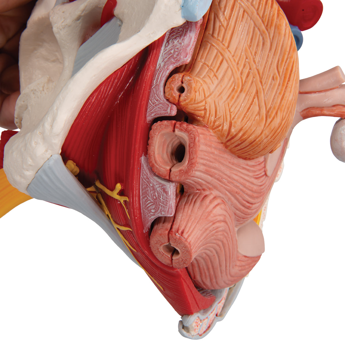
The internal genitalia include the ovaries (ovaries), the fallopian tubes (tubae uterinae), the uterus (uterus) and the vagina (vagina), all of which are seen in detail. Anatomically, it is located in the small pelvis.
The external genitalia are shown with the largest and most important details, which anatomically are located in front of and below the symphysis.
The genitals are seen in relation to the urinary bladder (vesica urinaria) with the urethra (urethra) and the rectum (rectum). Many of all these organs function as a channel or a reservoir (eg the vagina and the urinary bladder), which is why they are seen in a pedagogical way with an air-filled cavity (cavity).
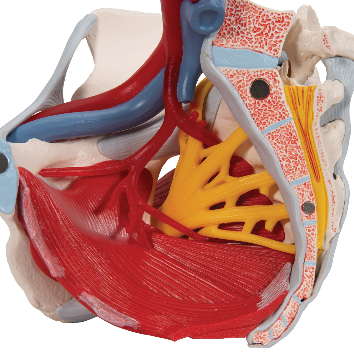
Ligaments, nerves and blood vessels are seen on the right side of the model.
Since the model i.a. can be divided in half, important details can be seen in a median section (including cauda equina). As for nerves, the sacral plexus, sciatic nerve (nervus ischiadicus) and other important nerves are also seen. As for blood vessels, the large artery and vein of the pelvis and parts of their branches are seen.