SKU:EA1-A552
Skin model of one skin area
Skin model of one skin area
10 in stock
Couldn't load pickup availability
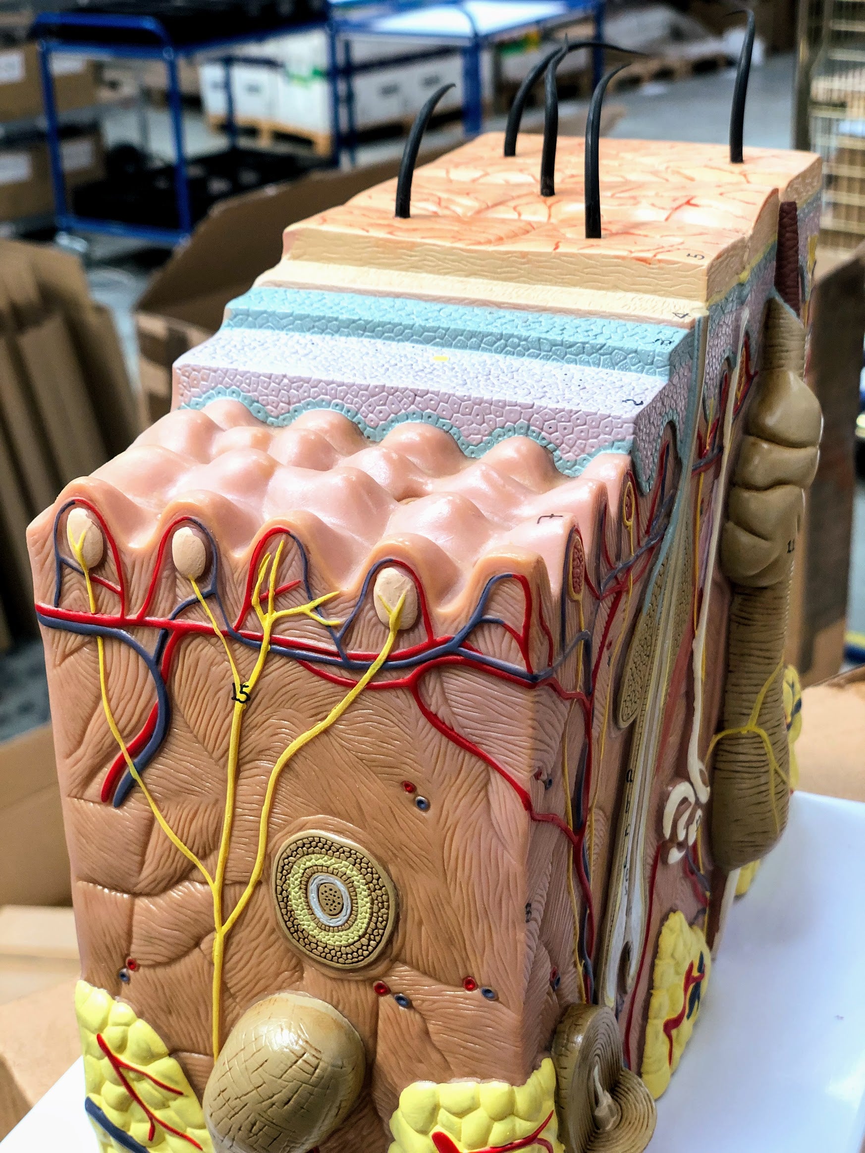
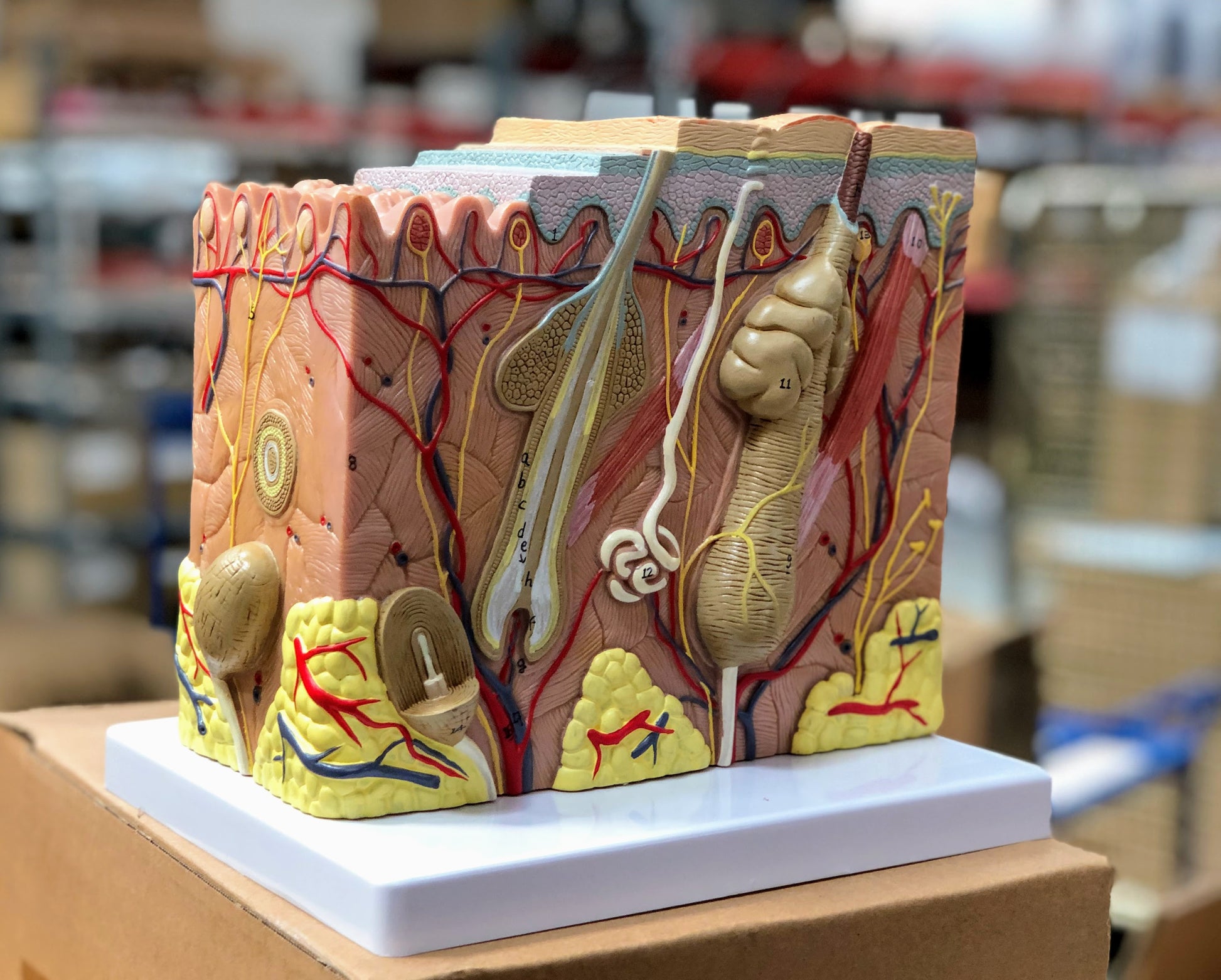
If you are looking for a skin model that shows all the most important details in the layers of the skin, we would highly recommend this one.
The model shows the skin at 70 times normal size. It measures 11 x 20 x 21 cm and is delivered without a stand.
Anatomical features
Anatomical features
Anatomically, the model shows some hair and the skin's two layers, the epidermis and the dermis. The skin (subcutis) is also seen.
The epidermis consists primarily of keratinocytes, and the model clearly shows the layered structure, which is caused by the displacement of these cells upwards towards the skin surface. This is particularly shown using different colors. Furthermore, the basement membrane can be seen at the bottom.
The thick connective tissue layer with blood vessels, hair follicles with attached smooth muscle (m. arrector pili) and sweat and sebaceous glands can be seen in the dermis.
Some of the subcutaneous tissue is also shown in yellow at the bottom, which symbolizes fat cells (since the subcutaneous tissue mainly consists of fat cells).
Product flexibility
Product flexibility
Clinical features
Clinical features
Clinically, the model can be used to understand skin disorders and skin diseases such as psoriasis, folliculitis, acne and skin tumors.
It can also be used to understand other things, such as burns, which are traditionally divided into degrees (1st degree, 2nd degree, etc.).
Share a link to this product


A safe deal
For 19 years I have been at the head of eAnatomi and sold anatomical models and posters to 'almost' everyone who has anything to do with anatomy in Denmark and abroad. When you shop at eAnatomi, you shop with me and I personally guarantee a safe deal.
Christian Birksø
Owner and founder of eAnatomi ApS


