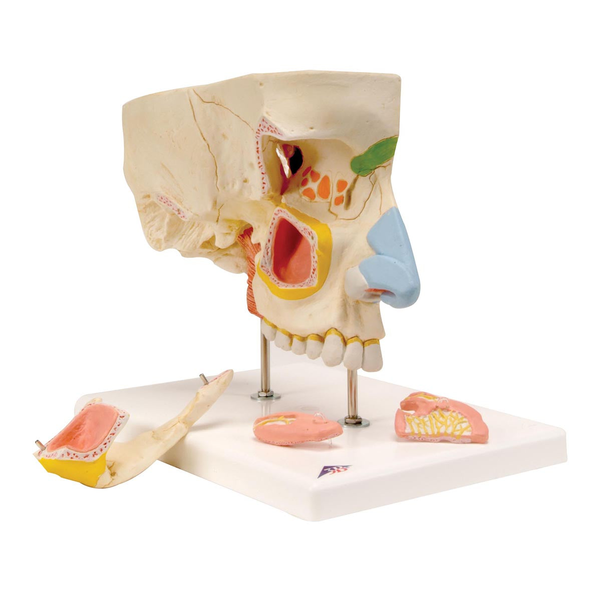
Anatomically, the model shows several structures on the right side of the facial skeleton and a bit of the braincase. The model can be separated into 5 parts, because the following 4 parts can be removed:
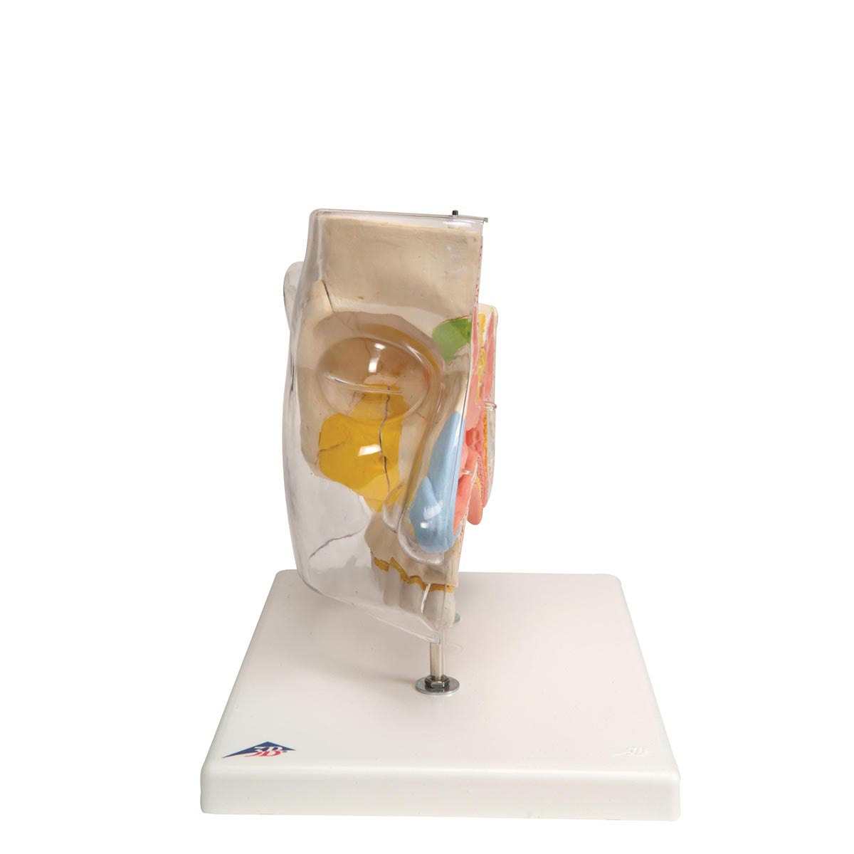
The transparent skin which i.a. covers the upper jaw and cheek
The cheekbone (os zygomaticum) with the arch of the cheek (arcus zygomaticus)
Concha nasalis inferior (the lower concha bone) incl. some nerves
Concha nasalis media (the middle concha bone) incl. some nerves
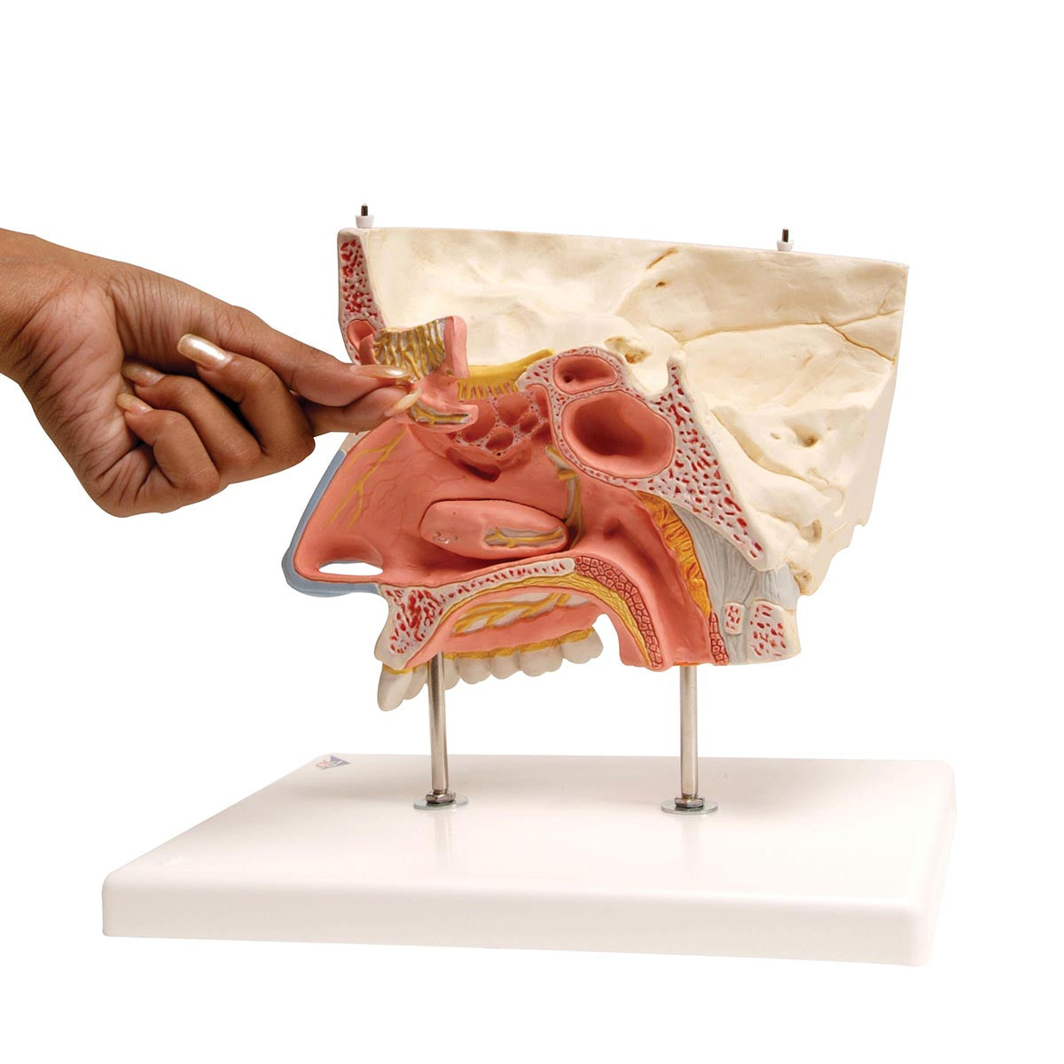
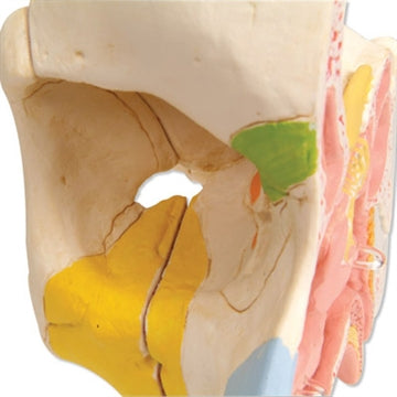
The model shows an outside and an inside. On the outside, several bones can be seen such as the frontal bone, the temporal bone, the zygomatic bone, the lacrimal bone, and the nasal cartilage. On the inside, you can see primarily the nasal cavity with clam bones, a bit of the oral cavity, a number of nerves (with the sense of smell), a bit of the pharynx, a bit of the upper cervical vertebra (atlas) and ligaments in this region.
The 4 sinuses are shown in a particularly educational manner. They are seen both on the inside and outside (but not the sinus frontalis/jaw cavity which is only seen on the outside). The sinuses are shown as follows:
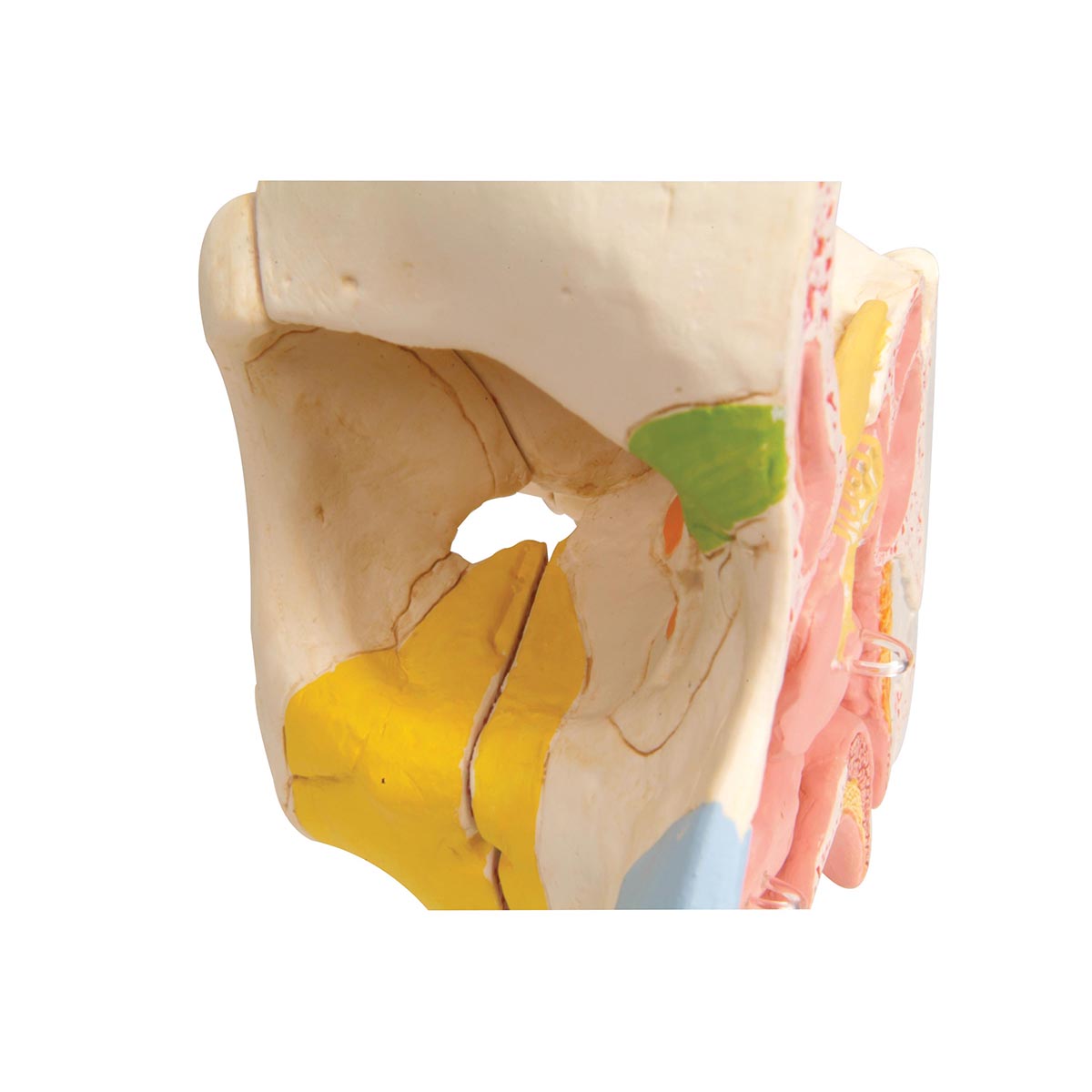
The sinus frontalis (forehead cavity) can be seen on the inside and in addition the location can be seen on the outside using green color
The sinus sphenoidalis (the sphenoid sinus) is seen on the inside and in addition the localization is seen on the outside using purple color
The maxillary sinus (jaw cavity) is only visible on the outside. If you remove the cheekbone and cheekbone, you can see the large hollow. If you don't take it off, the location can be identified using yellow color
The ethmoid sinus consists of thin-walled chambers, which can be divided into an anterior, a middle and a posterior set (also called labyrinthus ethmoidalis). On the inside of the model, you can see them all if you take out the middle clam bone. If the latter is not removed, the rear set of thin-walled chambers can still be seen. On the outside of the model, the location of all of them can be seen using orange color