Skip to product information
1 of 6

SKU:EA1-1000281

Model of the female internal and external genitalia seen in a median section

Model of the female internal and external genitalia seen in a median section

Regular price €276,95 EUR
Regular price Sale price €276,95 EUR
Sale Sold out
Not including taxes if ordering from outside EU. Shipping calculated at checkout.

Low stock: 1 left

This model shows the female pelvis in a median section. The model shows many different organs and tissues in an educational way.


It weighs approximately 1.9 kg and the dimensions are 41 x 31 x 20 cm. A continuous piece of the model can be detached. The piece includes, among other things part of the vagina, uterus, rectum and bladder. Therefore, you can see both their outside and their inside in a median section (see the pictures on the left). The model is delivered on a stand. Overall, the whole thing has been developed to be able to hang on a wall or similar.

Anatomical features

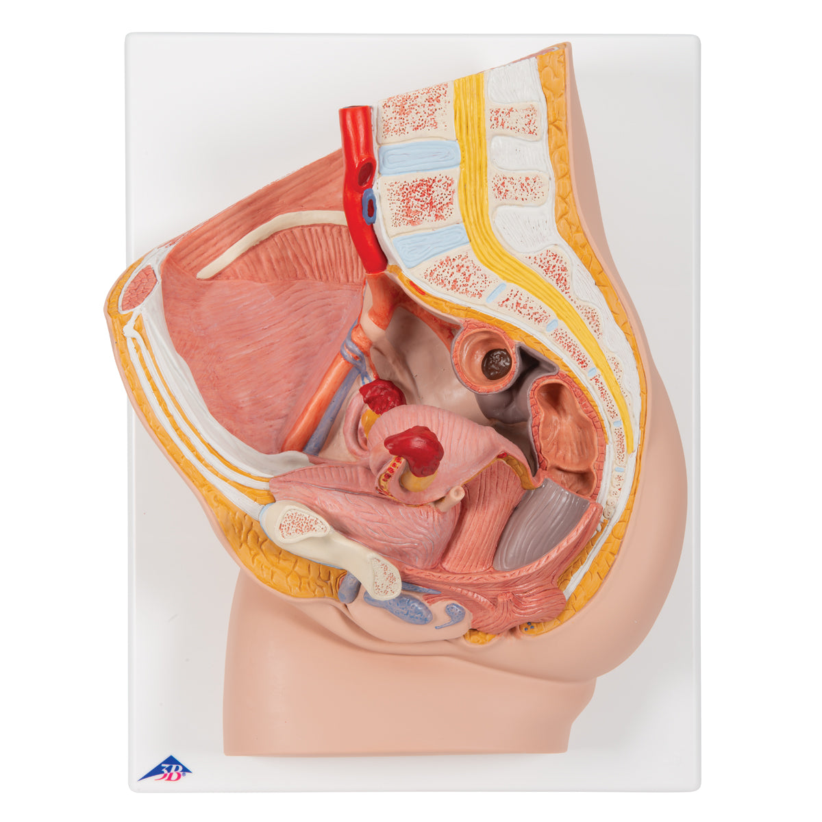
Anatomically, the model shows many different tissues and organs, some of which can be seen in 3 dimensions.


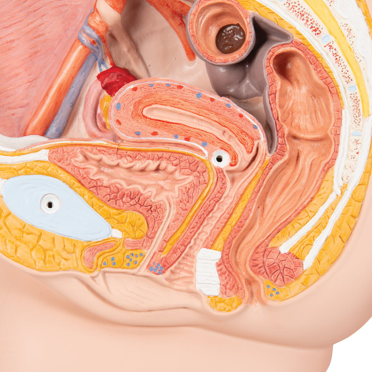
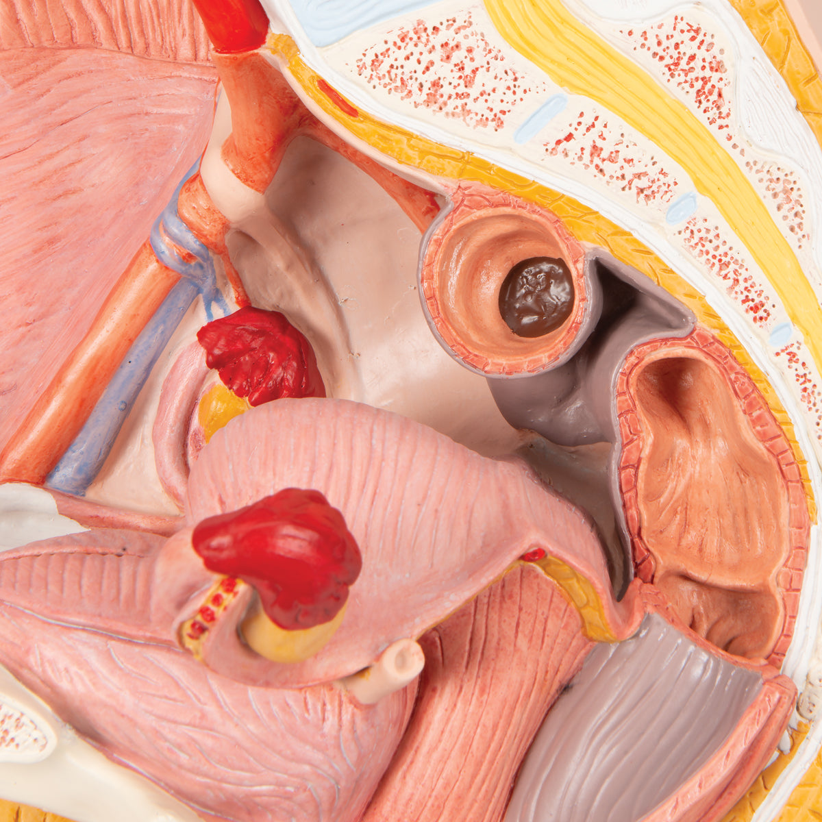
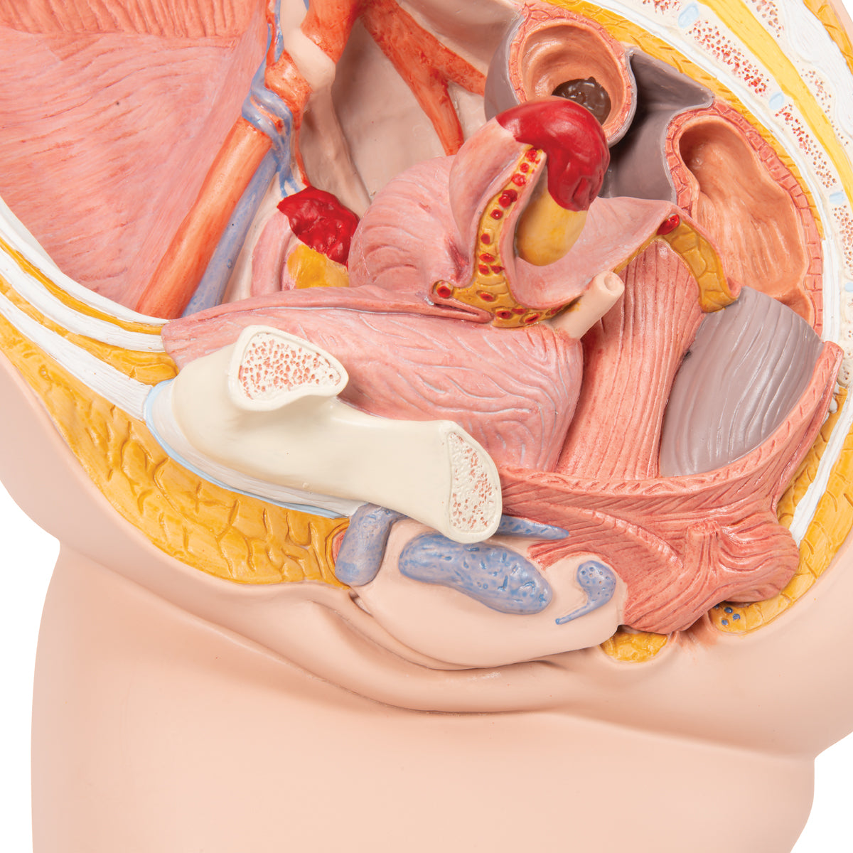
The pelvis (pelvic ring), which consists of bones and the pelvic floor, can only be seen on the model. For example, a cross-section of the symphysis (the front joint) is seen in the pelvic ring. On the other side, some of the large intestine (colon sigmoideum), the rectum (rectum), one ureter (ureter), the urinary bladder (vesica urinaria) with the urethra (urethra), the large artery and vein of the pelvis and parts of their branches can be seen. Ligaments are largely invisible, while nerves are not visible at all (however, the lower part of the spinal cord is visible).


The median section allows the organs to be seen in detail. In addition, the regions where the excavatio vesicouterina and excavatio rectouterina are formed are seen. On the model, however, you cannot see that they are formed by the peritoneum (peritoneum).


Some tissues/organs are also seen in the horizontal plane (also called the transverse plane) when viewing the model "from above". Here you can see i.a. back and abdominal muscles as well as the kidney tissue in the right kidney.


The internal genitalia include the ovaries (ovaries), the fallopian tubes (tubae uterinae), the uterus (uterus) and the vagina (vagina), all of which are seen in detail. Anatomically, it is located in the small pelvis.


The external genitalia are shown with the largest and most important details, which anatomically are located in front of and below the symphysis.


Many of the pelvic organs function as a channel or a reservoir (e.g. the rectum and the urinary bladder), which is why they are seen in a pedagogical way with an air-filled cavity (cavity).

Product flexibility

In terms of movement, the pelvis is not flexible. The few joints that are visible are not movable.

Clinical features

Clinically speaking, the model can be used to understand diseases and disorders in the woman's pelvis - for example in the genitals, the urinary bladder or the rectum. Examples are anal fistula, anal fissure, urinary tract infection and tumors.


Furthermore, the model can be used to understand disorders and diseases such as stones in the ureter and deep vein thrombosis (DVT) in the pelvis.

View full details

A safe deal

For 19 years I have been at the head of eAnatomi and sold anatomical models and posters to 'almost' everyone who has anything to do with anatomy in Denmark and abroad. When you shop at eAnatomi, you shop with me and I personally guarantee a safe deal.

Christian Birksø
Owner and founder of eAnatomi ApS

Read more about eAnatomi