Skip to product information
1 of 7

SKU:EA1-1000251

Ear model 3 times enlarged - can be separated into 5 parts

Ear model 3 times enlarged - can be separated into 5 parts

Regular price €231,95 EUR
Regular price Sale price €231,95 EUR
Sale Sold out
Not including taxes if ordering from outside EU. Shipping calculated at checkout.

Out of stock

this product is made to order. To place an order please call or write us.

This model shows in a high level of detail the structure of both the outer ear, the middle ear and the inner ear. Since the model can be separated into several parts, it is possible to examine even the smallest structures, thereby gaining an understanding of the ear's complex anatomy.


The model is enlarged 3 times compared to the size of a natural ear, has the dimensions 34 x 16 x 19 cm (width x height x length) and weighs approx. 1.4 kg.


In total, the model can be separated into 5 separate parts, and it is delivered on a white plastic stand.

Anatomical features

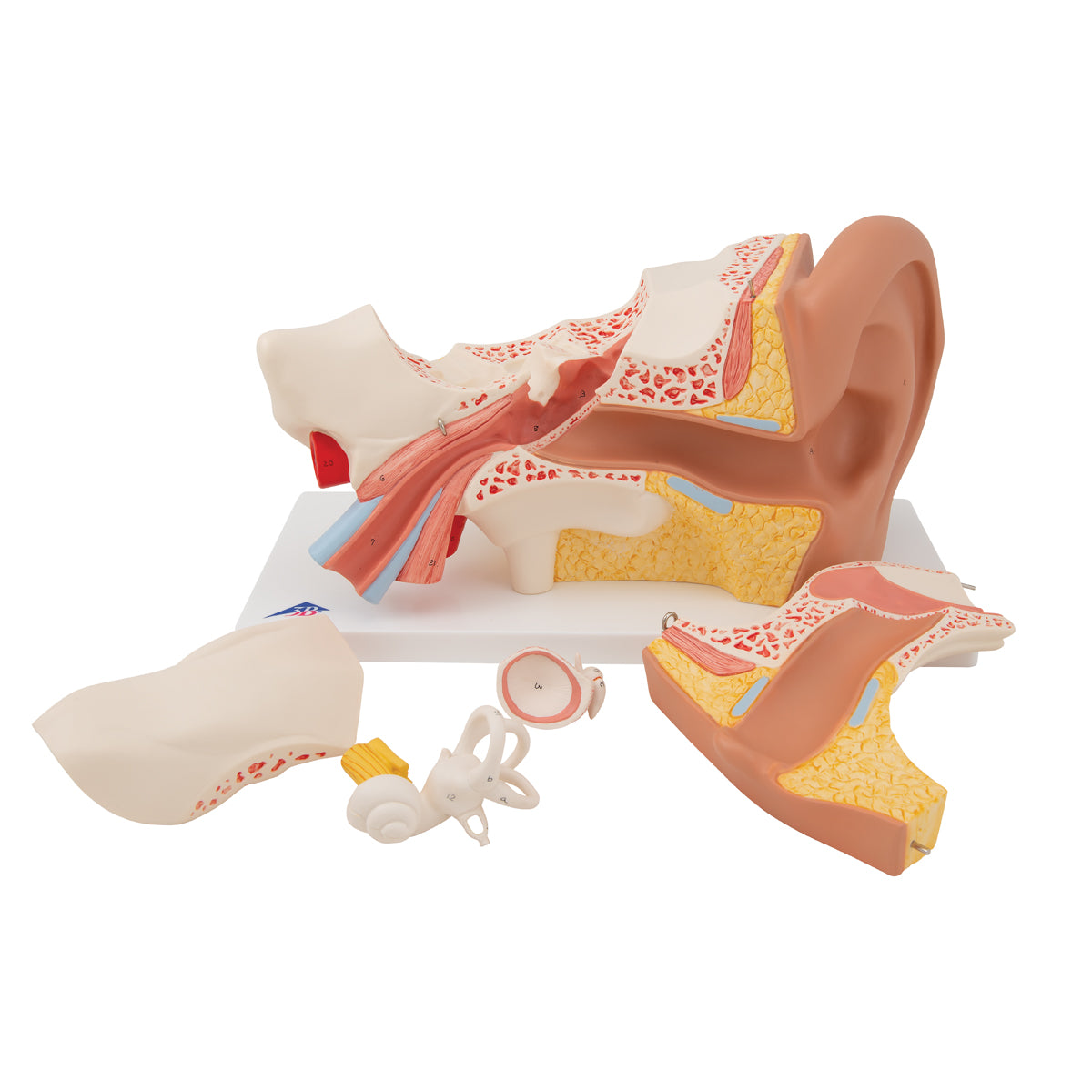
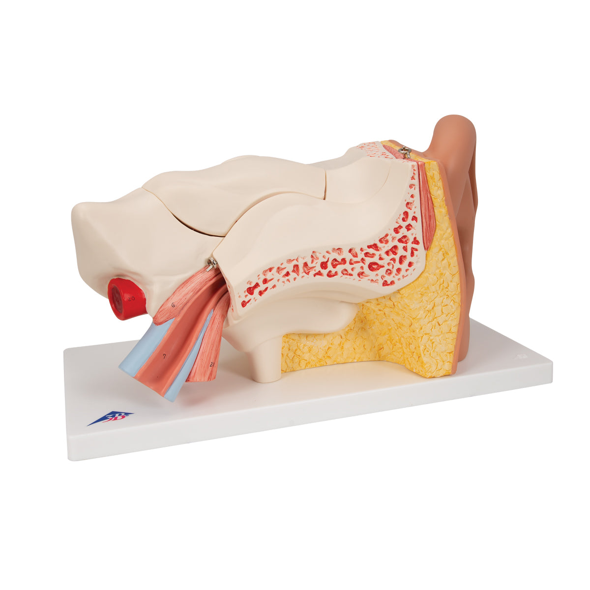
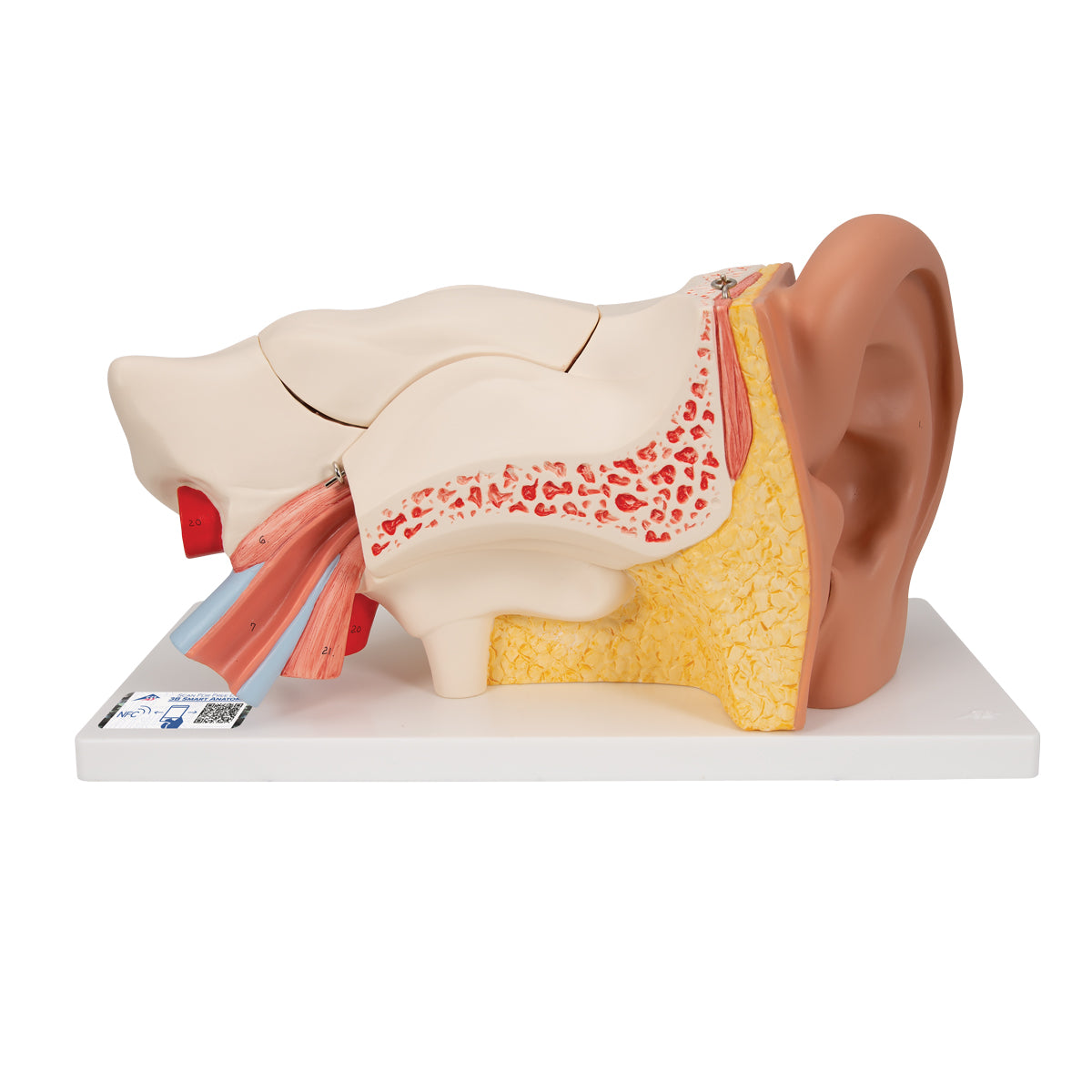
Anatomically speaking, the model can be used to understand the structure of the entire ear, consisting of the outer ear with the auricle and the external ear canal, the middle ear and the inner ear. In addition, the eustachian tube (tuba auditoria) is seen, which communicates with the nasopharynx, as well as the location of the ear in the temporal bone (os temporale).


The model can be separated into 5 separate parts as follows:


The front part of the temporal bone can be removed so that the inside of the external ear canal becomes visible.
The upper part of the temple bone can also be removed
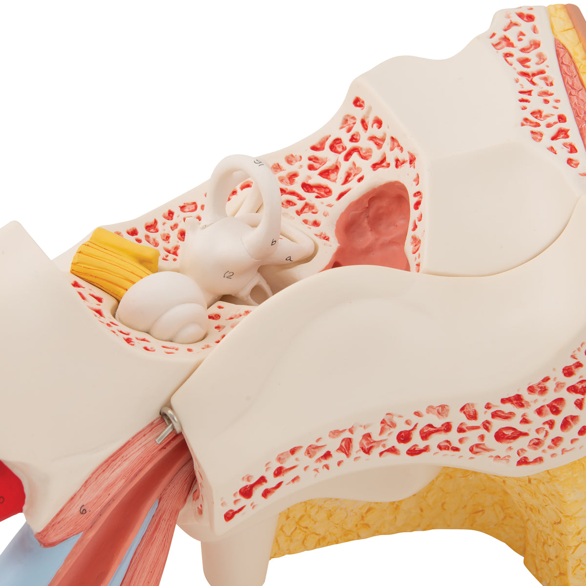
The eardrum (membrana tympanica) with the three middle ear bones - the hammer (malleus), the anvil (incus) and the stapes - can be removed and examined
The organ of hearing and balance can be removed


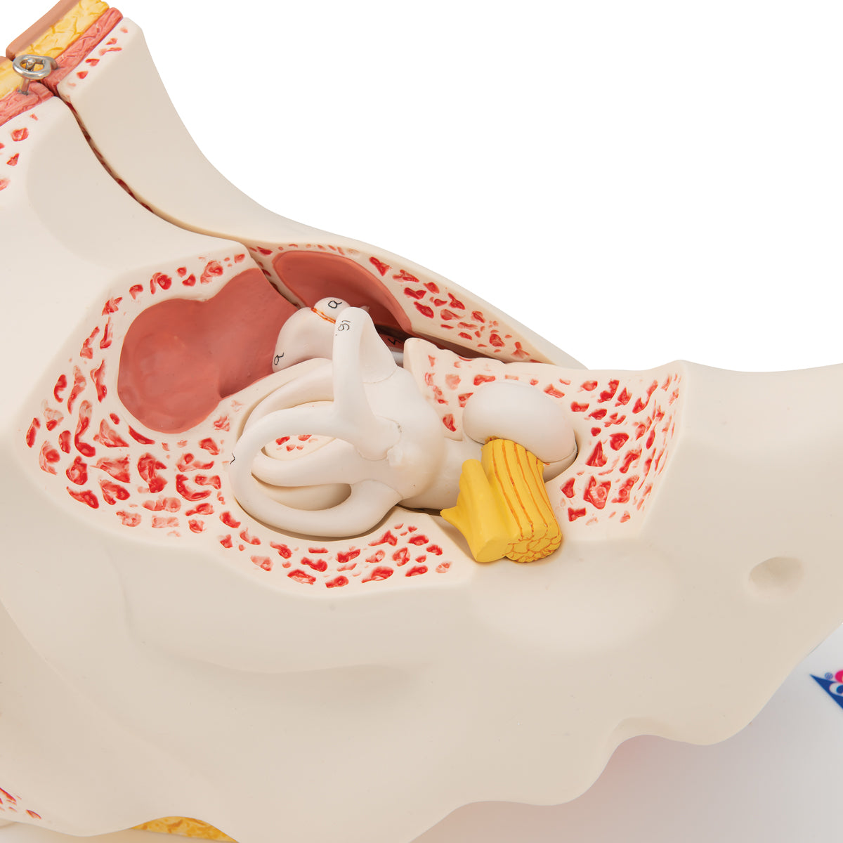
In the inner ear, the organs of hearing and balance are illustrated with associated nerves (N. vestibulocochlearis). The model shows both the arch canals (canales semicirculares), the vestibulum and the cochlea (the cochlea).


Of other structures, the oval and the round window (fenestra vestibuli and fenestra cochleae) can be seen on the back wall of the middle ear, and it is also seen how the middle ear is related to the A. carotis interna.

Product flexibility

Clinical features

Clinically speaking, the model can be used to understand ear diseases such as otitis media, cholesteatoma (bone eater), perforated eardrum and bullous myringitis.


Furthermore, the ear model can be used to understand examination methods and treatments such as the installation of a drain (tympanic membrane drain).

View full details

A safe deal

For 19 years I have been at the head of eAnatomi and sold anatomical models and posters to 'almost' everyone who has anything to do with anatomy in Denmark and abroad. When you shop at eAnatomi, you shop with me and I personally guarantee a safe deal.

Christian Birksø
Owner and founder of eAnatomi ApS

Read more about eAnatomi