SKU:EA1-1013282
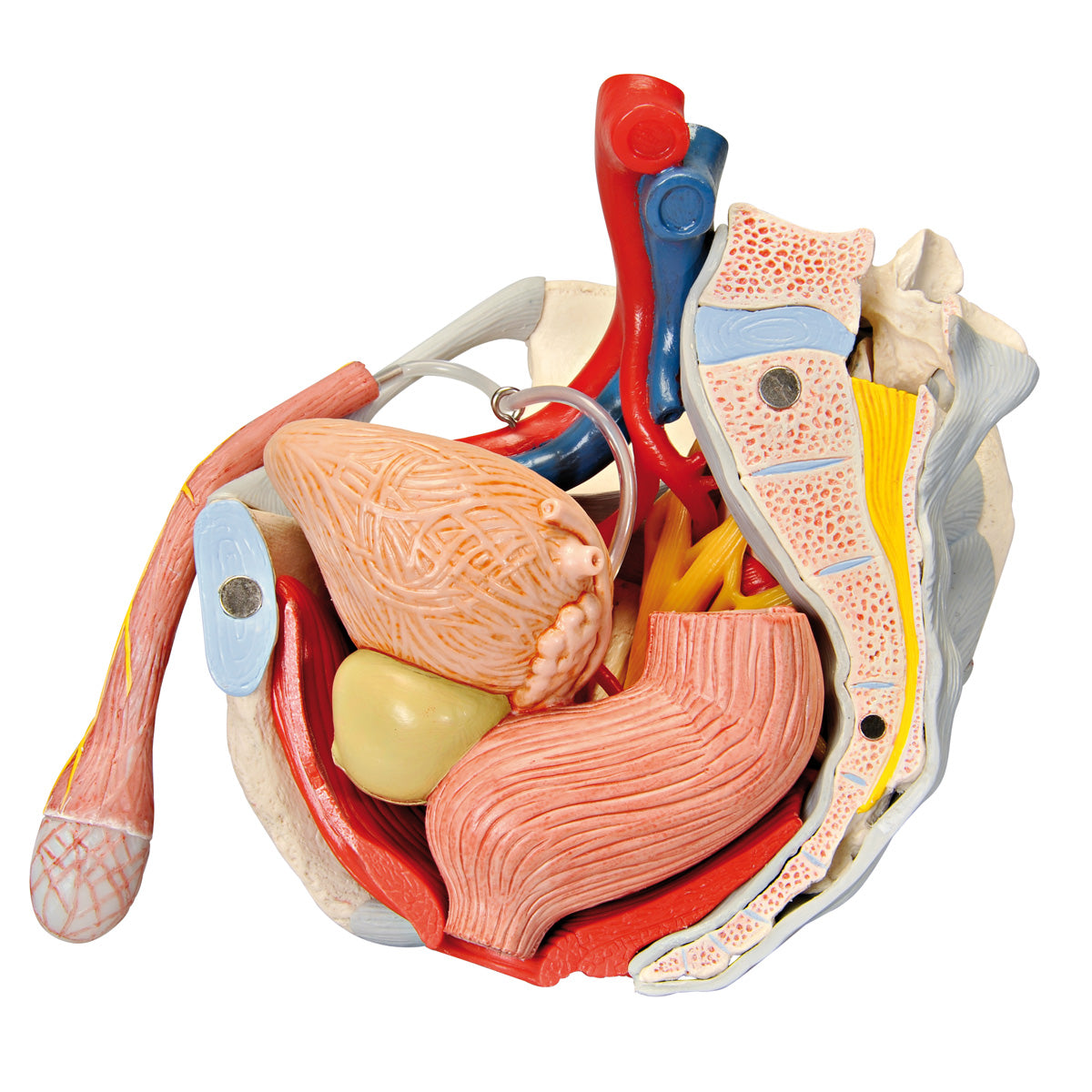
Pelvic model showing the pelvic floor, genitals, ligaments, nerves and blood vessels in the man
Pelvic model showing the pelvic floor, genitals, ligaments, nerves and blood vessels in the man
ATTENTION! This item ships separately. The delivery time may vary.
Couldn't load pickup availability
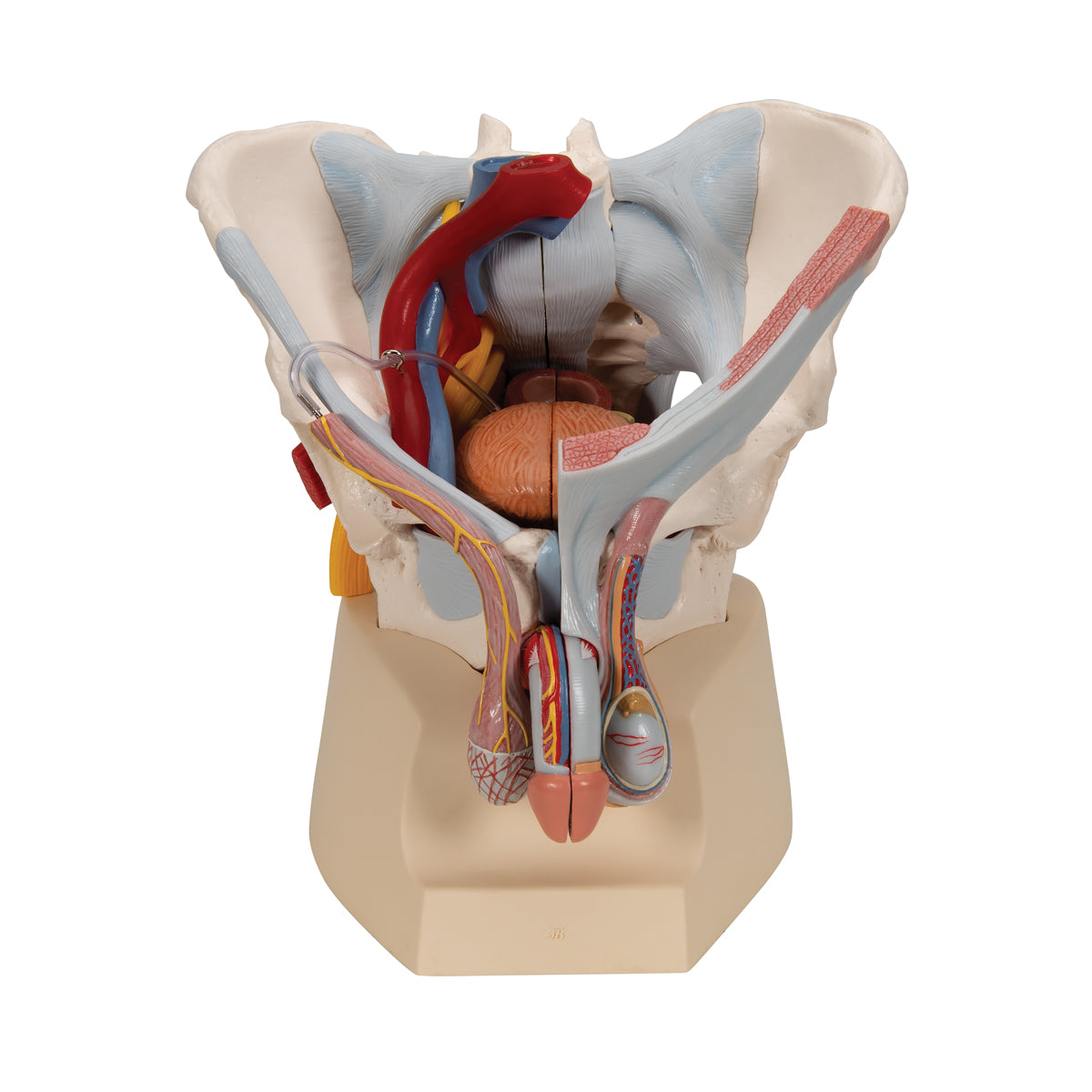
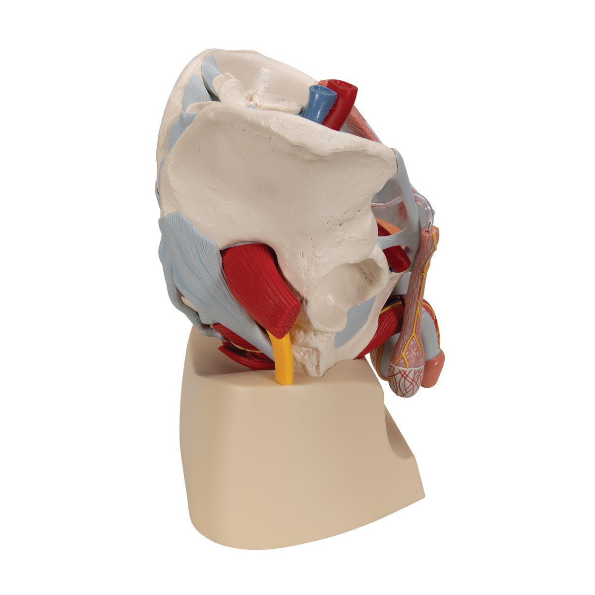
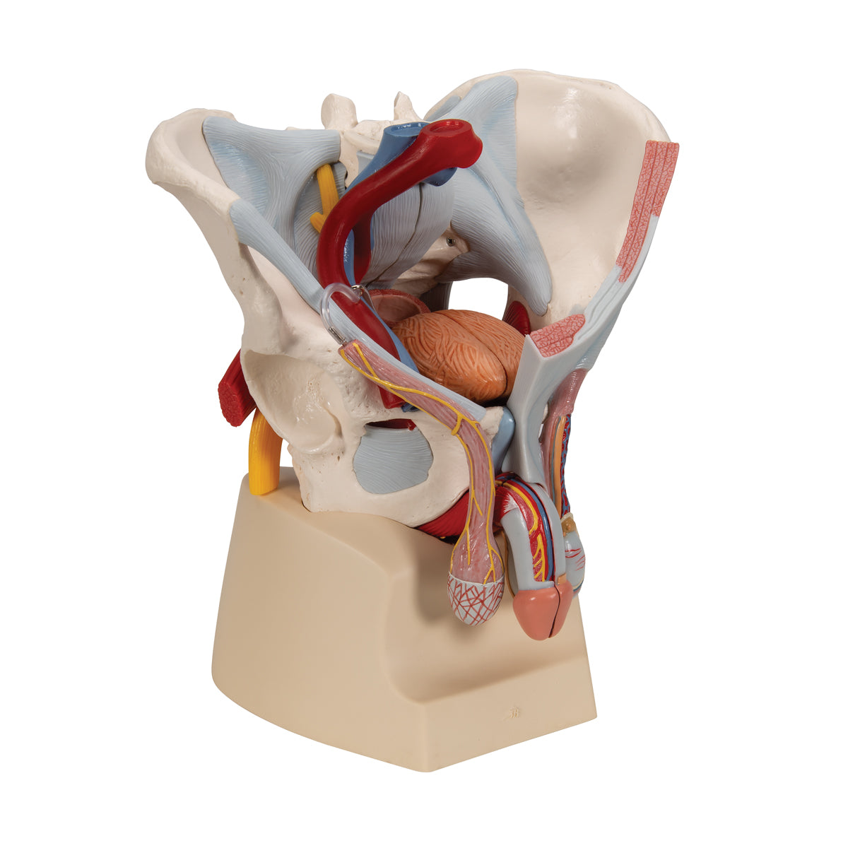
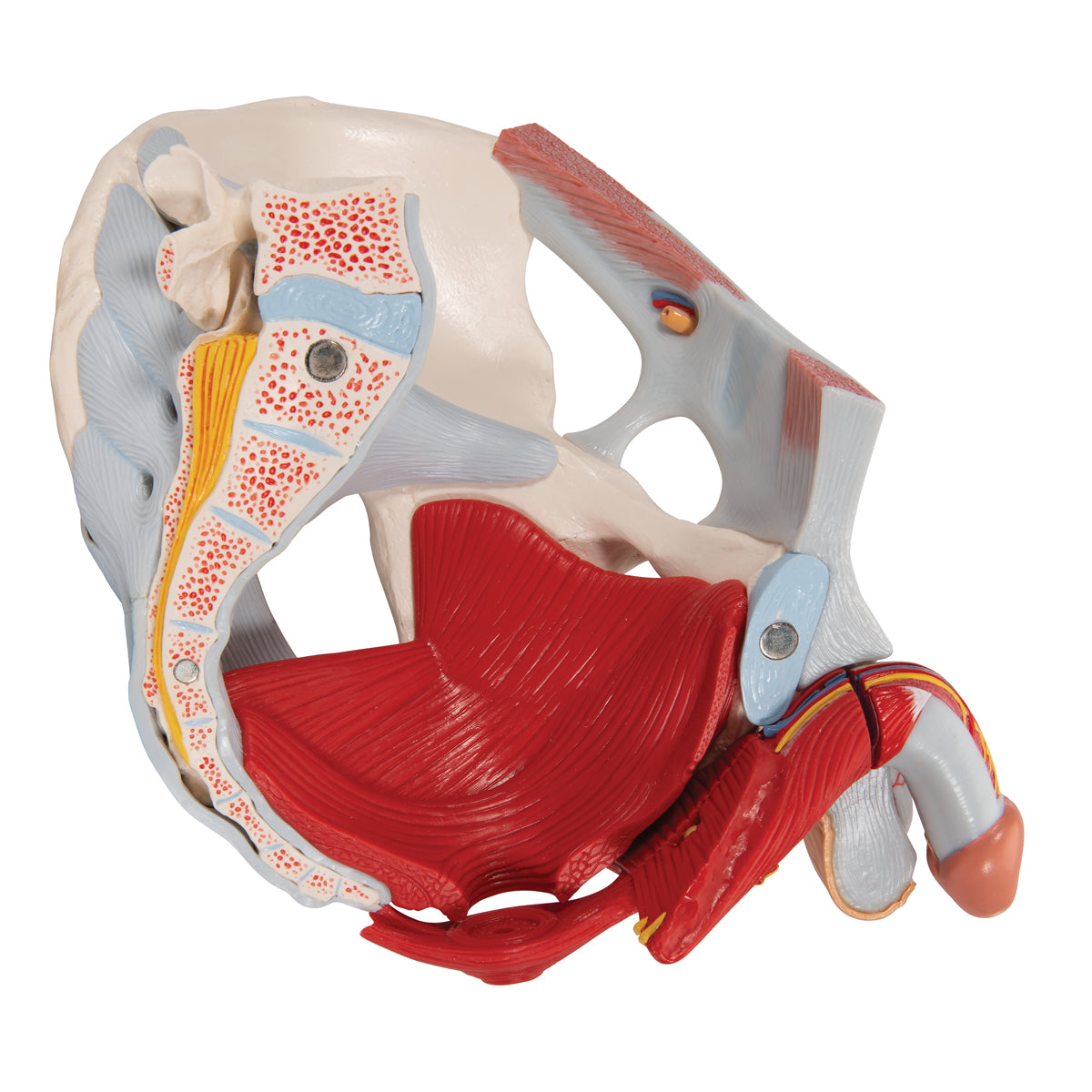
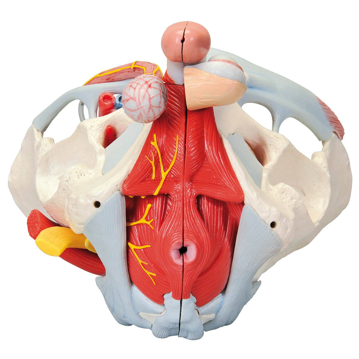
This pelvis model shows the male pelvis with the following anatomical structures:
- The pelvis (pelvic ring)
- Pelvic floor (pelvic floor)
- The internal genitalia
- The spermatic cords
- Some of the accessory gonads
- The external genitalia
- Ligaments, blood vessels and nerves in the pelvis
- A bit of the abdominal muscles
The model is developed in natural adult size. It weighs approximately 3 kg and the dimensions are
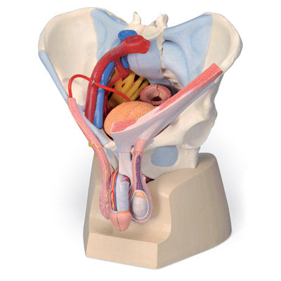
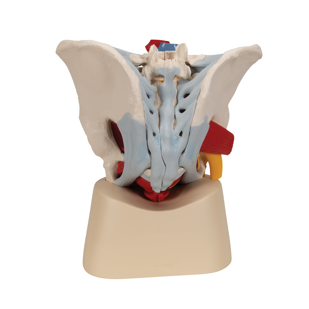
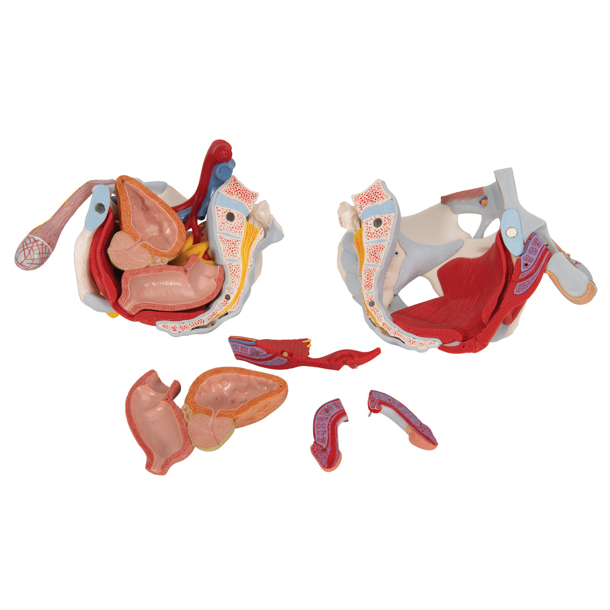
21 x 28 x 31 cm. It can be separated into 7 parts (see the pictures on the left), which are held together using magnets and metal pins. The degree of detail on the bones is good, but there is no movement in the pelvic joints (e.g. the SI joints and the symphysis), although it e.g. can be split in half. The model is delivered on a stand.
Anatomically speaking
Anatomically speaking
Anatomically speaking, the model shows many different tissues and organs, several of which can be seen in 3 dimensions because the model can be separated into 7 parts.
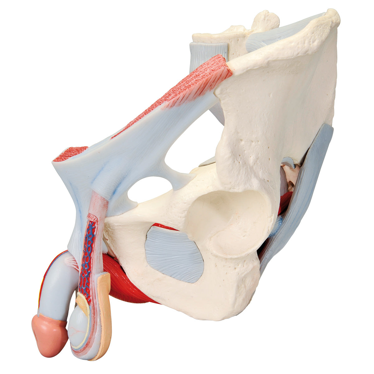
1) The pelvis consists of the 3 building blocks, which are the 2 hip bones and the sacrum. The model also shows the lower lumbar vertebra. In the bone tissue, the most important details can be seen, such as large nodules. Smaller details such as the linea glutaea are omitted. Overall, the model shows the following bones:
Os coxae (the 2 hip bones) of which both are composed
- Os ilium (iliac bone/hip bone)
- Os ischii (seat bone)
- Os pubis (pubic bone)
Us sacrum (sacrum) incl. os coccygis (coccyx)
5th lumbar vertebra (lumbar vertebra)
2) The pelvic floor (also called the pelvic floor or diaphragma pelvis in Latin) consists of the following muscles, which are clearly visible:
- M. levator ani (consisting of m. pubococcygeus and m. iliococcygeus)
- M. coccygeus
Other muscles are also seen such as the ischiocavernosus muscle, the bulbospongiosus muscle, the obturatorius internus muscle and the sphincter ani externus muscle. In addition, the relationships to the urinary bladder (vesica urinaria) and the rectum (rectum) are seen in detail.
3) The internal genitalia include the testicles (testes) and the epididymides, which are seen in relation to the scrotum and the spermatic cord (funicle). It is not possible to see the internal structures of the testicles (tubules, etc.). Both the contents of the spermatic cord and its barrier/leaves are clearly seen.
NB: In our assortment of pelvic models, we have 2 models that show the internal structures of the testicles: A large model with many details that can be viewed via THIS LINK and a miniature model that can be viewed via THIS LINK
4) The seminal ducts include the ductus deferens (the vas deferens), the ductus ejaculatorius and the urethra masculinum (the urethra), all of which are visible.
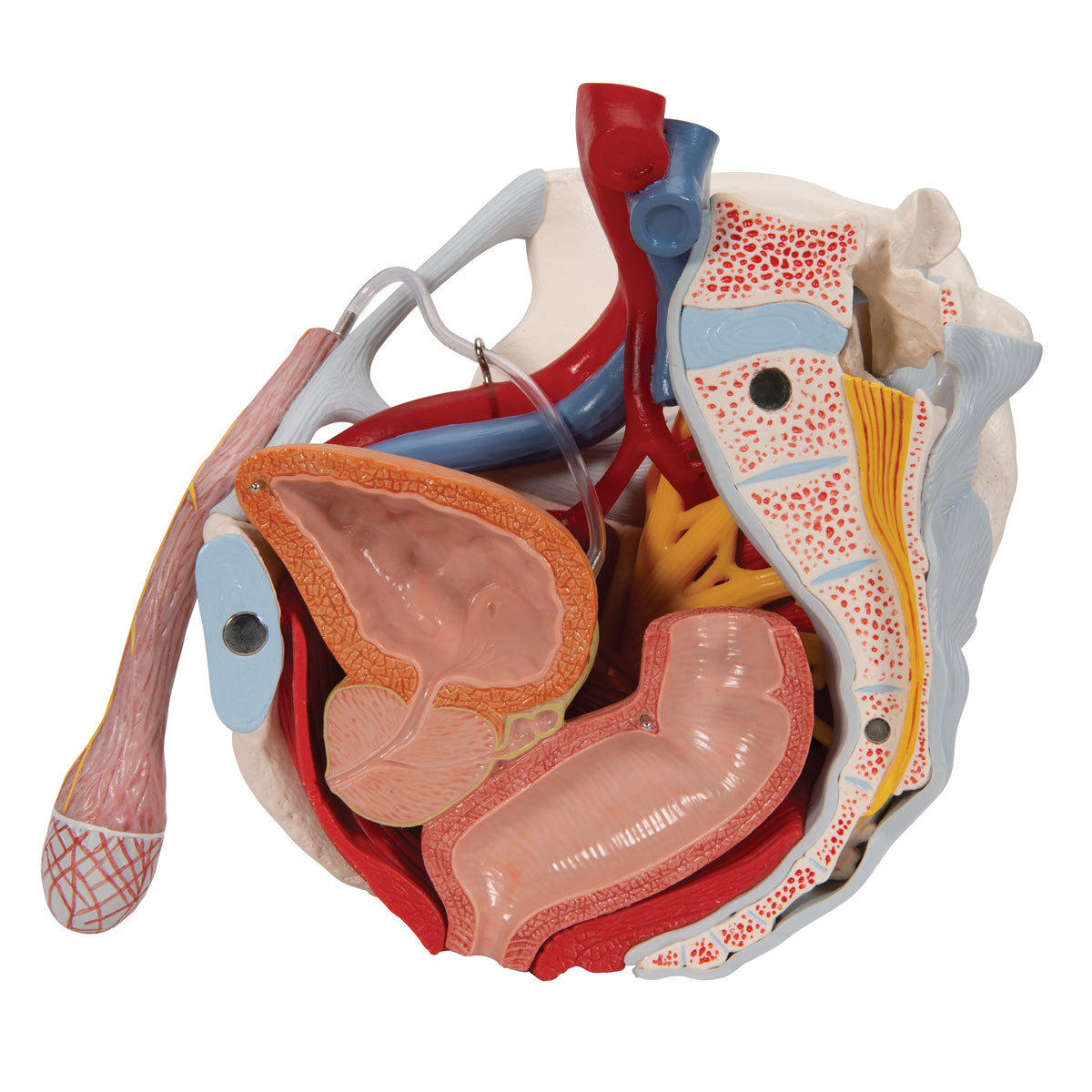
5) In relation to the accessory gonads, the prostate (bladder neck gland) and vesiculae seminales (seminal vesicles) are seen, but not gll. bulbourethrales and etc. urethral. The 2 first-mentioned glands are seen in cross-section, and therefore the excretory ducts and their connection are seen in detail.
6) The external genitalia include the penis and the scrotum, which are also seen. Opposite the testicles, the internal structure of the penis can be seen.
7) Ligaments, blood vessels and nerves are seen in detail. Ligaments are seen on both sides of the pelvic model, while blood vessels and nerves are mainly seen on the right side of the model.
Since the model i.a. can be divided in half, important details can be seen in a median section (including cauda equina). As for nerves, the sacral plexus, sciatic nerve (nervus ischiadicus) and other important nerves are also seen. As for blood vessels, the large artery and vein of the pelvis and parts of their branches are seen.
Many of the pelvic organs function as a channel or a reservoir (e.g. the rectum and the urinary bladder), which is why they are seen in a pedagogical way with an air-filled cavity (cavity).
8) Some of the abdominal muscles can also be seen on the model, so that the connection between the testes, the spermatic cords and the abdominal wall can be better seen.
Flexibility
Flexibility
In terms of movement, the pelvis is not flexible. Although the various joints are visible (e.g. the SI joints and the symphysis), they are not movable.
Clinically speaking
Clinically speaking
Clinically speaking, the model can be used to understand diseases and disorders in the man's pelvis - for example in the genitals, the urinary bladder or the rectum. Examples are torsio testis, benign space-filling processes in the scrotum, urethral stricture, anal fissure, BPH (benign prostatic hyperplasia) and prostate cancer.
Furthermore, the model can be used to understand disorders and diseases such as cauda equina syndrome and deep vein thrombosis (DVT) in the pelvis.
Share a link to this product

A safe transaction
For 19 years I have been managing eAnatomi and sold anatomical models and posters to 'almost everyone' who has anything to do with anatomi in Scandinavia and abroad. When you place your order with eAnatomi, you place your order with me and I personally guarantee a safe transaction.
Christian Birksø
Owner and founder of eAnatomi and Anatomic Aesthetics

