
SKU:EA1-A159II
Skull model Incl. the dural septa e.g. falx cerebri from dura mater
Skull model Incl. the dural septa e.g. falx cerebri from dura mater
5 in stock
Couldn't load pickup availability
This skull model covers most needs, is produced in adult size and is sold at the lowest possible price. As something special, it also shows the dural septa from the dura mater encephali.
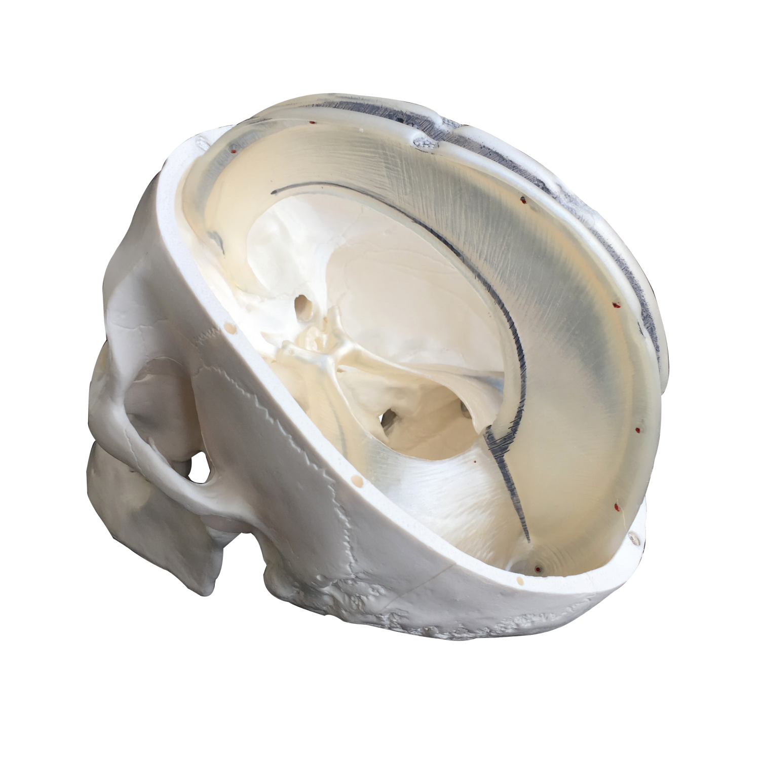
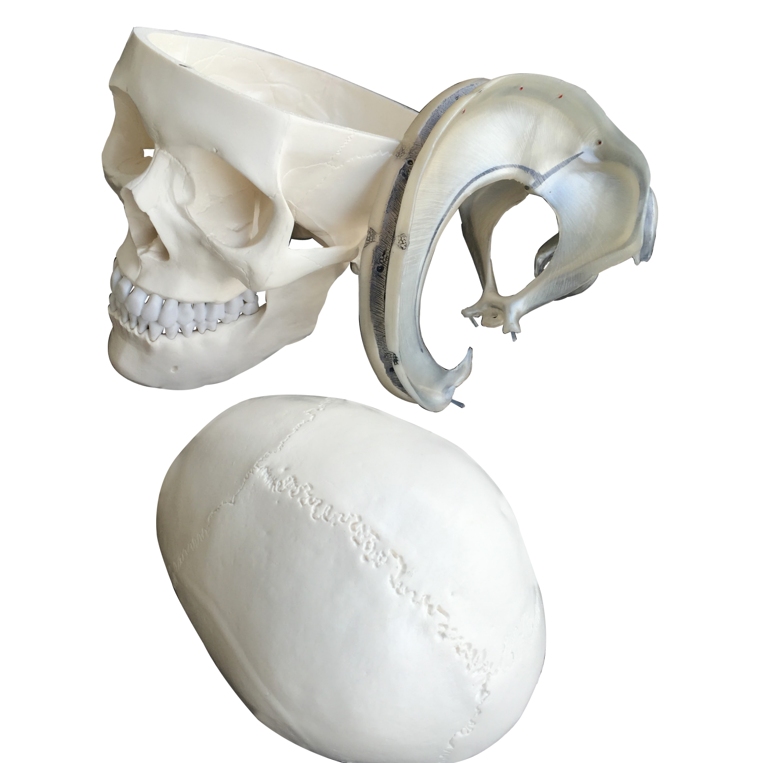
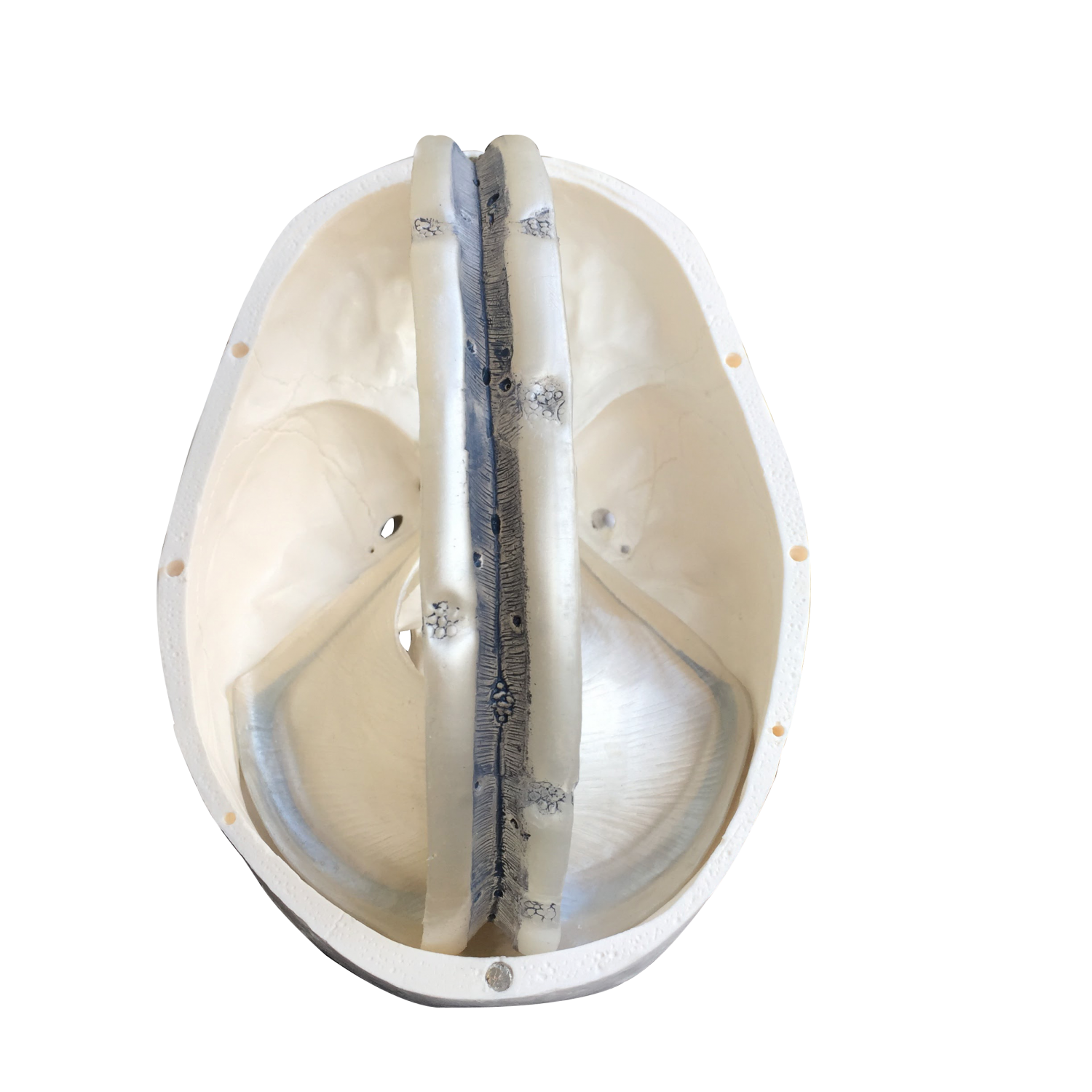
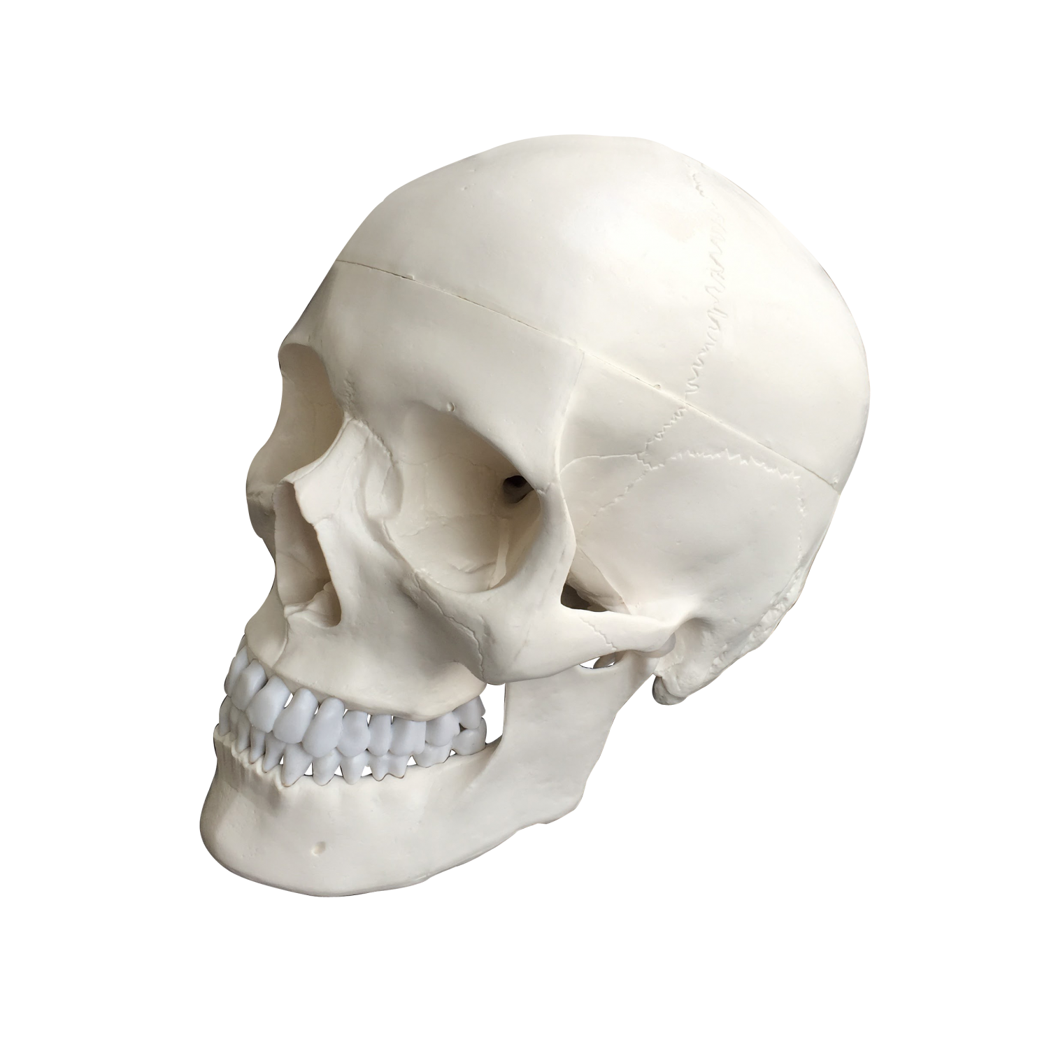
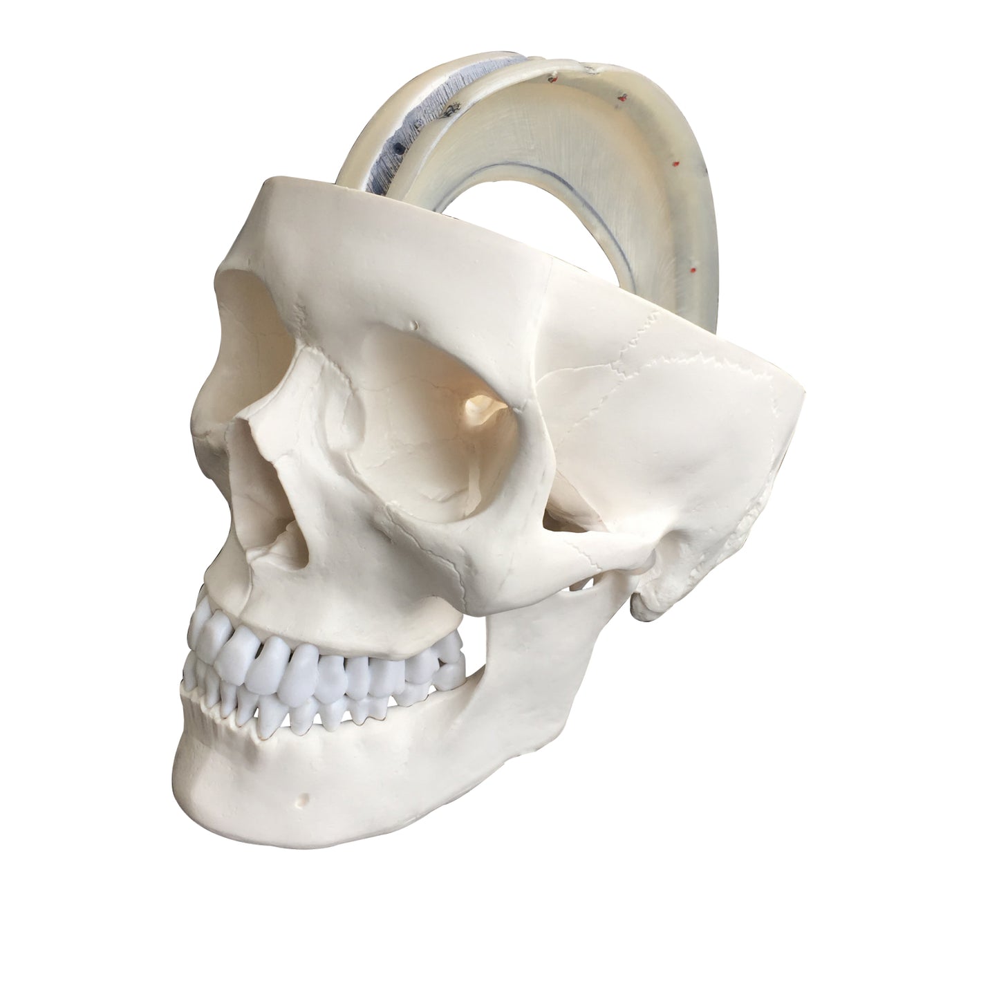
As described for the second variant of the skull model (without the dural septa), it is molded in white plastic and comes in a size corresponding to an adult person. The skull cap ("the top") can be removed, so i.a. the base of the skull (basis cranii interna) can be studied. The skull cap is held in place by small magnets and plastic pins, which are only visible when removed. It should be mentioned that this joint appears all round with an uneven/frayed edge when the skull cap is fitted. This lack of finishing must be compared to the skull model's low price.
Anatomical features
Anatomical features
Anatomically, it must be emphasized that this variant of the skull model offers an educational and practical opportunity to study the dural septa, which are duplicates from the inner layer of the dura mater encephali. These septa function i.a. as support structures for the brain.
We have many skull models in our range and this variant offers the most detailed representation of the dural septa.
They are made of a nice, light and transparent plastic, which is a bit flexible. They can be taken out of the skull in a coherent unit (see the pictures on the left) and therefore allow for a better understanding of the venous sinus of the brain.
The following dural septa are seen:
Falx cerebri (cerebral seal)
Tentorium cerebelli (cerebellar tent)
Falx cerebelli (cerebellar seal)
Diaphragma sellae (which forms a lid over the hypophysial fossa)
As described by the second variant of the skull model (without the dural septa), the human skull can be divided into 2 parts, and the skull model therefore shows the following:
1) The braincase (neurocranium), which is intended to enclose the brain and the hearing-equilibrium organ
2) The facial skeleton (viscerocranium) which surrounds the nasal cavity and forms the tooth-bearing framework around the oral cavity. The 32 teeth are also included
The braincase consists of 8 bones. There are 4 unpaired (the frontal, sphenoid, sphenoid and occipital bones) and 2 paired (the occipital and temporal bones). All these bones as well as sutures can be identified on the skull model.
The facial skeleton includes 6 paired bones (the maxilla, palatine, cheekbone, nasal bone, lacrimal bone, and lower conchbone) and 3 unpaired bones called the mandible, the ploughshare, and the zygomatic bone (some do not count the zygomatic bone as part of the facial skeleton). All these bones and sutures can also be identified on the skull model. NB: The hyoid bone is also included in the facial skeleton but cannot be seen on this skull model.
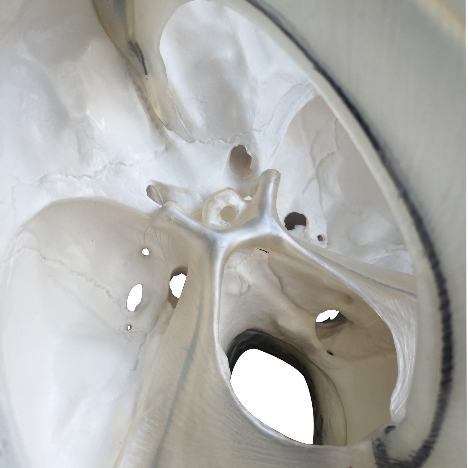
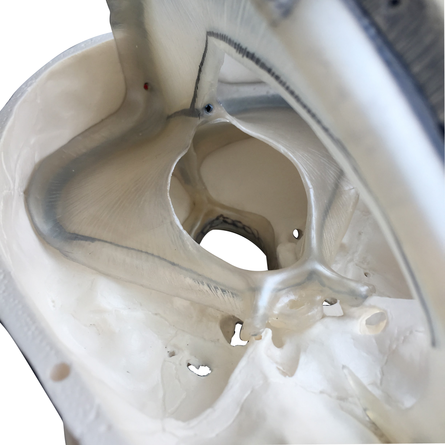
The human skull contains many holes and channels containing vessels and nerves. Overall, there are connections between the braincase and the neck, to and from the eye socket, to and from the pterygo-palatine fossa and to and from the nasal cavity.
This skull model shows many of the most important holes and canals, but not all. Furthermore, the level of detail on the bones is good. As for "osseous landmarks" such as the processus styloideus, many of the most important are seen, while some minor participants are omitted.
NB: If you are looking for a more detailed skull model, we would recommend our extremely lifelike skull model. On the skull model, the number of holes and canals and the level of detail in the bone structure are as anatomically complete as possible. The price of the skull model is of course higher, and it is bought e.g. of medical students. See it HERE
Product flexibility
Product flexibility
In terms of movement, it is only relevant to mention the jaw joint. The lower jaw bone ("mandible") is held in place via a relatively long metal spring, which is attached via screws to the mandible and palatine bone. In addition, large pieces of rubber are mounted in the joint bowls (fossa mandibularis) of the jaw joints, which hold the joint head (caput mandibulae) "like in a rubber bowl" (see the pictures on the left).
The model's jaw joints are not very flexible. Natural movements in the jaw joint require that, among other things, the articular head can slide forward and downward until the articular head is forward on the tuberculum articulare. The latter is the bony part with a weak guiding groove in front of the depression (fossa mandibularis). This can only be demonstrated on the model if the rubber in the joint cups is removed, which the model is not designed for.
If you take a firm hold of the mandible, you can however move it up and down and to the sides, although the natural sliding joint mechanism as described above cannot be demonstrated at all.
The mandible can be completely removed, but it requires at least one screw to be removed with a screwdriver.
Clinical features
Clinical features
Clinically, the skull model can be used to understand specific conditions such as calcification of the falx cerebri.
The model is also ideal for understanding diseases, disorders and disorders in this part of the skeleton.
Share a link to this product


A safe deal
For 19 years I have been at the head of eAnatomi and sold anatomical models and posters to 'almost' everyone who has anything to do with anatomy in Denmark and abroad. When you shop at eAnatomi, you shop with me and I personally guarantee a safe deal.
Christian Birksø
Owner and founder of eAnatomi ApS