Skip to product information
1 of 4

SKU:EA-1000121

Flexible spine model in adult size incl. pelvis

Flexible spine model in adult size incl. pelvis

Regular price €177,95 EUR
Regular price Sale price €177,95 EUR
Sale Sold out
Not including taxes if ordering from outside EU. Shipping calculated at checkout.
Variant

Low stock: 1 left

This classic spine model illustrates the structure of the spine and is ideal for demonstrating which movements can be performed in the different segments of the spine.

In addition, the model shows the neck bone (os occipitale), a male pelvis, A. vertebralis, spinal nerves and a herniated disc between L3 and L4.

The model measures 74 cm in height and weighs 1.79 kg. The spine is held together by a thick copper wire and a rubber tube that runs behind the spinal nerves, which ensures flexibility, and the individual discs and vertebrae are held together by a pin/hole system.

The model is delivered with or without femoral heads / upper part of the femur.

Stand not included.

Anatomical features

Anatomically speaking, the model shows the entire vertebral column, which consists of a neck part, a chest part, a lumbar part, a pelvic part and the coccyx. The neck bone is also included.

The model thus shows the two most important joint types in the spinal column, which are:

The symphyses (discs) between the vertebral bodies

The facet joints, which are sliding joints between the pivots of the vertebrae.

In addition, both the upper and lower neck joint and the sacrococcygeal joint are seen.


In addition, the model shows how the pelvis as a whole forms a ring structure which ensures that the axial skeleton is held together with the lower limbs. The model also shows the SI joint between the sacrum and the pubic bone, as well as the symphysis between the pubic bones.

Furthermore, the model shows the cervical part of the vertebral artery, the 31 pairs of spinal nerves and a herniated disc in the lumbar part.

Product flexibility

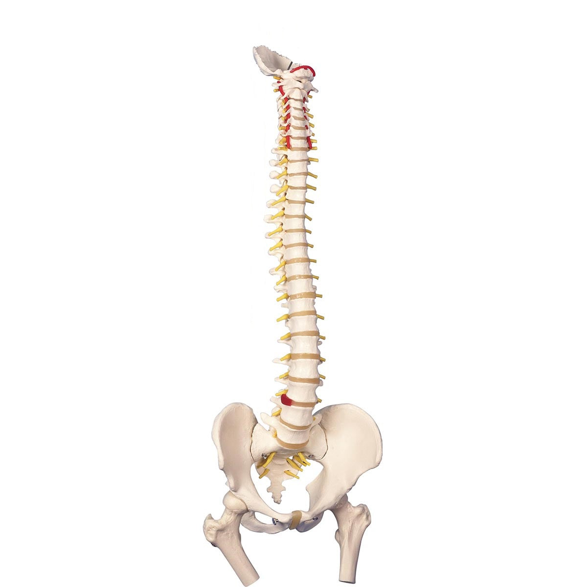
In terms of movement, the model is very flexible. The mobility of the human spine is greatest in the cervical and lumbar regions (which are lordotic), while the thoracic region (which is kyphotic) is less mobile. This model can be used to demonstrate the most important movements of the spine, which are flexion-extension, lateral flexion and rotation in the neck and lumbar region.


The above-mentioned copper wire and rubber hose both ensure that you can detach the disc from the vertebrae without it falling apart, and that stability is felt when moving the spine.


The neck bone (os occipitale), upper cervical vertebra (atlas) and second upper cervical vertebra (axis) are gathered together with elastic string, which causes a special elastic effect, where the vertebrae both follow each other during movement and come together again in a natural way. You can thus demonstrate the most important movements in the neck joints, which are the nodding and rocking of the head, which takes place in the upper neck joint, as well as the shaking of the head, which takes place in the lower neck joint.

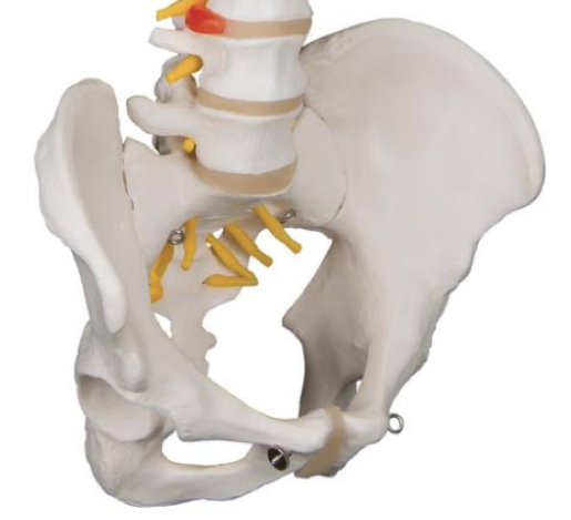
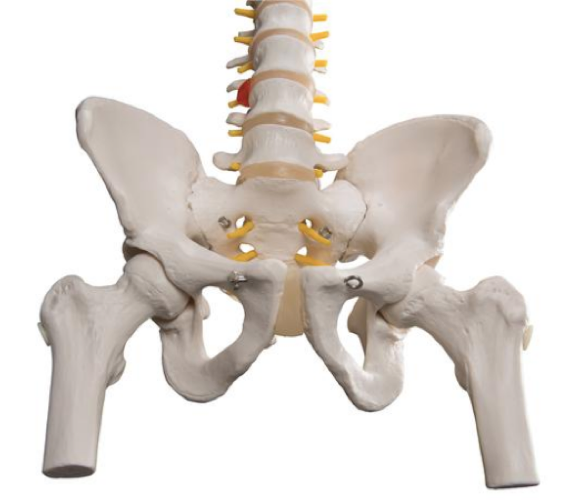
For the variant with femoral heads: In the hip joints, the femoral head is held firmly in the socket via elastic. Therefore, different movements of the hip joint, such as abduction and rotation, can be demonstrated. However, the femoral head cannot be removed.

Clinical features

Clinically, this model is ideal for understanding disc herniation, root involvement and spondylolisthesis. It can also be used to understand disorders such as back pain, scoliosis, spondylosis, spinal stenosis and osteoarthritis.


The model can of course also be used to understand fractures, a. vertebralis dissection and many different disorders related to the pelvis.

View full details

A safe deal

For 19 years I have been at the head of eAnatomi and sold anatomical models and posters to 'almost' everyone who has anything to do with anatomy in Denmark and abroad. When you shop at eAnatomi, you shop with me and I personally guarantee a safe deal.

Christian Birksø
Owner and founder of eAnatomi ApS

Read more about eAnatomi