Collection: Digestive system
-
Denture model enlarged incl. large toothbrush
Regular price 495,00 DKKRegular priceUnit price / per850,00 DKKSale price 495,00 DKKSale -
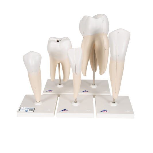
5 enlarged and different teeth (incl. caries) presented on separate stands
Regular price 3.925,00 DKKRegular priceUnit price / per -
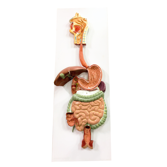
Model of the entire digestive system at 91 cm in height
Regular price 2.450,00 DKKRegular priceUnit price / per -
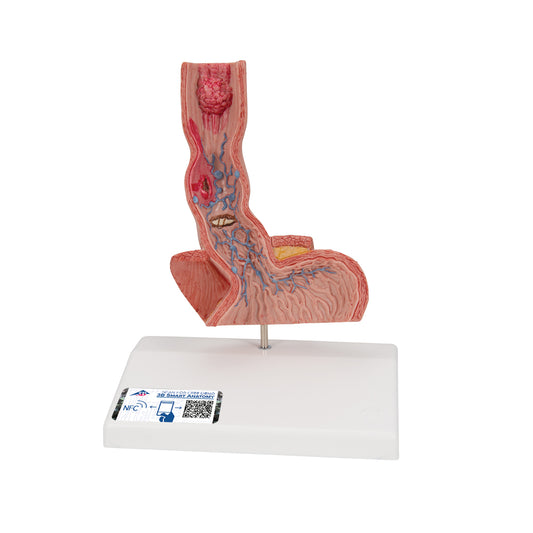
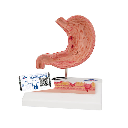
Model showing the esophagus and a bit of the inside of the stomach with various diseases
Regular price 730,00 DKKRegular priceUnit price / per -
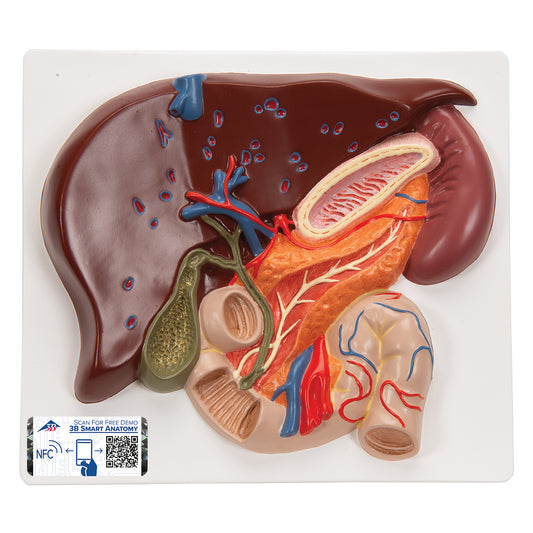
Model of the liver, gallbladder, bile ducts and related organs
Regular price 1.405,00 DKKRegular priceUnit price / per -
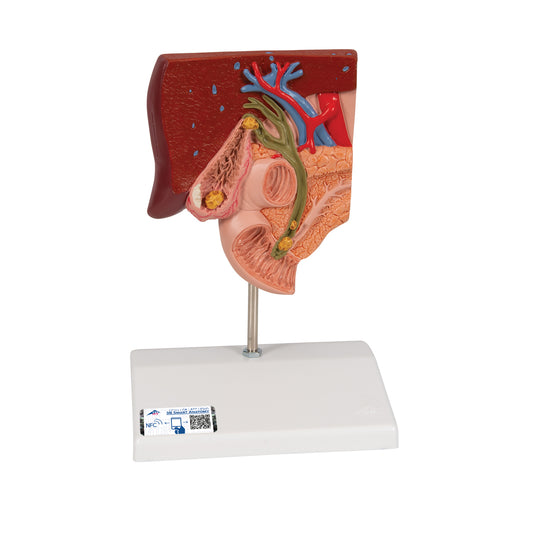
Reduced scale model of the biliary tract showing gall bladder and gallstones
Regular price 770,00 DKKRegular priceUnit price / per -
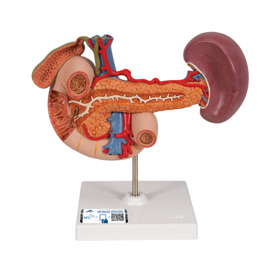
Model of the duodenum and the relationship of the pancreas to other organs
Regular price 1.640,00 DKKRegular priceUnit price / per -
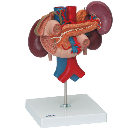
Detailed model of the duodenum and the relationship of the pancreas to other organs - can be separated into 3 parts
Regular price 2.510,00 DKKRegular priceUnit price / per -
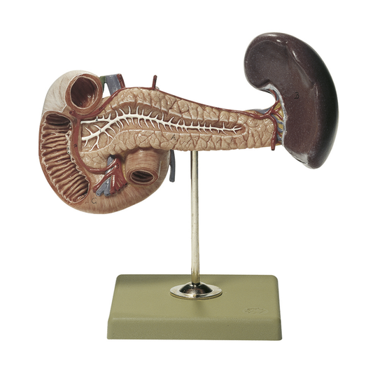
Model of pancreas with spleen and duodenum
Regular price 3.135,00 DKKRegular priceUnit price / per -
Reduced and detailed model of the stomach showing inflammation and ulcers
Regular price 790,00 DKKRegular priceUnit price / per -
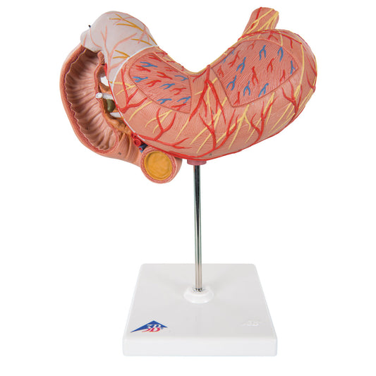
Detailed model of the stomach, duodenum and pancreas
Regular price 2.510,00 DKKRegular priceUnit price / per -
 Sold out
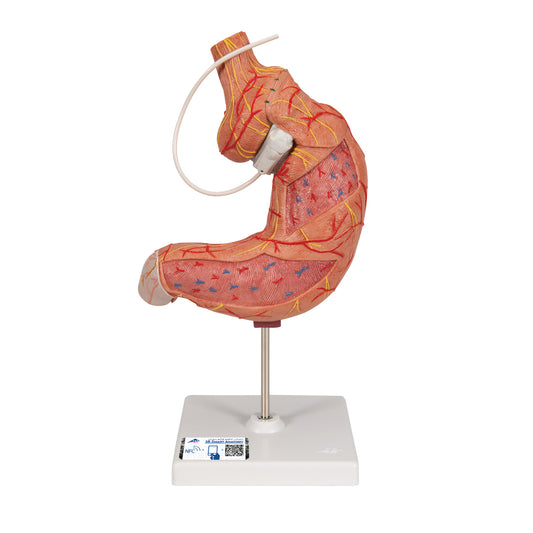
Gastric Banding model
Regular price 1.090,00 DKKRegular priceUnit price / per -
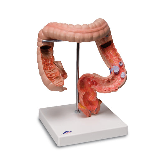
Detailed model of the colon showing several diseases
Regular price 1.165,00 DKKRegular priceUnit price / per -
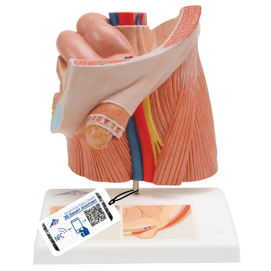
Anatomical model illustrating inguinal hernia
Regular price 780,00 DKKRegular priceUnit price / per -
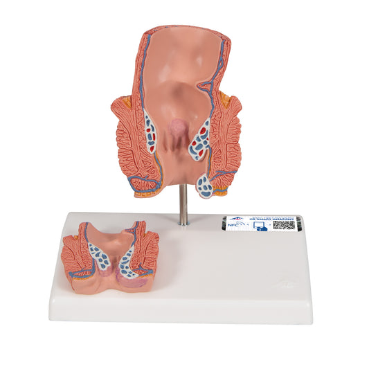
Model of the rectum showing hemorrhoids
Regular price 730,00 DKKRegular priceUnit price / per
Collapsible content
Read more about the product category here
In our selection of models of the digestive system, you can choose between models that are reduced, made in natural size or enlarged. There are both models with and without diseases. The selection includes a model of the entire digestive system, dental models (teeth), models of the esophagus, models of the stomach, models of the duodenum with related organs such as the pancreas, liver models with the gallbladder and bile ducts, and models of the large intestine including the rectum.
Models of the digestive system are used especially for understanding anatomy as well as clinical aspects such as diseases, examinations and treatment.
Anatomically speaking, the digestive tract/gastrointestinal tract includes the mouth, esophagus, stomach and small and large intestine. If the liver, gallbladder and pancreas are included, it is called the digestive system.
With our model of the entire digestive system at hand, all its organs (and the spleen) can be studied. Openings into the intestinal lumen (where food passes) ensure that you can also study the mucous membrane with folds and other things in e.g. stomach and rectum.
There are 2 tooth models in the selection. One is a bite/tooth model which is enlarged. On the dental model, you can see the overall structures such as the tongue, teeth and upper and lower jaw. The second model is a collection of different teeth that have been enlarged. With these tooth models in hand, you can study structures such as the tooth crown, tooth neck and tooth root.
With our model of the esophagus/oesophagus in hand, one can study its passage through the diaphragm, the closing function (the lower esophageal sphincter) and the many diseases seen on the model. In the selection we also have 2 models of the stomach (ventriculus) with and without diseases. The model of the stomach without diseases is very detailed and shows its relationship to the duodenum and pancreas.
With one of our 2 liver models in hand, you can also study the gallbladder (vesica biliaris, vesica fellea) and bile ducts, because these tissues are seen on both liver models. You can also see the right and left lobes (lobus hepatis dexter and sinister), blood vessels such as v. portae (the portal vein) and much more.
Our 2 models of the colon show respectively the entire large intestine (intestinum crassum) and rectum (rectum) in isolation. Both models show diseases, but can also be used to study internal folds in the mucosa as well as other characteristics. The model of the entire large intestine shows both the caecum (the actual appendix), the appendix vermiformis (the worm-shaped appendix/cecum), the colon and the rectum with the canalis analis.
Clinically speaking, a model of the digestive system/alimentary canal/gastrointestinal tract or just a single organ from this can be used to understand diseases and disorders. These can be, for example, caries and periodontitis, reflux and ulcers in the oesophagus, oesophageal varices, diaphragmatic hernia, inflammation of the stomach, peptic ulcer, stones in the gallbladder and bile ducts, inflammation of the gallbladder, pancreatitis, pancreatic cancer, chronic inflammatory diseases (ulcerative colitis and Crohn's disease), colon cancer, diverticulosis, polyps, hemorrhoids, fistulas and fissures.
Furthermore, the models can be used to understand examinations such as ultrasound examination and MRCP as well as treatment such as cholecystectomy.

A window to a world of anatomy
Whatever you're looking for
Then we can procure or produce it. eAnatomi is more than just a retailer of existing products. We have our own development department, where we create unique and original products that are used for training, guidance and inspiration.

19 years of anatomy
A safe transaction
For 19 years I have been at the head of eAnatomi and sold anatomical models and posters to 'almost everyone' working with anatomy in Denmark and abroad. When you do business with eAnatomi, you do business with me and I personally guarantee a safe transaction.
Christian Birksø
Owner and founder of eAnatomi
Latest blog news
View all-

2 meter tall anatomy figures - we call it anato...
For more than a decade, eAnatomy has produced our own anatomical illustrations, performed by the best illustrators. Where most illustrations are used on classic printed media such as posters, selected...
2 meter tall anatomy figures - we call it anato...
For more than a decade, eAnatomy has produced our own anatomical illustrations, performed by the best illustrators. Where most illustrations are used on classic printed media such as posters, selected...
-

Anatomical Chart Company - changes the format
Anatomical Chart Company har i 2023 besluttet af udfase papirvarianten for fremover kun at levere den let laminerede version med ringhuller. Dette betyder at det ikke længere bliver muligt, at...
Anatomical Chart Company - changes the format
Anatomical Chart Company har i 2023 besluttet af udfase papirvarianten for fremover kun at levere den let laminerede version med ringhuller. Dette betyder at det ikke længere bliver muligt, at...
-

The heart model's place in teaching & expla...
Teachers, professionals and other mediators can easily be challenged when they have to explain the anatomy and diseases of the heart.
The heart model's place in teaching & expla...
Teachers, professionals and other mediators can easily be challenged when they have to explain the anatomy and diseases of the heart.


