-
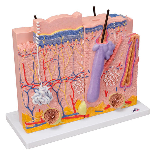
Skin model of 3 skin areas. Can be separated into 3 parts
Regular price 2.195,00 DKKRegular priceUnit price per -
Skin model of one skin area
Regular price 650,00 DKKRegular priceUnit price per1.125,00 DKKSale price 650,00 DKKSale -
Skin model of 2 skin areas, a nail and a hair root assembled on a stand
Regular price 780,00 DKKRegular priceUnit price per -
Very enlarged and detailed ear model which can be separated into 4 parts
Regular price 495,00 DKKRegular priceUnit price per975,00 DKKSale price 495,00 DKKSale -
Very enlarged and detailed ear model which can be separated into 4 parts
Regular price 1.495,00 DKKRegular priceUnit price per -
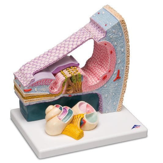
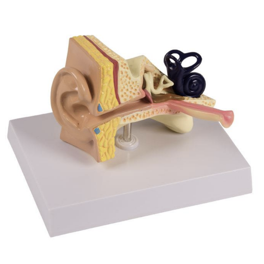
Detailed ear model that shows both the entire cochlea and a cross-section with three-dimensional details
Regular price 2.375,00 DKKRegular priceUnit price per -
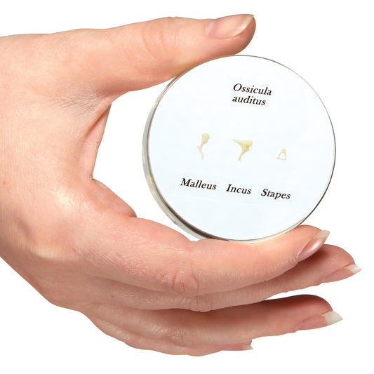
Life-size model of the 3 small bones of the middle ear
Regular price 450,00 DKKRegular priceUnit price per1.025,00 DKKSale price 450,00 DKKSale -
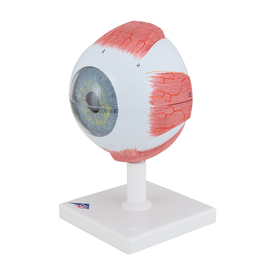
Classic eye model which is enlarged and can be separated into 6 parts
Regular price 1.575,00 DKKRegular priceUnit price per -
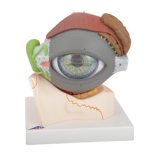
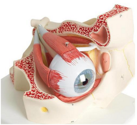
Complete eye model which is enlarged and can be separated into 8 parts
Regular price 3.245,00 DKKRegular priceUnit price per0,00 DKKSale price 3.245,00 DKK -
Practical eye model which is enlarged and shows 11 eye diseases/disorders
Regular price 2.970,00 DKKRegular priceUnit price per -
Classic model of the 4 sinuses as well as oral cavity, nasal cavity and pharynx
Regular price 395,00 DKKRegular priceUnit price per -
Nose and throat model
Regular price 1.475,00 DKKRegular priceUnit price per -
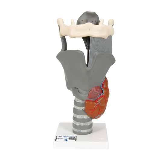
Larynx model with vocal folds and several other tissues. Can be separated into 5 parts
Regular price 1.475,00 DKKRegular priceUnit price per -
Enlarged larynx model with vocal folds and several other tissues. Can be separated into 5 parts
Regular price 975,00 DKKRegular priceUnit price per -

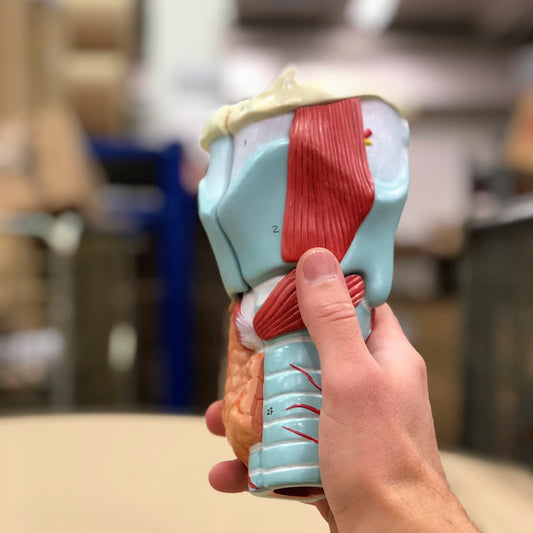
Enlarged larynx model with vocal folds and several other tissues. Can be separated into 7 parts
Regular price 1.580,00 DKKRegular priceUnit price per2.565,00 DKKSale price 1.580,00 DKKSale -
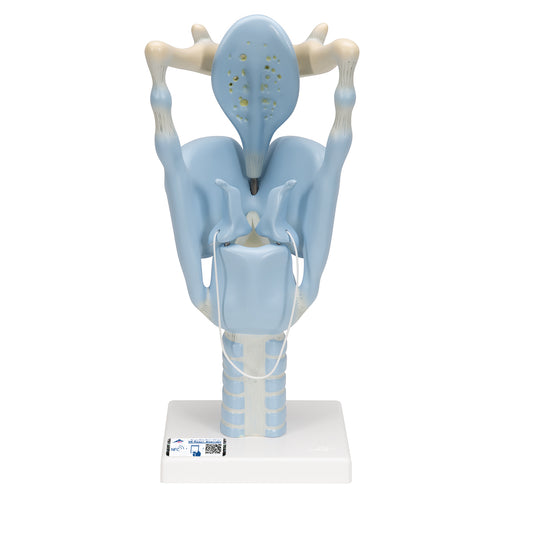
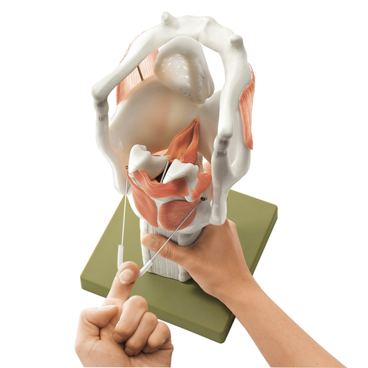
Model of the larynx with movable nasal cartilages and epiglottis
Regular price 2.015,00 DKKRegular priceUnit price per -
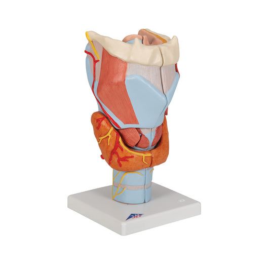
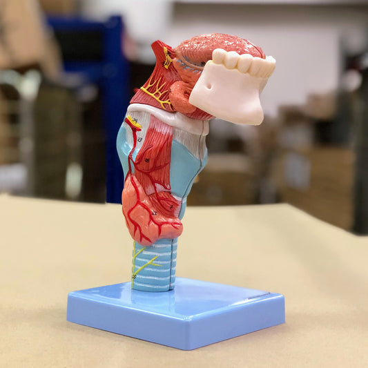
Enlarged and particularly movable larynx model with vocal folds and relationships to several other tissues
Regular price 5.950,00 DKKRegular priceUnit price per -
 Sold out
Functional larynx in 3 x normal size
Regular price 2.690,00 DKKRegular priceUnit price per -
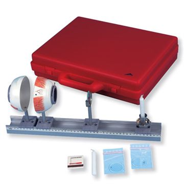
Practical eye model for demonstrating the refraction and refractive errors of the eye
Regular price 7.260,00 DKKRegular priceUnit price per -
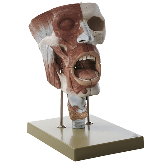
Detailed model of nose, throat and throat in strong magnification
Regular price 28.295,00 DKKRegular priceUnit price per -
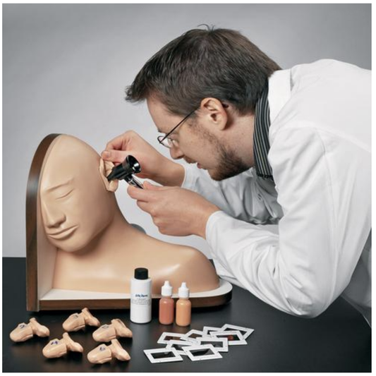
Simulator for training in ear examination
Regular price 14.975,00 DKKRegular priceUnit price per -
Educational glasses that simulate the visual changes caused by alcohol intoxication
Regular price 2.830,00 DKKRegular priceUnit price per -
Ear model of a child's ear showing otitis media
Regular price 750,00 DKKRegular priceUnit price per -
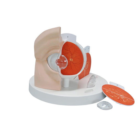
Eye model 3 x magnification in 7 parts
Regular price 2.615,00 DKKRegular priceUnit price per


A window to a world of anatomy
Whatever you're looking for
Then we can procure or produce it. eAnatomi is more than just a retailer of existing products. We have our own development department, where we create unique and original products that are used for training, guidance and inspiration.

19 years of anatomy
A safe transaction
For 19 years I have been at the head of eAnatomi and sold anatomical models and posters to 'almost everyone' working with anatomy in Denmark and abroad. When you do business with eAnatomi, you do business with me and I personally guarantee a safe transaction.
Christian Birksø
Owner and founder of eAnatomi
Latest blog news
View all-

2 meter tall anatomy figures - we call it anato...
For more than a decade, eAnatomy has produced our own anatomical illustrations, performed by the best illustrators. Where most illustrations are used on classic printed media such as posters, selected...
2 meter tall anatomy figures - we call it anato...
For more than a decade, eAnatomy has produced our own anatomical illustrations, performed by the best illustrators. Where most illustrations are used on classic printed media such as posters, selected...
-

Anatomical Chart Company - changes the format
Anatomical Chart Company har i 2023 besluttet af udfase papirvarianten for fremover kun at levere den let laminerede version med ringhuller. Dette betyder at det ikke længere bliver muligt, at...
Anatomical Chart Company - changes the format
Anatomical Chart Company har i 2023 besluttet af udfase papirvarianten for fremover kun at levere den let laminerede version med ringhuller. Dette betyder at det ikke længere bliver muligt, at...
-

The heart model's place in teaching & expla...
Teachers, professionals and other mediators can easily be challenged when they have to explain the anatomy and diseases of the heart.
The heart model's place in teaching & expla...
Teachers, professionals and other mediators can easily be challenged when they have to explain the anatomy and diseases of the heart.


