SKU:EA1-1000258
Eye model 3 x magnification in 7 parts
ATTENTION! This item ships separately. The delivery time may vary.
Couldn't load pickup availability
Are you looking for a model that produces the eye in an educational way? On this model you can see both the structures that surround the eyeball, but in addition the eyeball can be separated to gain an understanding of the different layers of the eye.
The model is produced in 3 times natural size, can be separated into 7 parts and has the dimensions 18 x 26 x 19 cm (height x length x width). The model weighs 1.35 kg and is delivered on a white stand.
Anatomically speaking
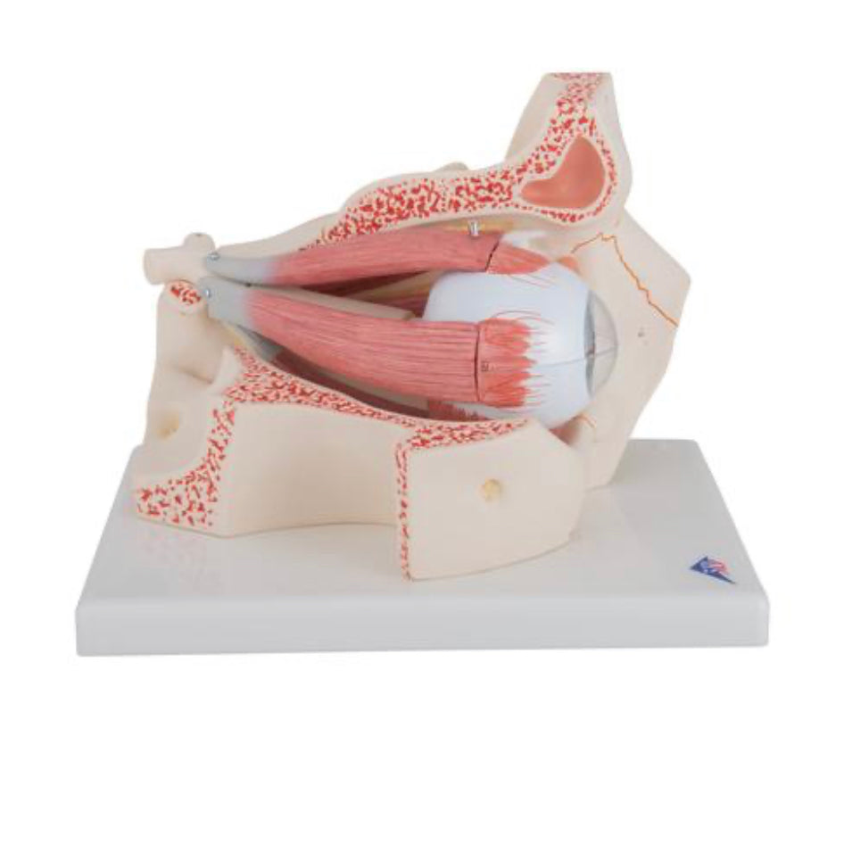
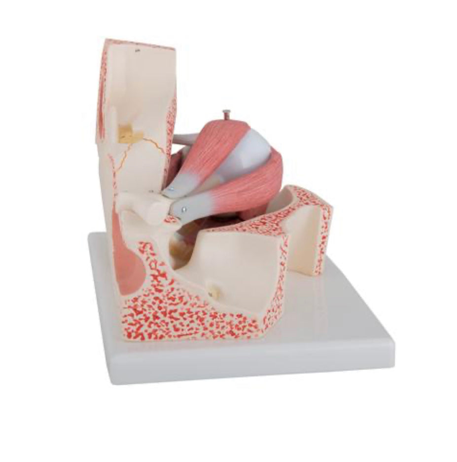
Anatomically, the model gives both an insight into many of the structures found in the orbit, but can also be used to understand the structure of the orbit, with its many bony components.
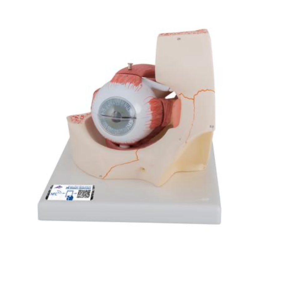
The model shows the structure of the eyeball with its different layers:
The outermost layer is made up of the transparent cornea, which at the limbus is connected to the sclera (the white tendon that makes up the back 5/6).
The middle layer (called the uvea) which consists of the iris (rainbow) incl. the pupil, the corpus ciliare (the ray body) of which the ciliary muscle is primarily seen and the choroid (the choroid).
The innermost layer is called the retina.
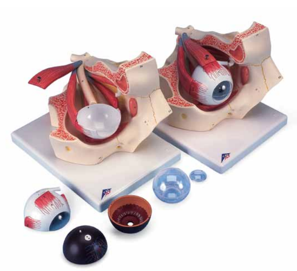
The outermost layer of the eyeball can be halved and removed, so that the vitreous body (corpus vitreum) and the lens become visible.
Furthermore, it can be seen how the optic nerve (N. opticus) leaves the eyeball at the back, as well as the crossing of the optic nerve (chiasma opticus).
On the outside of the white tendon membrane (sclera) you can see the 6 striated muscles that control eyeball movements. These are divided into the 4 straight muscles (musculi recti) and the 2 oblique muscles (musculi obliqui).
Flexibility
Flexibility
Clinically speaking
Clinically speaking
Clinically, the model can be used to understand diseases of the eye. It can be, for example, "red eye", retinal detachment, vitreous collapse, diabetic eye disease, uveitis, scleritis as well as metastases and primary tumors such as malignant melanomas (birthmark cancer) in the uvea.
Share a link to this product


A safe transaction
For 19 years I have been managing eAnatomi and sold anatomical models and posters to 'almost everyone' who has anything to do with anatomi in Scandinavia and abroad. When you place your order with eAnatomi, you place your order with me and I personally guarantee a safe transaction.
Christian Birksø
Owner and founder of eAnatomi and Anatomic Aesthetics