SKU:EA1-1020162
5-part brain model in skull
5-part brain model in skull
ATTENTION! This item ships separately. The delivery time may vary.
Couldn't load pickup availability
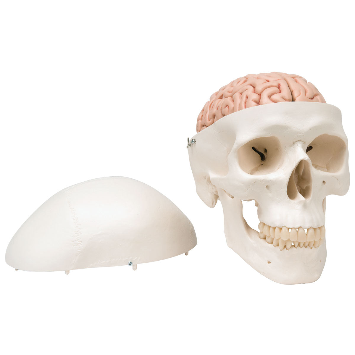
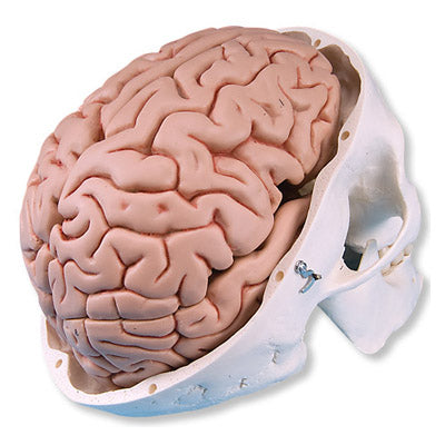
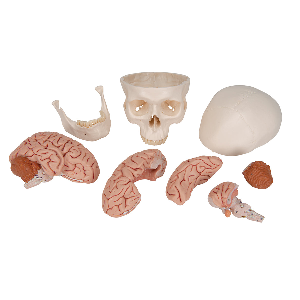
With this model, you get a skull that can be separated into three parts. This reveals its contents in the form of a classic five-part brain model, cast after a real preparation. Thus, the model becomes an ideal tool for anyone dealing with brain anatomy, brain injuries and patient education.
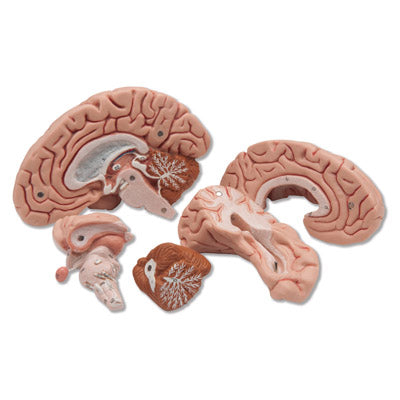
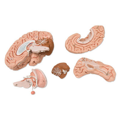
The skull can be separated into three parts, as the top of the skull and the lower jaw can be removed. The brain can be separated into two hemispheres, one of which can be further divided into:
- Combined temporal and occipital lobe
- Combined frontal and parietal lobe
- The brainstem
- cerebellum
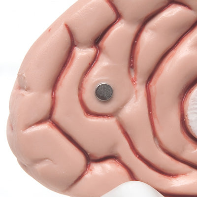
The individual brain parts are held together by discrete magnets, and are thus easy to handle when the model needs to be taken apart and reassembled.
The model weighs 1.58 kg and has the dimensions 20 x 13.5 x 15.5 cm (height x width x length). Since the model is molded from a real brain preparation, it will be slightly smaller than the average brain.
Important anatomical structures are numbered on the brain, but not on the skull. The names of the individual structures appear in the product manual, which can be downloaded as a pdf via our website - see under the product images on the left. This model is referred to as C18 in the manual.
Anatomically speaking
Anatomically speaking
Anatomically, the model shows both the human brain and the skull that surrounds it. The model thus provides a good insight into how the various structures that run from the brain pass through various holes in the skull. For example, you can see how the brainstem runs through the forearm magnum at the base of the skull.
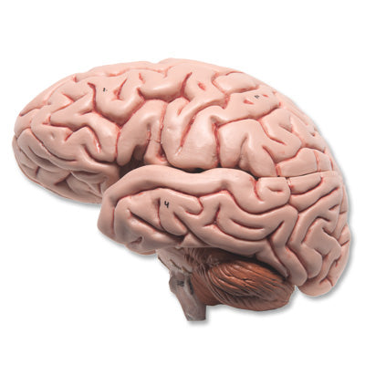
The brain can generally be divided into the cerebrum (cerebrum), the cerebellum (cerebellum) and the brainstem (truncus encephali).
These 3 structures are clearly separated via different color tones, and the difference between gray and white matter can be clearly seen on this model.
In the cerebrum (telencephalon and diencephalon), the lobes of the brain, as well as the thalamus and hypothalamus (and the pituitary gland) are primarily seen
In the cerebellum, the vermis cerebelli and the cerebellar hemispheres (hemisperium cerebelli) are seen
In the brainstem you can see its 3 parts (the midbrain, the pons and the medulla oblongata) as well as the apparent origin of the cranial nerves (also called the cranial nerves)
Other structures such as the brain stem, fornix, ventricular system and the first 2 cranial nerves (the olfactory and optic nerves) are also seen, which do not originate from the brainstem
The pictures on the left show how the model can be separated into 5 parts. This makes it possible to study the internal structures of the brain, and several of these are seen in 3 dimensions.
The limbic system
Many of our customers ask about the limbic system in connection with the purchase of brain models. Hence this description.
The limbic system includes various anatomical structures in the central nervous system (CNS), and is primarily responsible for emotional functions such as anxiety, aggressiveness, mood, memory and social adaptability. Clinically, it is therefore often related to psychiatric disorders.
The limbic system includes, among other things amygdala, hippocampus, gyrus parahippocampalis, hypothalamus, fornix, corpus mammillare, the prefrontal cerebral cortex and the monoaminergic systems of the brainstem. The list is quite a bit longer - especially because numerous fiber connections connect the limbic structures. Many customers ask in particular about the amygdala and hippocampus (which is why they are mentioned first in this section).
NB: In this brain model, the hippocampus can be seen as well as some of the other limbic structures such as the fornix - but not the amygdala.
The amygdala is involved in anxiety and emotional coloring of sensory impressions. It lies as an almond-shaped nucleus IN FRONT of the hippocampus in the anterior pole of the temporal lobe (amygdala and hippocampus are therefore separate).
The hippocampus is involved in memory. It lies as an irregular twisted structure in the medial part of the temporal lobe.
As the amygdala lies IN FRONT of the hippocampus (roughly speaking further "toward the forehead"), both of these structures can only be seen on a brain model if the model includes at least 2 frontal/coronal sections through the temporal lobe - or if the brain model is partially transparent (frontal/ coronal incisions roughly correspond to the incision direction "from ear to ear").
We have not yet seen a brain model that shows 2 cuts through the temporal lobe so that both the amygdala and the hippocampus are seen. In our range, on the other hand, we have a partially see-through brain model in the highest price range, which shows both structures. The model can be viewed via THIS LINK.
All brain models in our range can be separated into different parts. All models (both with and without educational colors) that can be separated into 4 or more parts show the hippocampus. On almost all of these models, the hippocampus is also numbered and named on an overview that can be downloaded from the product descriptions of the brain models. This also applies to this brain model.
Flexibility
Flexibility
Clinically speaking
Clinically speaking
Clinically, the model can be used to understand lesions and disorders in specific areas of the brain. Examples are epilepsy, brain tumors, hydrocephalus, lesions involving cranial nerves and sclerosis (multiple sclerosis).
Although the brain's blood supply is not visible on the model, it can also be used to understand apoplexy (stroke).
Share a link to this product

A safe transaction
For 19 years I have been managing eAnatomi and sold anatomical models and posters to 'almost everyone' who has anything to do with anatomi in Scandinavia and abroad. When you place your order with eAnatomi, you place your order with me and I personally guarantee a safe transaction.
Christian Birksø
Owner and founder of eAnatomi and Anatomic Aesthetics