Skip to product information
1 of 7

SKU:EA1-1000275

CT bronchi with larynx 3D model via tomographic data with lungs

CT bronchi with larynx 3D model via tomographic data with lungs

Regular price 5.940,00 DKK
Regular price Sale price 5.940,00 DKK
Sale Sold out
Not including taxes if ordering from outside EU. Shipping calculated at checkout.

Out of stock

this product is made to order. To place an order please call or write us.

If you are looking for a model that uniquely reproduces the realistic structure of the bronchial tree, right down to the segmental bronchi, we recommend this one. Since the model also includes transparent lungs, the model is ideal for obtaining a spatial understanding of the location of the individual lung segments.


The model was created from computer tomographic data from a human (male, 40 years old) and is therefore able to depict the actual relationships between the components of the bronchial tree, and the location of the individual segmental bronchi, very precisely.


Of structures, the model shows the larynx, trachea and bronchial tree. The larynx can be removed at the level of the second tracheal cartilage and can be further divided in the saggital plane so that the inside of the larynx becomes visible. The lungs are also removable.


The larynx (epiglottis) is movable, and the bronchial tree itself is made of an elastic material that makes it flexible.


The model has the dimensions 22 x 18 x 37 cm (length x width x height) and weighs 1.905 kg. The model is delivered on a white stand.

Anatomical features

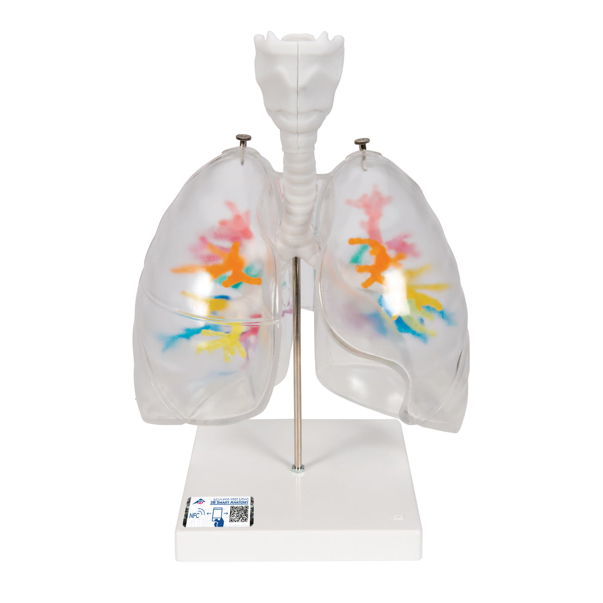
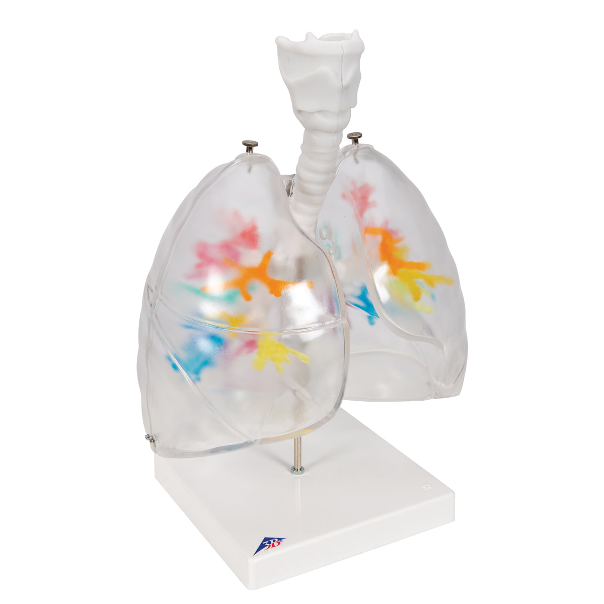
Anatomically, this model focuses on the location of the various segmental bronchi, but the model also shows the lungs, trachea and larynx with the hyoid bone (os hyoideum).


The trachea divides into a left and right main bronchus, each of which divides into 2 and 3 lobed bronchi, respectively. These, along with the throat, are pictured in white.


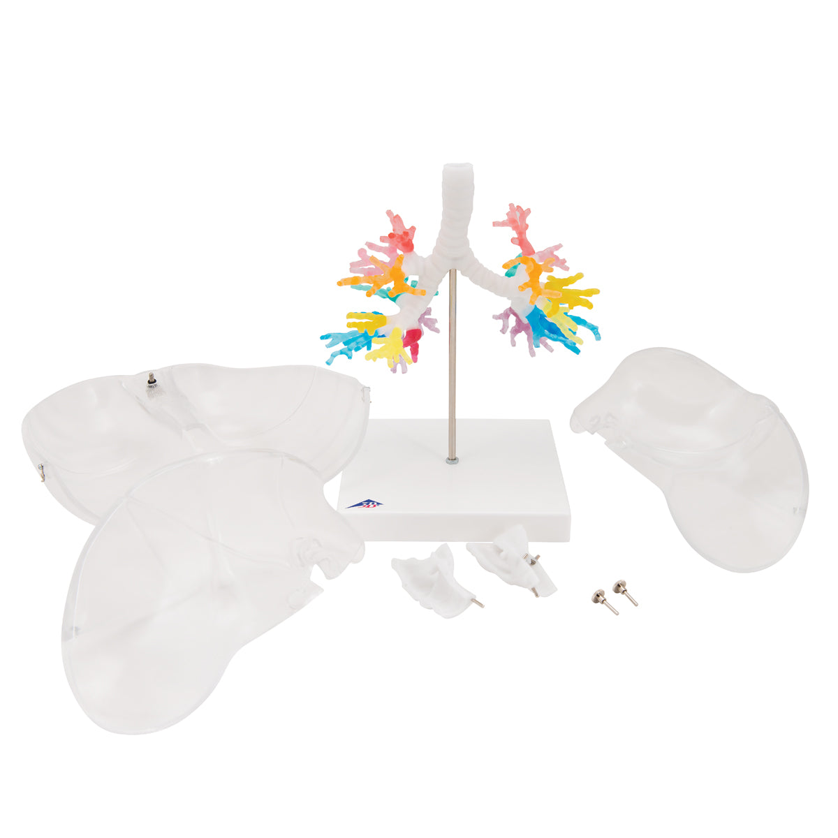
The lap bronchi divide into segmental bronchi, each of which supplies an area (segment) of the lungs with air. On the model, the individual segmental bronchi are depicted in different transparent colors to make it easier to distinguish them from each other.


On the model, the lungs are reproduced in a transparent material, which makes it possible to see the different segmental bronchi from the outside. In addition, the various furrows that divide the lung tissue into lobes (fissura obliqua and fissura horizontalis) can be seen. The lungs can be detached so that the bronchial tree can be studied in detail.

Product flexibility

Clinical features

Clinically, the model can be used to understand disease states involving the larynx, trachea and bronchial tree. For example, bronchiectasis, chronic bronchitis or epiglottitis.


Furthermore, the model can be used to understand examination methods such as bronchoscopy or the installation of a tracheostomy.

View full details

A safe deal

For 19 years I have been at the head of eAnatomi and sold anatomical models and posters to 'almost' everyone who has anything to do with anatomy in Denmark and abroad. When you shop at eAnatomi, you shop with me and I personally guarantee a safe deal.

Christian Birksø
Owner and founder of eAnatomi ApS

Read more about eAnatomi