SKU:EA1-1013869
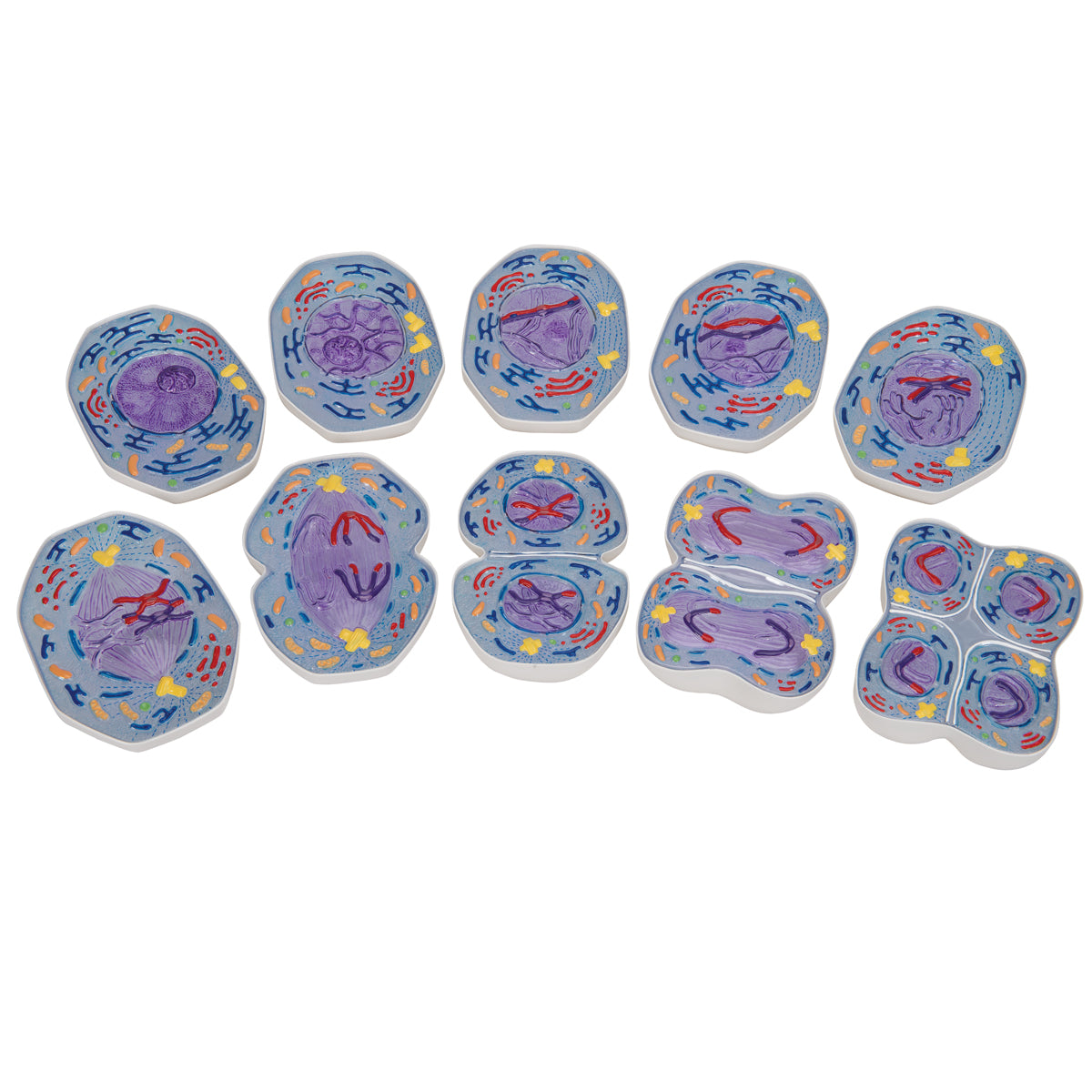
Flexible model of the phases of meiosis
Flexible model of the phases of meiosis
Out of stock
this product is made to order. To place an order please call or write us.Couldn't load pickup availability
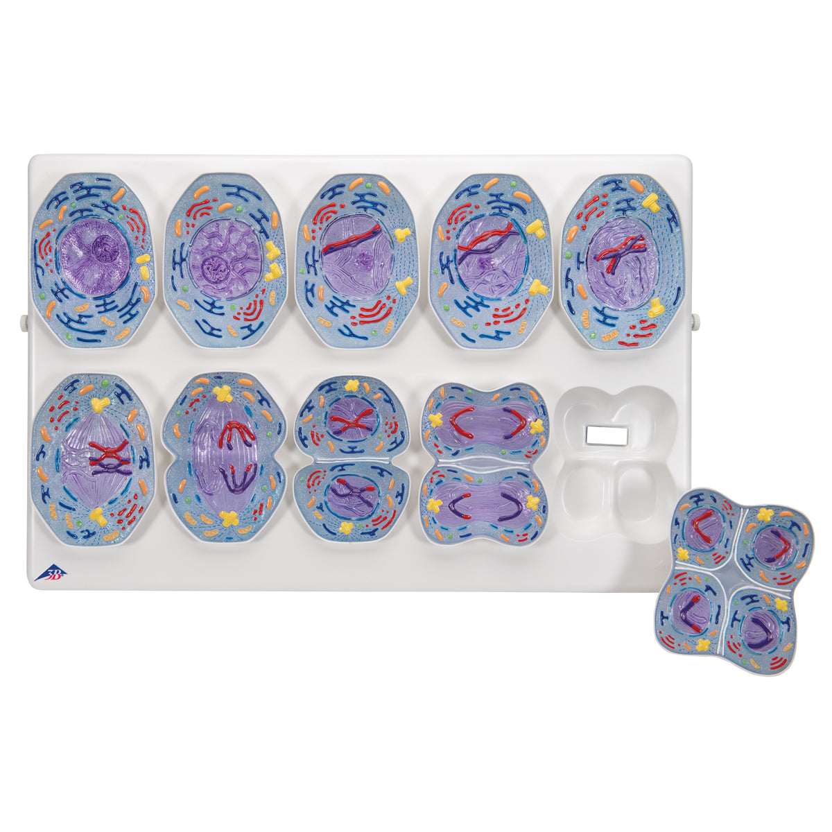
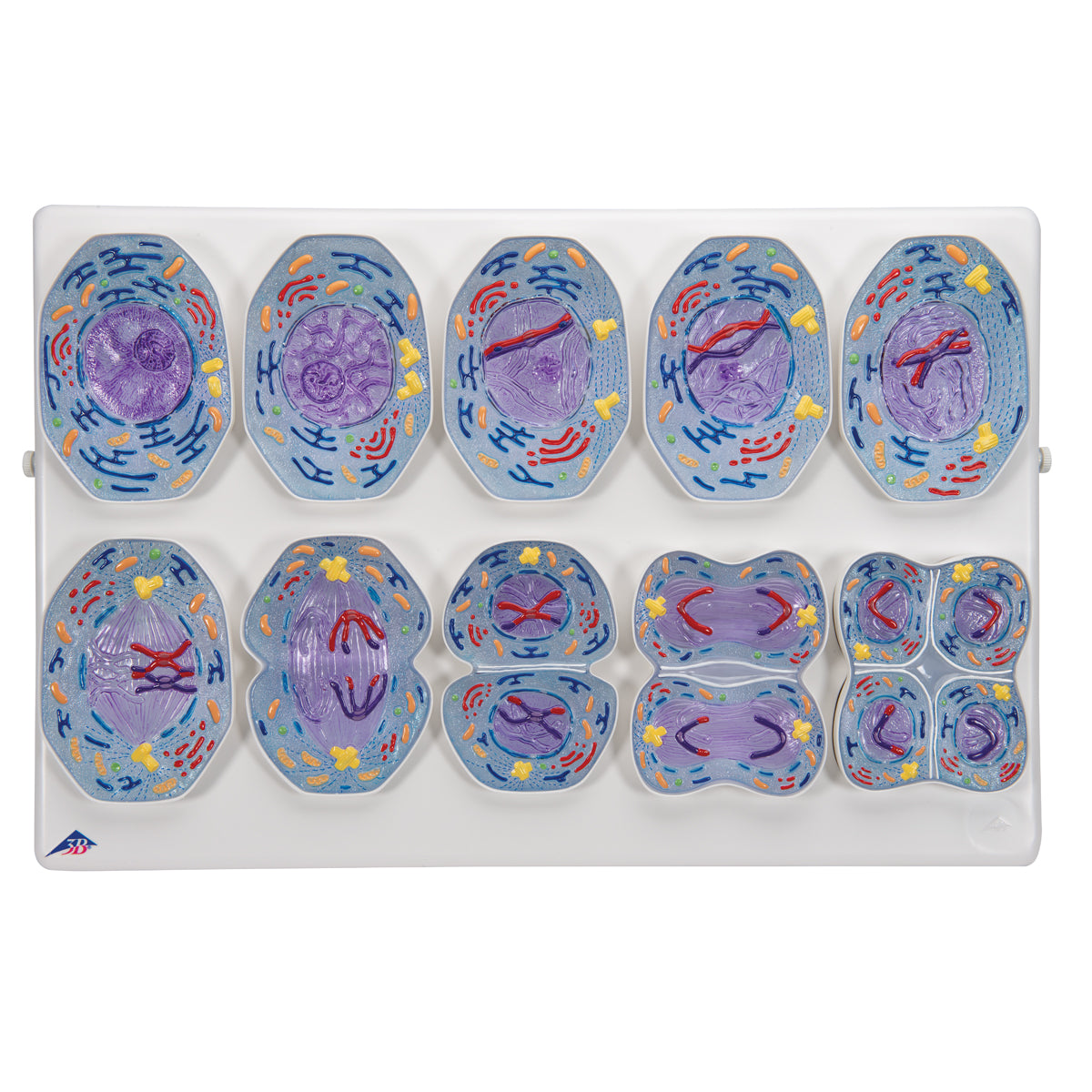
This flexible model shows the phases of meiosis using educational coloring.
The model weighs approximately 1.7 kg and the dimensions are 60 x 40 x 6 cm. Compared to the natural meiosis, the model is enlarged 10,000 times.
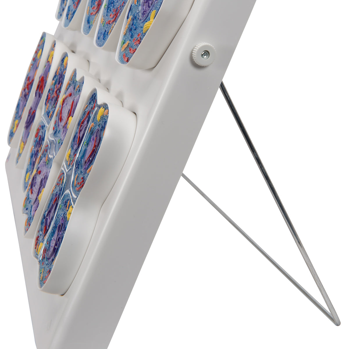
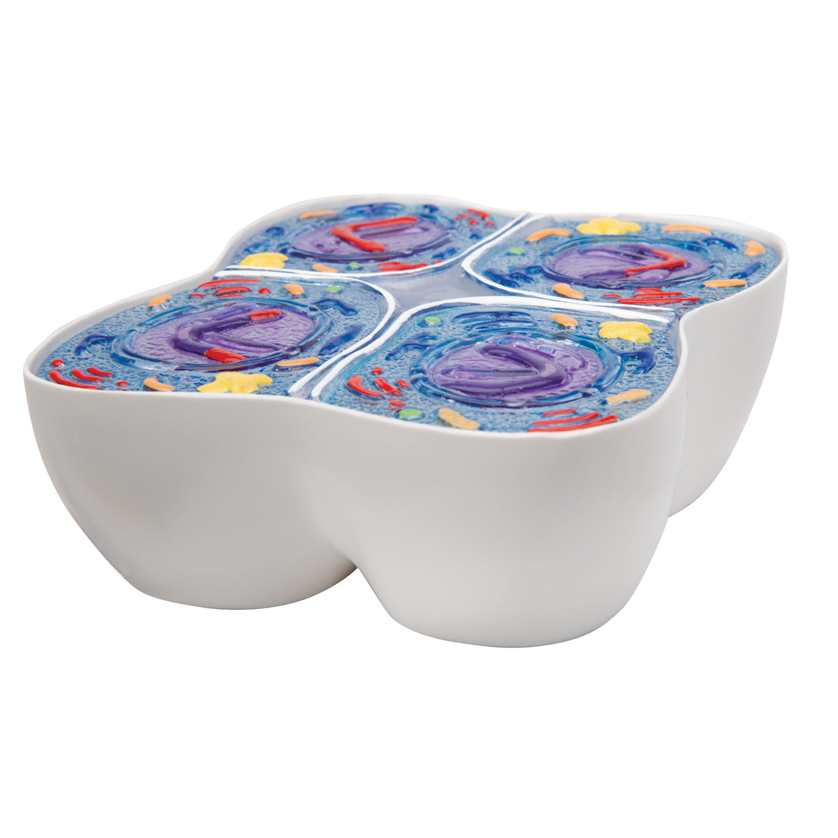
The model's 10 parts (the phases) are delivered on a stand, which can both stand on a table or hang on the wall. Furthermore, all 10 parts are provided with a magnet on the back, so that they can be removed from the stand and placed on a magnetic surface.
Anatomical features
Anatomical features
Anatomically, the model shows meiosis, which is the cell division (reduction division) in the germ cells (in the ovaries and testes). The chromosomes are colored according to a color standard, and organelles are colored in an educational way.
The following 10 phases are displayed:
1. Interphase (stage in the G1 phase)
2. Prophase I (leptotene stage)
3. Prophase I (zygote stage and pachytene)
4. Prophase I (diplotene stage)
5. Prophase I (diakinesis stage)
6. Metaphase I
7. Anaphase I
8. Telophase I, cytokinesis I, interkinesis, prophase II and metaphase II
9. Anaphase II
10. Telophase II and cytokinesis II
Product flexibility
Product flexibility
Clinical features
Clinical features
Clinically speaking, the model can for example be used to understand chromosomal diseases.
Share a link to this product


A safe deal
For 19 years I have been at the head of eAnatomi and sold anatomical models and posters to 'almost' everyone who has anything to do with anatomy in Denmark and abroad. When you shop at eAnatomi, you shop with me and I personally guarantee a safe deal.
Christian Birksø
Owner and founder of eAnatomi ApS

