SKU:EA1-1000523
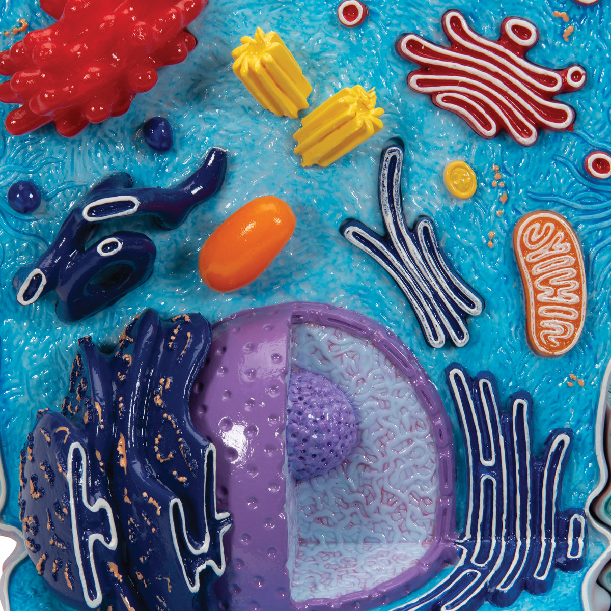
Highly detailed model of a cell in an electron microscopic perspective
Highly detailed model of a cell in an electron microscopic perspective
Out of stock
this product is made to order. To place an order please call or write us.Couldn't load pickup availability
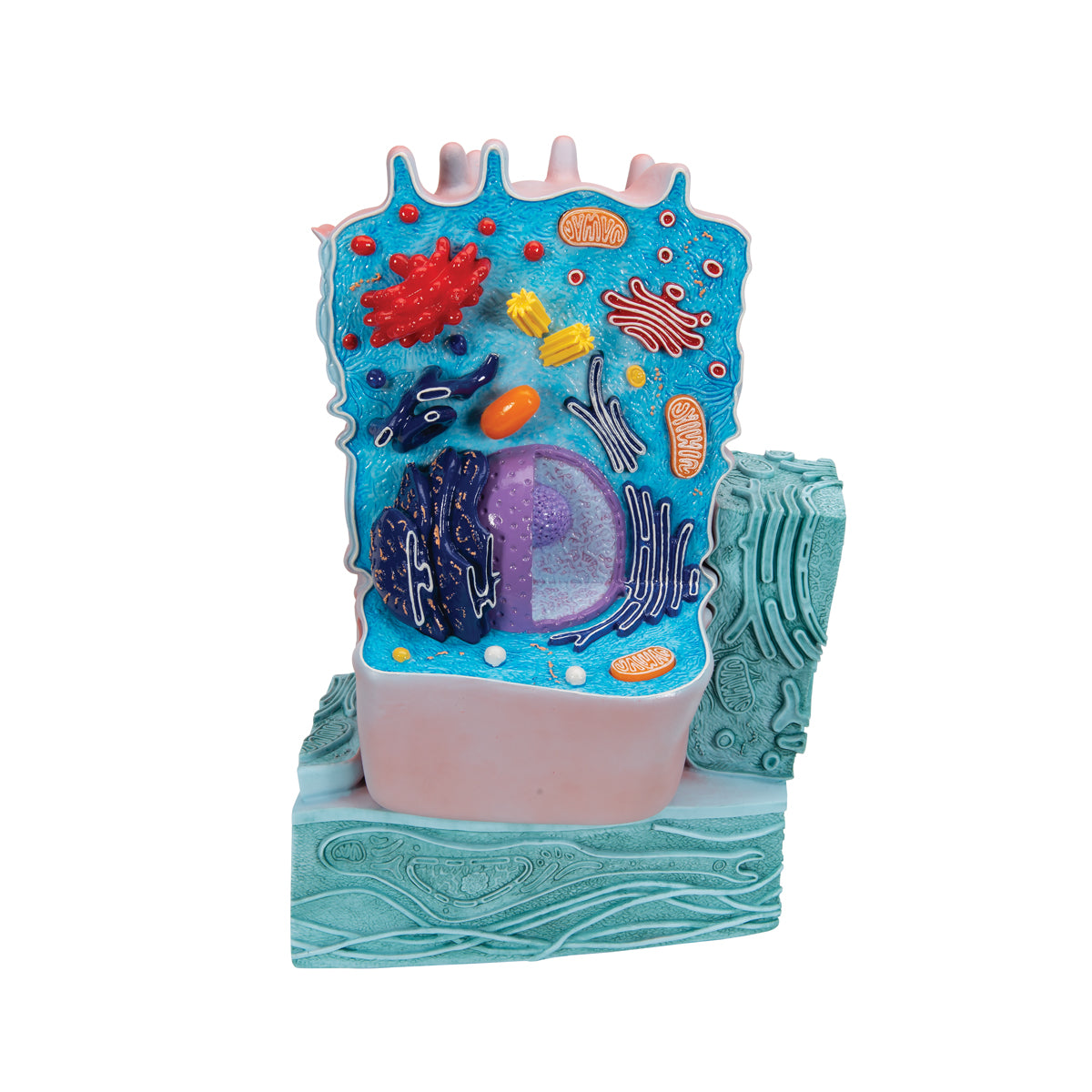
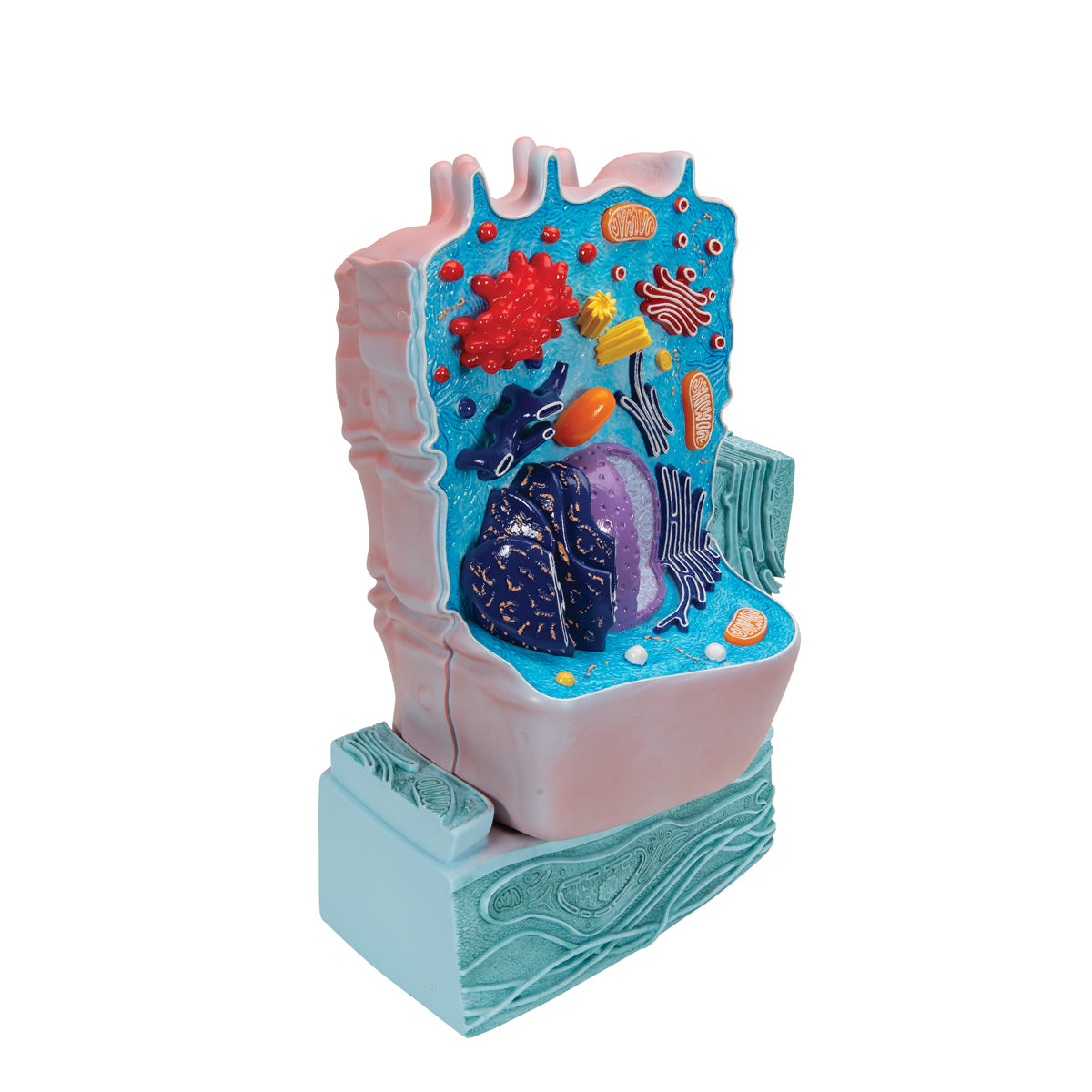
This model of an animal cell shows the main organelles in an electron microscopic perspective.
The model weighs approximately 1.3 kg and the dimensions are 21 x 11 x 31 cm.
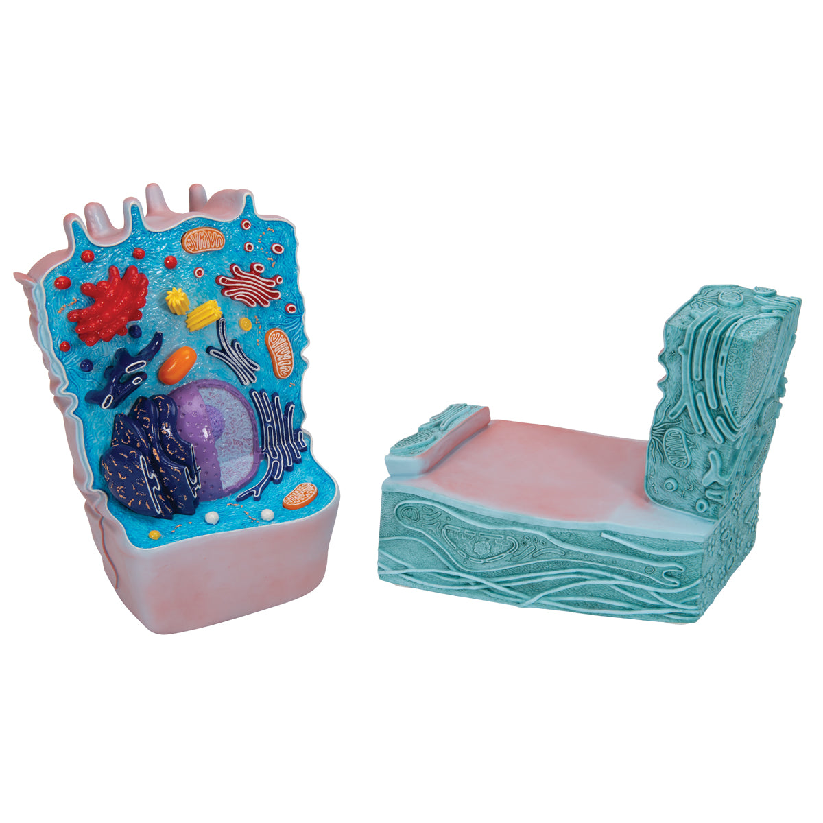
The animal cell rests on a structure in green, which illustrates a basement membrane with an underlying extracellular matrix. At the same time, this works as a stand.
Anatomical features
Anatomical features
Anatomically, the model shows organelles in a general animal cell, its relationship to another cell and a basement membrane with an underlying extracellular matrix.
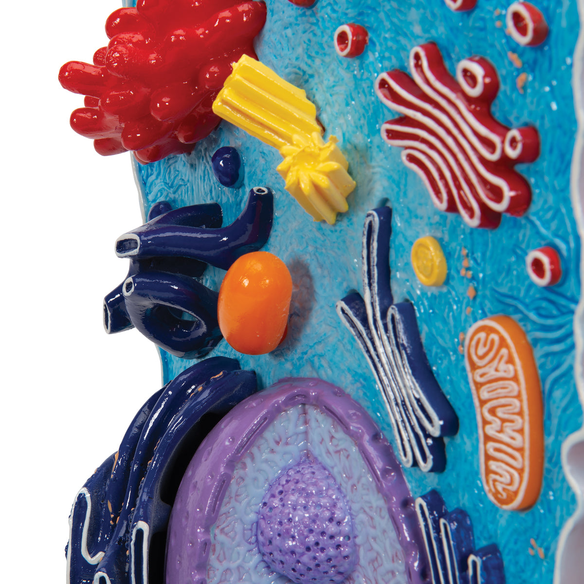
In the animal cell, for example, you see:
Nucleus with chromatin and nucleolus
Ribosomes
Rough and smooth IS
The Golgi apparatus
Mitochondria
Lysosomes and peroxisomes
And much more
Furthermore, the following relationships are seen:
The connection of the animal cell to another cell via a desmosome (a hemidesmosome also seen)
A basement membrane on which the animal cell rests. "Below" can be seen extracellular matrix with collagen and a fibroblast
Product flexibility
Product flexibility
Clinical features
Clinical features
Clinically, the model can be used to understand diseases, although the model is most suitable for studying the cell and its components.
Examples of diseases could be mitochondrial diseases, lysosomal diseases and connective tissue diseases in the skin where too much connective tissue (especially collagen) is produced.
Share a link to this product


A safe deal
For 19 years I have been at the head of eAnatomi and sold anatomical models and posters to 'almost' everyone who has anything to do with anatomy in Denmark and abroad. When you shop at eAnatomi, you shop with me and I personally guarantee a safe deal.
Christian Birksø
Owner and founder of eAnatomi ApS

