Skip to product information
1 of 7

SKU:EA1-1008494

Lung model with heart and bronchi, divided into segments

Lung model with heart and bronchi, divided into segments

Regular price 8.310,00 DKK
Regular price Sale price 8.310,00 DKK
Sale Sold out
Not including taxes if ordering from outside EU. Shipping calculated at checkout.

Out of stock

this product is made to order. To place an order please call or write us.

This model shows in a pedagogical way the division of the two lungs into segments, and the supply of the individual segments from the segmental bronchi. In addition, the model shows the contents of the mediastinum, and thus provides a good overview of the relationships between the lungs and the segments.


Each lung is divided into lobes, which are further divided into segments. Segments from the same lobe are depicted in the same color to make it easy to assemble and separate the lungs. In addition, the individual parts are mounted with magnets to make it easy to handle the model.


The model has the dimensions 25 x 25 x 35 cm (length x width x height), weighs 4.291 kg and is delivered on a white stand.

Anatomical features

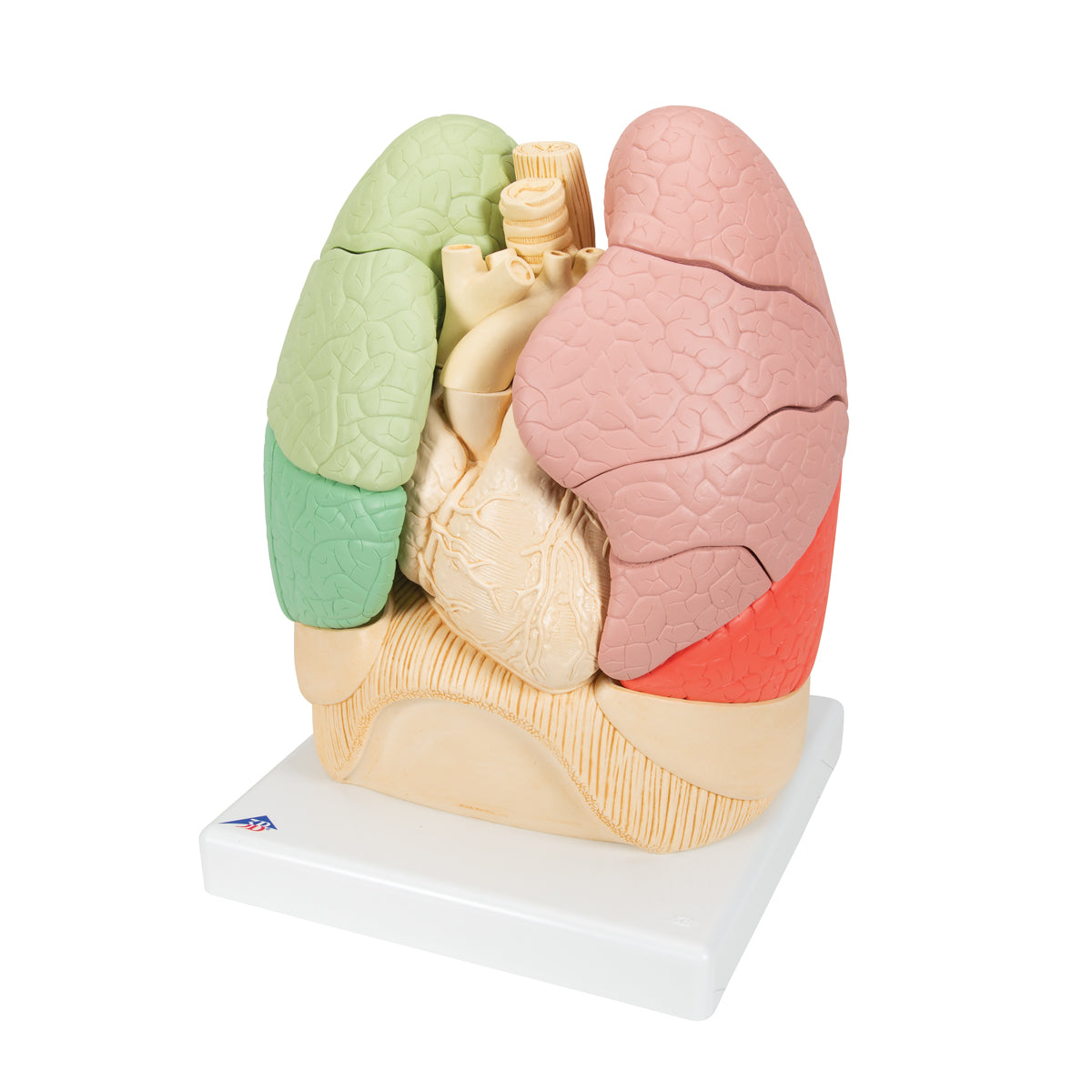
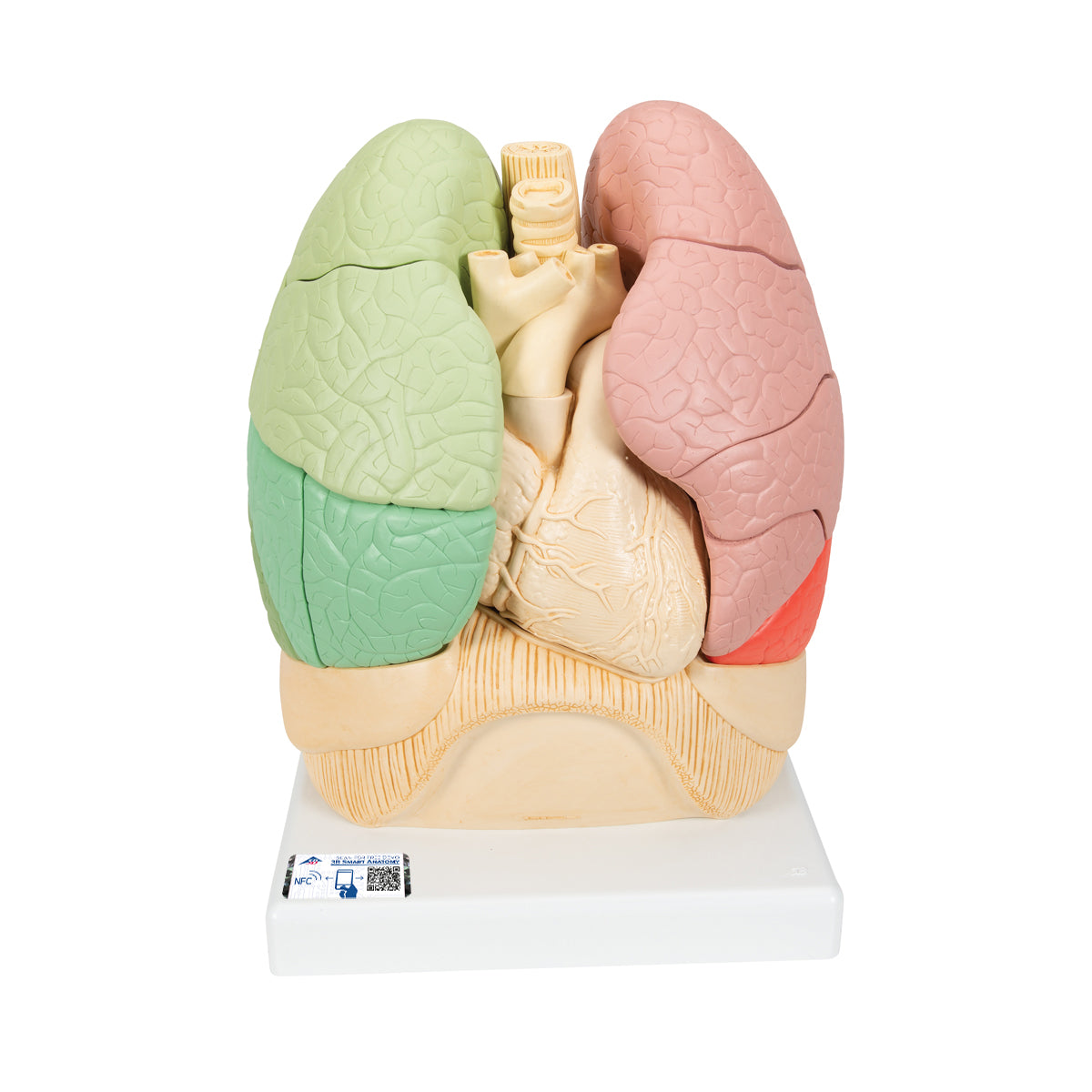
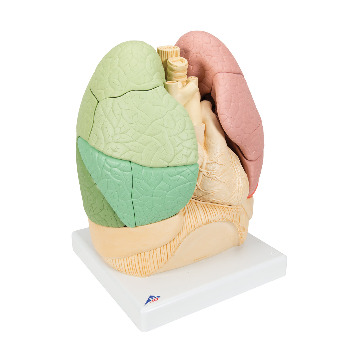
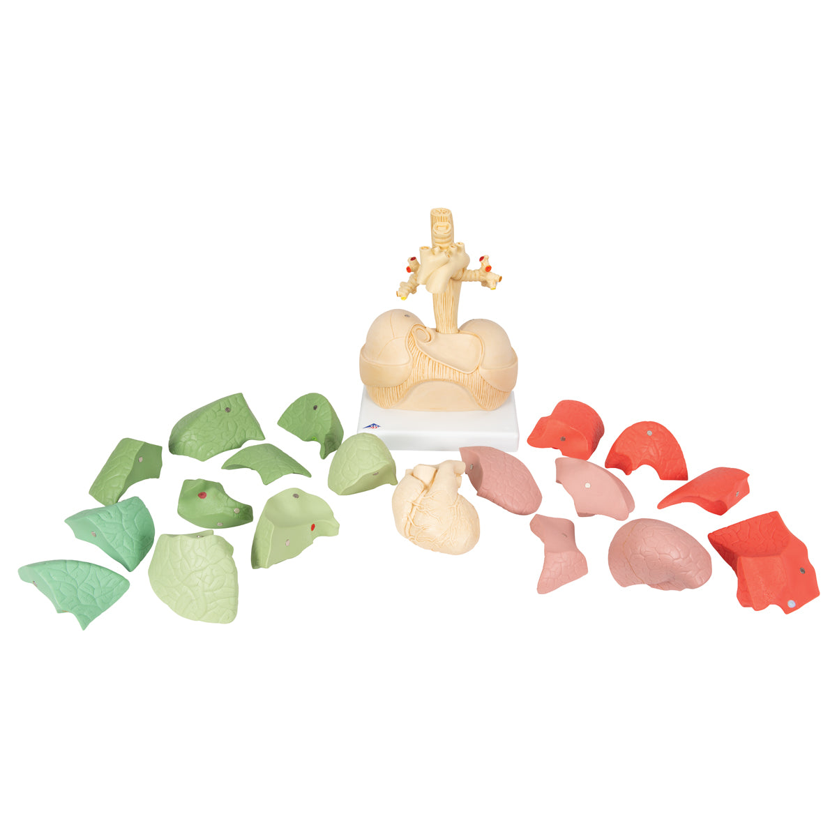
Anatomically, the model can be used to understand the structure of the bronchial tree and the division of the lungs into lobes and segments.


The bronchial tree begins where the trachea divides into two main bronchi. Each main bronchus then divides into lobe bronchi. Since the right lung consists of 3 lobes, while the left lung only consists of 2 lobes, there are 3 lobe bronchi on the right side, while on the left side there are 2.


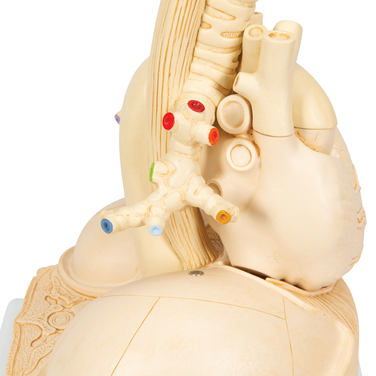
Each lobe bronchus subsequently divides into segmental bronchi that supply the individual lung segments.


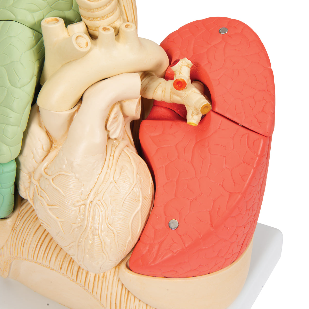
On the model, the individual segment bronchi are cut where the segment has been removed. To easily match segment with segmental bronchus, the two cut ends of the segmental bronchus are marked with the same color (see picture on the left).


When the lungs are assembled, the furrows that divide the lungs into segments (respectively fissura horizontalis and fissura obliqua) are visible.


Between the two lungs is the mediastinum, as well as some of the structures found here. Among other things, you can see the heart, which is removable on the model, as well as the large vessels that run to and from the heart.


Behind the trachea is the esophagus which, together with the aorta, can be followed down to the diaphragm, through which they both pass, on their way down into the abdominal cavity.

Product flexibility

Clinical features

Clinically speaking, the model can be used to understand diseases in the lungs. Examples are blood clots in the lungs, pneumothorax, obstructive lung diseases, bronchiectasis and cancer of the lungs.


The model can also be used to understand disease elsewhere in the chest cavity. Examples of heart disease are valvular disease, inflammation and blood clots.


Furthermore, the model can be used to understand examination methods such as bronchoscopy.

View full details

A safe deal

For 19 years I have been at the head of eAnatomi and sold anatomical models and posters to 'almost' everyone who has anything to do with anatomy in Denmark and abroad. When you shop at eAnatomi, you shop with me and I personally guarantee a safe deal.

Christian Birksø
Owner and founder of eAnatomi ApS

Read more about eAnatomi