Skip to product information
1 of 3

SKU:EA1-1000232

Nerve cell anatomy and physiology - 5 models on magnetic board

Nerve cell anatomy and physiology - 5 models on magnetic board

Regular price 6.305,00 DKK
Regular price Sale price 6.305,00 DKK
Sale Sold out
Not including taxes if ordering from outside EU. Shipping calculated at checkout.

Out of stock

this product is made to order. To place an order please call or write us.

Are you looking for a model that both gives an overview of how the individual nerve cell is structured, but also shows how the different parts of the nerve cell communicate with the environment? Then we recommend this model.


The model weighs 4.379 kg and has the dimensions 68 x 51 x 3 cm (length x width x height).


Overall, the model is divided into 2 parts. The larger part (left) shows a large illustration of the cell body of the nerve cell, while the smaller part (right) shows a drawing of a nerve cell along its entire length, with associated magnetic elements showing the underlying anatomical structures in relief.

Anatomical features

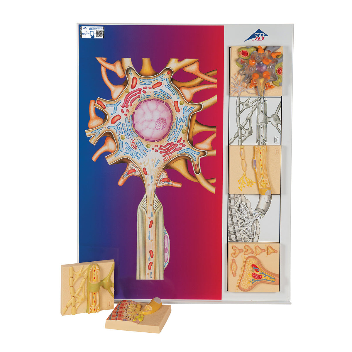
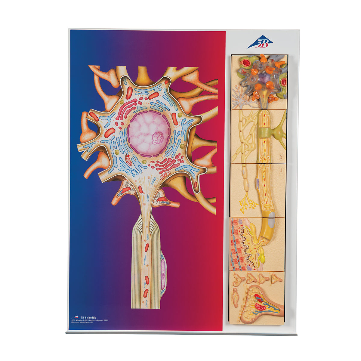
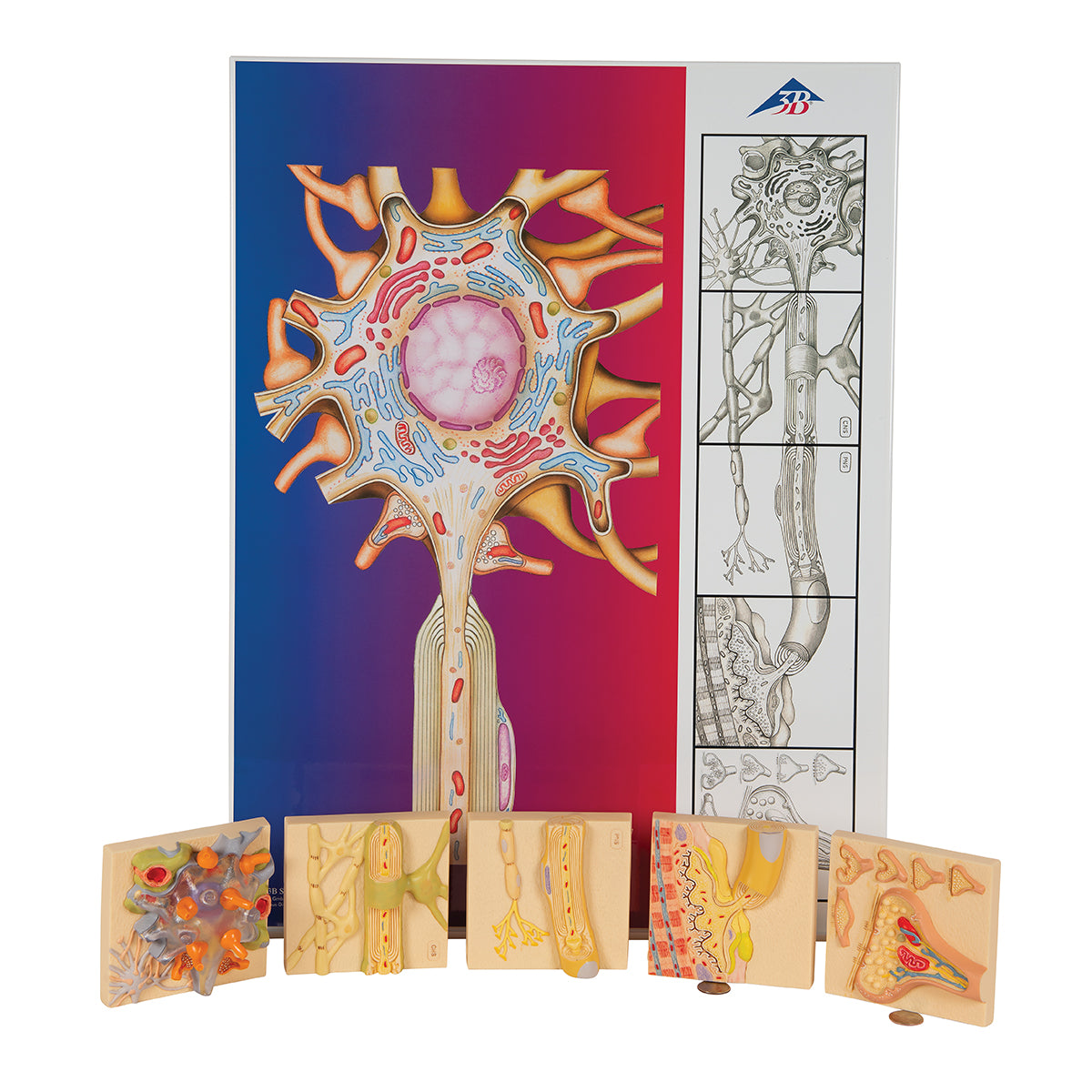
Anatomically speaking, each of the 5 magnet reliefs shows a section of the different segments of the nerve cell. Notice here that it varies from relief to relief, whether it is the central nervous system or the peripheral nervous system that is shown.


In the following, a description of each of the 5 segments is given:


1. Shows the cell body of the nerve cell with associated organelles. The cytoplasm is made transparent so that, among other things, the cell nucleus (nucleus) and the mitochondria can be seen through. Furthermore, it can be seen how nerve terminals (in orange) from surrounding nerve cells synapse along the edge of the cell body.


2. Shows how the nerve cell's axon, in the central nervous system, is surrounded by a myelin sheath, which here is formed by astrocytes (green cell).


3. Shows how the nerve cell's axon, in the peripheral nervous system, is surrounded by a myelin sheath, which here is formed by Schwann cells.


4. Shows the motor end plate, which is the area on a skeletal muscle cell where the nerve cell's axon terminal synapses. Here is the neuromuscular junction, where communication between the two cells takes place.


5. Shows a synapse with a synaptic cleft. In the axon terminal you can see the synaptic vesicles, which contain neurotransmitter substances. In addition, 5 smaller images are shown which show how the design of the synapse can vary.


On the left in segments 2 and 3, it is illustrated how a single astrocyte in the central nervous system forms several myelin sheaths, while each Schwann cell in the peripheral nervous system forms a single myelin sheath.

Product flexibility

Clinical features

Clinically speaking, the model can be used to describe and understand diseases that affect the neurons in the central nervous system as well as in the peripheral nervous system. This can for example be diseases that affect the myelin sheaths (such as ALS) or inflammatory conditions in the nerves (neuropathies).

View full details

A safe deal

For 19 years I have been at the head of eAnatomi and sold anatomical models and posters to 'almost' everyone who has anything to do with anatomy in Denmark and abroad. When you shop at eAnatomi, you shop with me and I personally guarantee a safe deal.

Christian Birksø
Owner and founder of eAnatomi ApS

Read more about eAnatomi