SKU:EA1-A589IIIM
Pelvic model showing the pelvic floor, the internal and external genitalia and the female bladder
Pelvic model showing the pelvic floor, the internal and external genitalia and the female bladder
2 in stock
Pickup available at Frederiksberg
Usually ready in 4 hours
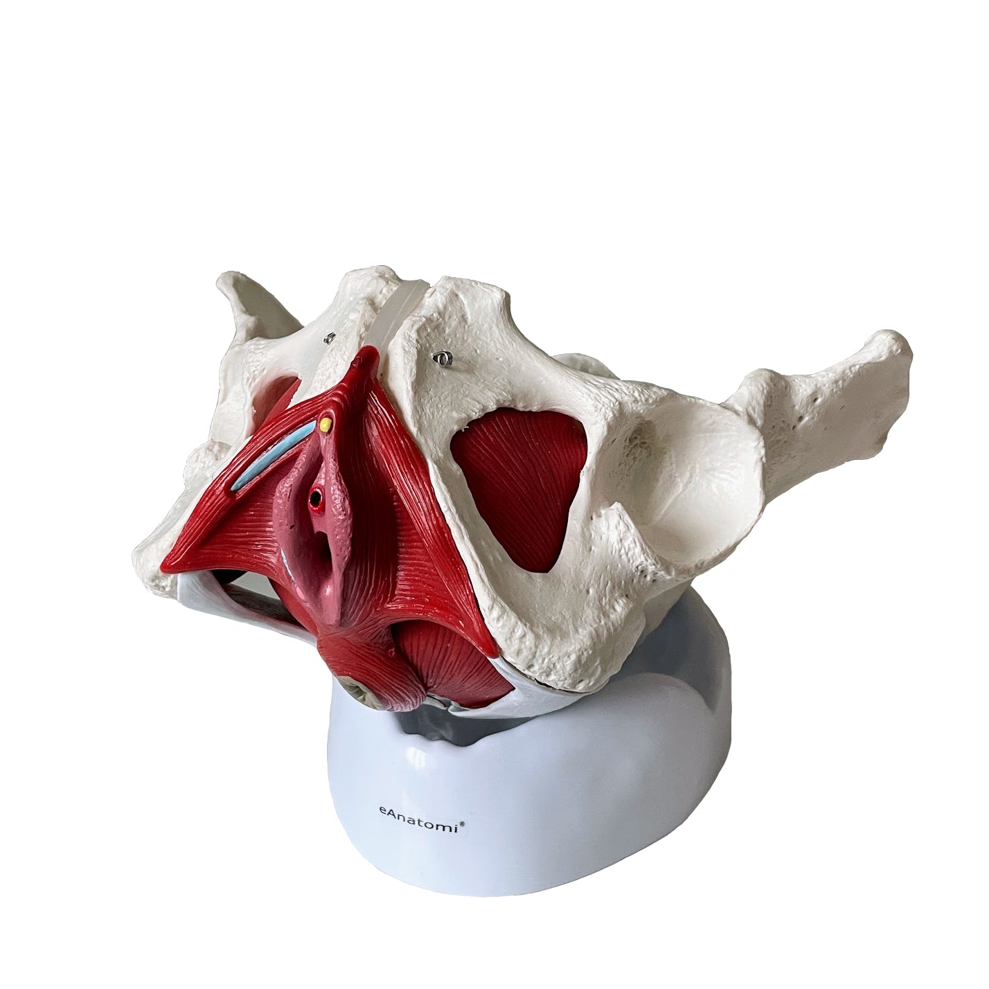
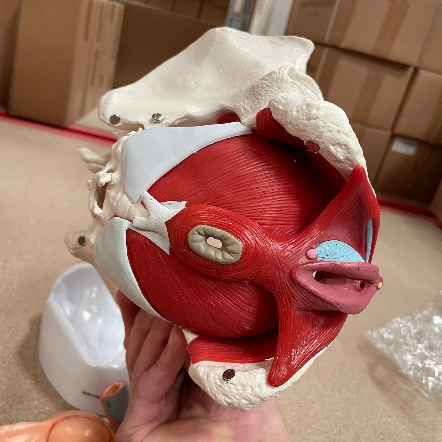
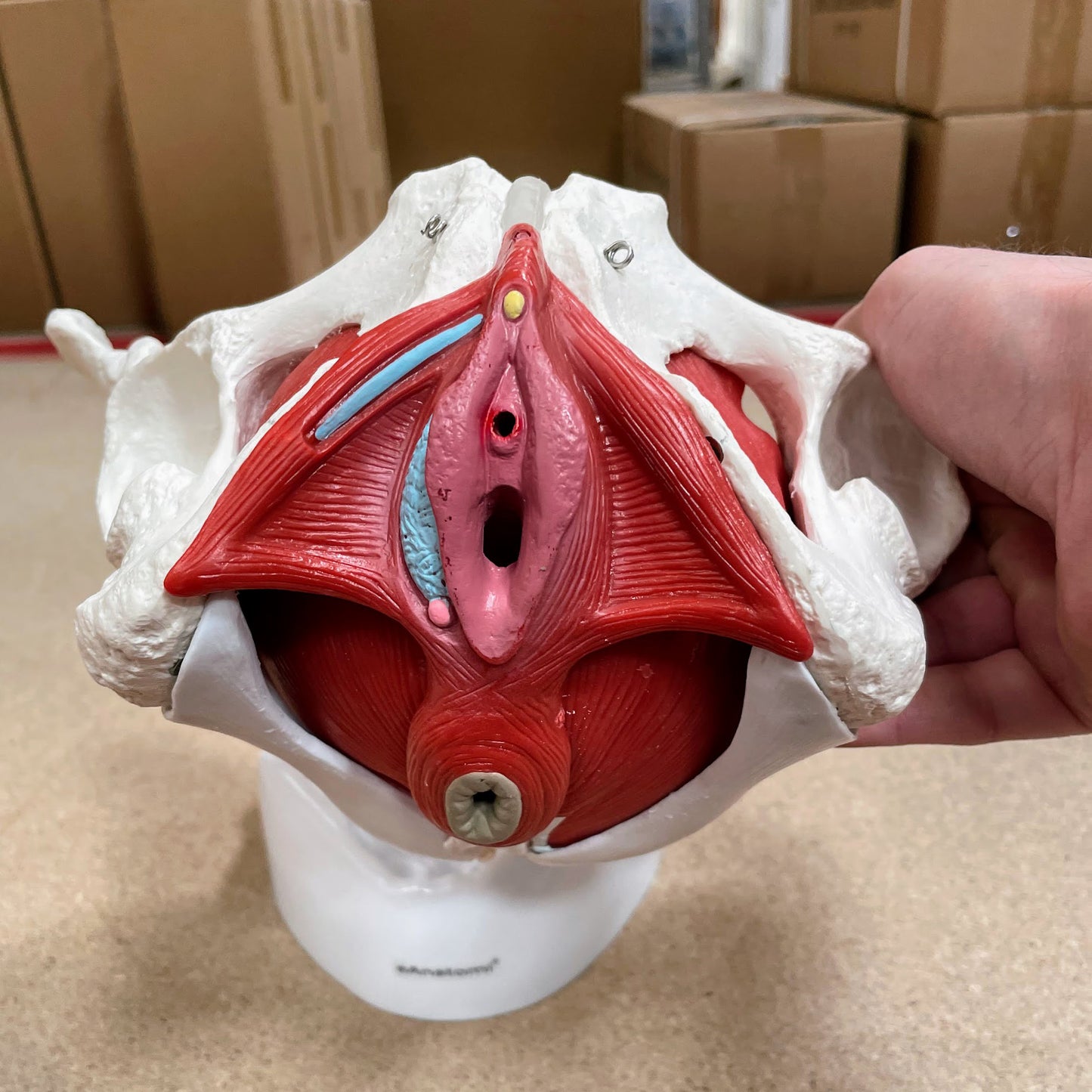
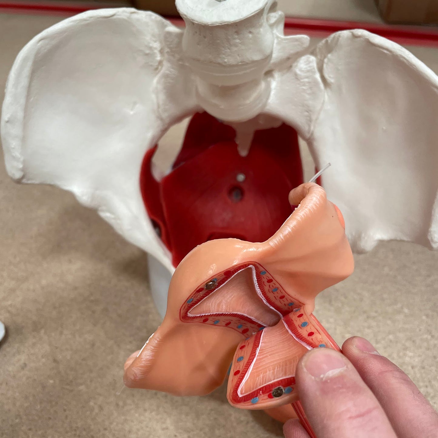
This cymbal model is particularly educational, practical and decorative. It shows the woman's pelvis with a focus on the pelvic floor and the internal and external genitalia.
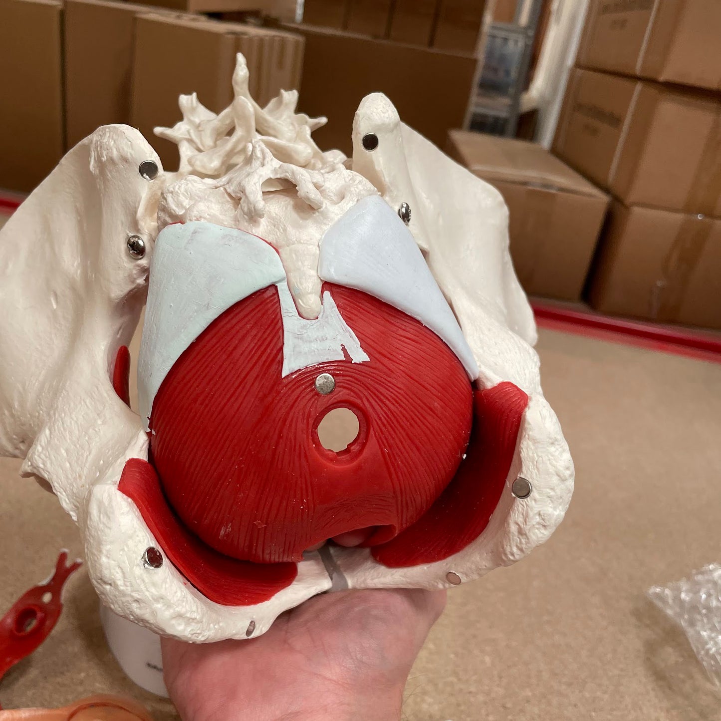
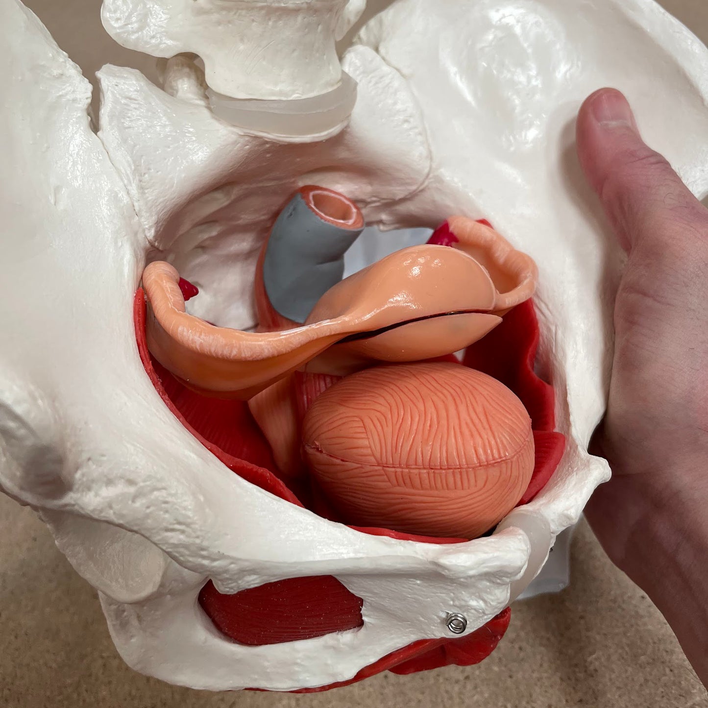
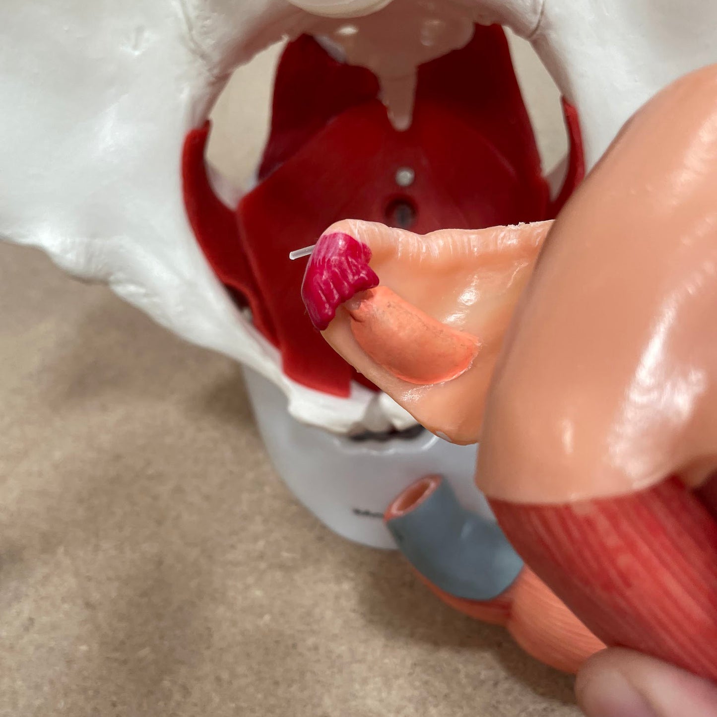
The model is slightly reduced compared to an adult and corresponds in size to a 14-year-old. The pelvic ring, which is made up of bones, cannot be separated at all, although the sacrum can be opened. On the other hand, all the other organs can be taken out of the model and some of them can be separated because they are held together by discrete magnets (see the pictures on the left).
NB: As the model has been developed to be able to be separated in an easy and educational way as mentioned above. If you want for practical reasons that fewer parts can be easily removed, you can use a little glue to fix the parts that you do not need to remove - for example the ligaments on the back of the model.
Anatomical features
Anatomical features
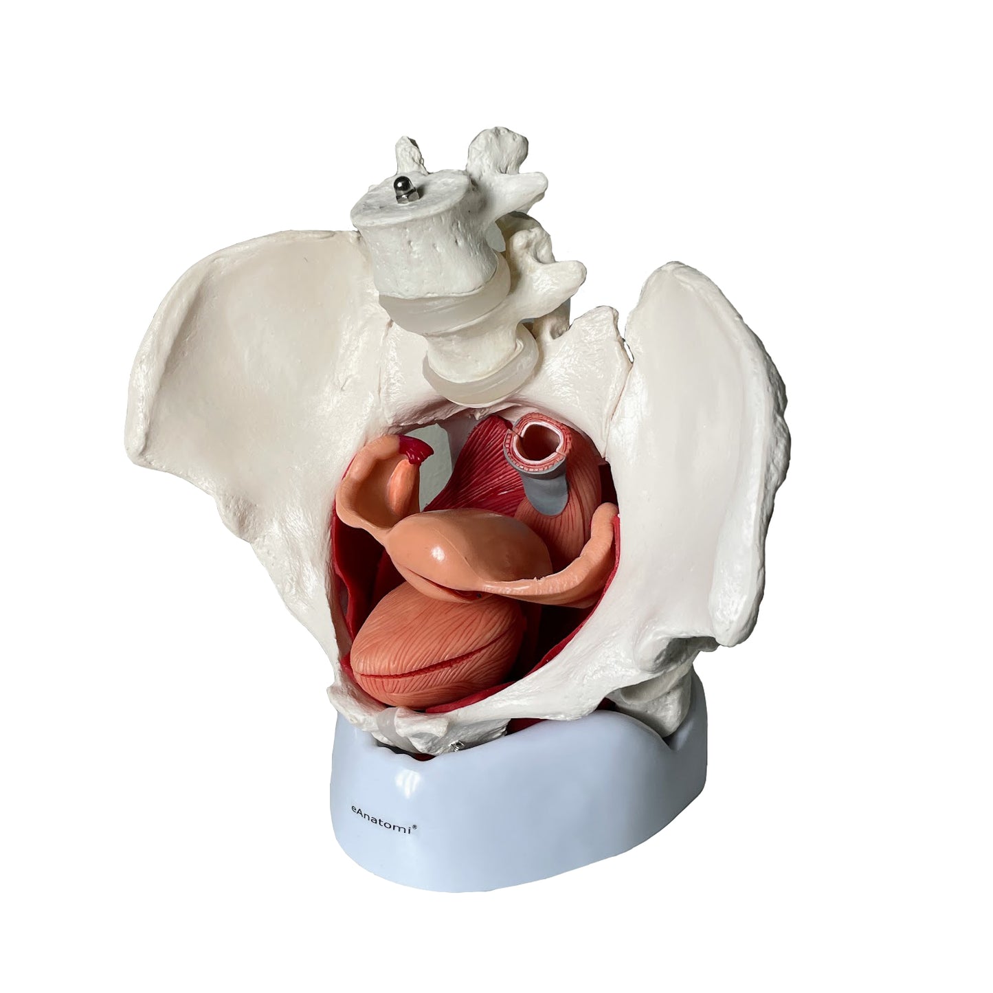
Anatomically speaking, the model shows both the woman's pelvis, the muscles in the pelvic floor, the internal and external genitalia as well as the relationships of the urinary bladder with the urethra and the rectum.
The pelvis consists of the 3 building blocks, which are the 2 hip bones and the sacrum. The model also shows the 2 lower lumbar vertebrae. Overall, the model shows the following bones:
Os coxae (the 2 hip bones) of which both are composed
- Os ilium (iliac bone/hip bone)
- Os ischii (seat bone)
- Os pubis (pubic bone)
Us sacrum (sacrum) incl. os coccygis (coccyx)
4th and 5th lumbar vertebra (lumbar vertebra)
In the bone tissue, the most important details can be seen, such as large nodules. Smaller details such as the linea glutaea are omitted. As something special, the sacrum can be opened. It makes it possible to see the canalis sacralis as well as the Y-shaped canals in its lateral wall (where nerves run). Furthermore, some ligaments are seen. It is first and foremost equal. sacrospinal and lig. sacrotuberal.
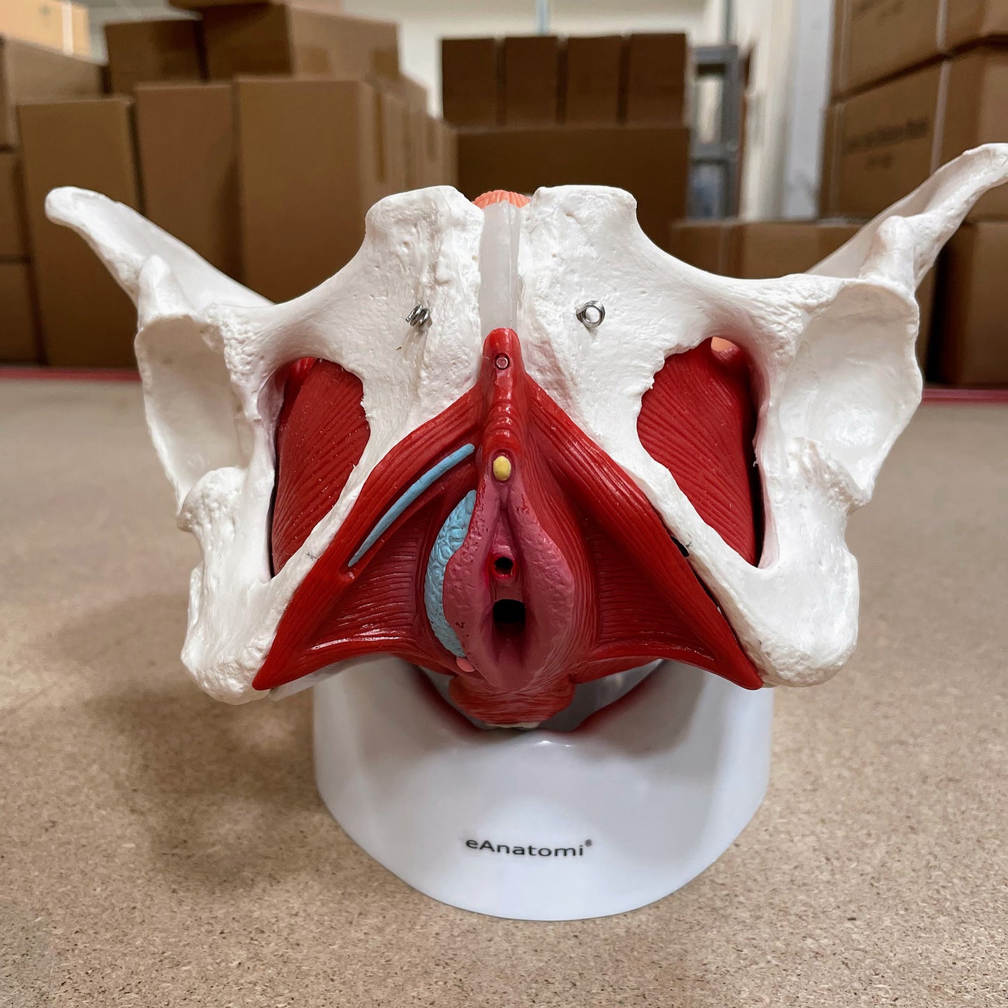
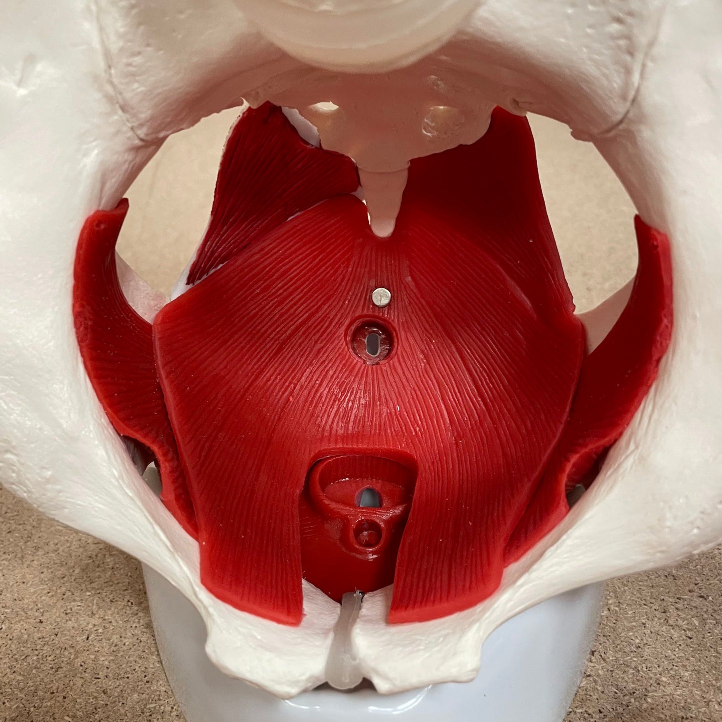
The pelvic floor (also called the pelvic floor or diaphragma pelvis in Latin) consists of the following muscles, which are clearly visible:
- M. levator ani (consisting of m. pubococcygeus and m. iliococcygeus)
- M. coccygeus
Other muscles are also seen such as the ischiocavernosus muscle, the bulbospongiosus muscle and the obturator internus muscle.
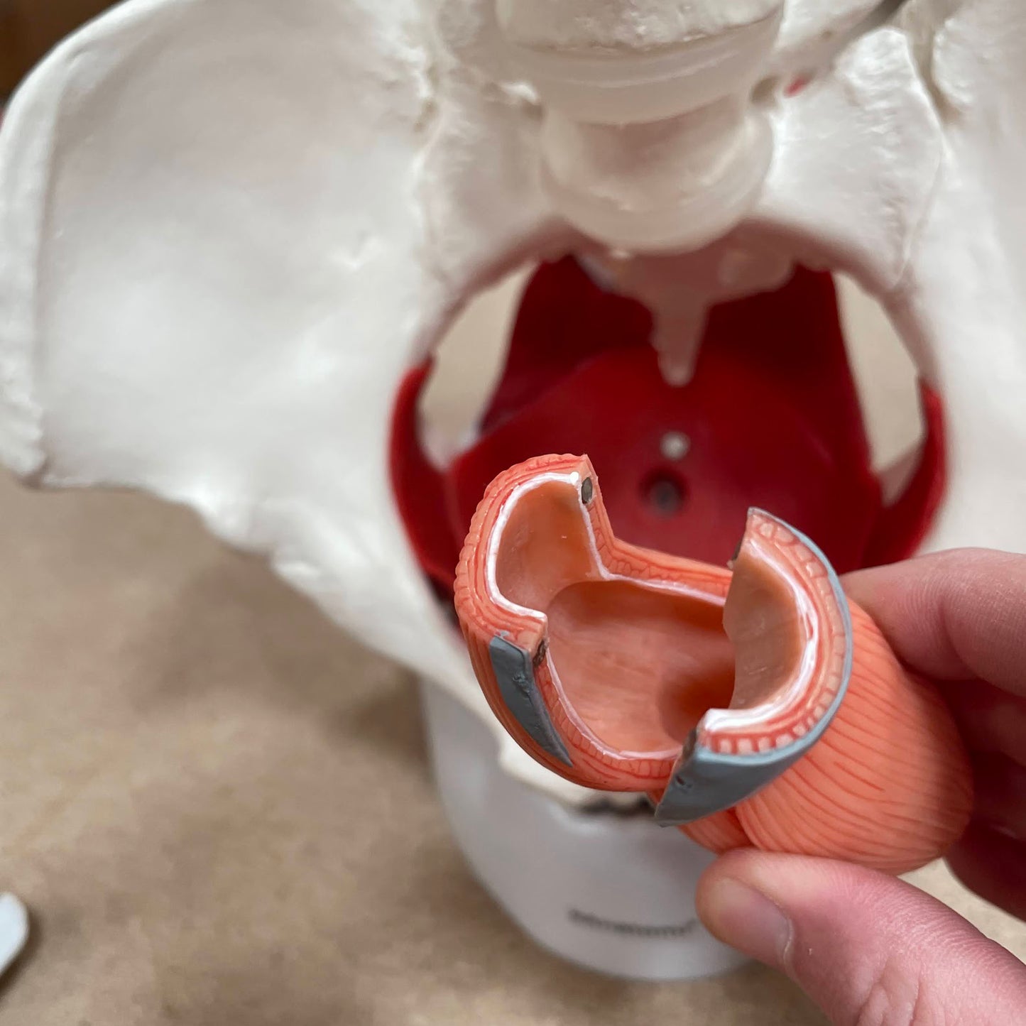
The internal genitalia include the ovaries (ovaries), the fallopian tubes (tubae uterinae), the uterus (uterus) and the vagina (vagina), all of which are seen in the greatest detail. Anatomically, it is located in the small pelvis. They are seen in relation to the urinary bladder (vesica urinaria) with the urethra (urethra) and the rectum (rectum).
The external genitalia are shown with the largest and most important details, which anatomically are located in front of and below the symphysis.
Many of these organs function as a channel or a reservoir (e.g. the vagina and the urinary bladder), which is why they are seen in a pedagogical way with an air-filled cavity (cavity). Special areas are shown in an educational color - for example glans clitoridis in yellow and fimbriae in dark red.
The pelvis as a birth canal
Normally the following 3 connection lines are specified:
- Conjugata vera (normally measures 11 cm in the female pelvis)
- Diameter obliqua (usually measures 12.5 cm in the woman's pelvis)
- Diameter transversa (usually measures 13 cm in the woman's pelvis)
On this pelvis model, all 3 above connecting lines measure 11 cm. This is due, among other things, to the fact that the size of the model corresponds to a 14-year-old (as mentioned at the beginning of the description above).
Product flexibility
Product flexibility
In terms of movement, the pelvis is not flexible, which is why joints (e.g. the SI joints and the symphysis) cannot be separated or moved. The same applies to the joints in the 2 lower lumbar vertebrae.
When the internal organs of the model are separated, they are generally a bit flexible. For example, the urinary bladder can be squeezed quite a bit when you hold it. The pelvic floor, on the other hand, has a firmer consistency and is cast in one piece with the external genitalia and the rectal opening (can be seen in one of the pictures on the left).
Clinical features
Clinical features
Clinically speaking, the model can be used in connection with rehabilitation after childbirth, operative interventions, etc. It can also be used to understand diseases and disorders in the female genital organs, the urinary bladder and the rectum. Examples are urinary tract infection and tumors.
Share a link to this product

A safe deal
For 19 years I have been at the head of eAnatomi and sold anatomical models and posters to 'almost' everyone who has anything to do with anatomy in Denmark and abroad. When you shop at eAnatomi, you shop with me and I personally guarantee a safe deal.
Christian Birksø
Owner and founder of eAnatomi ApS