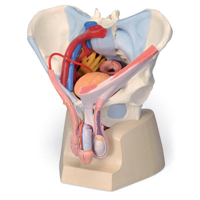
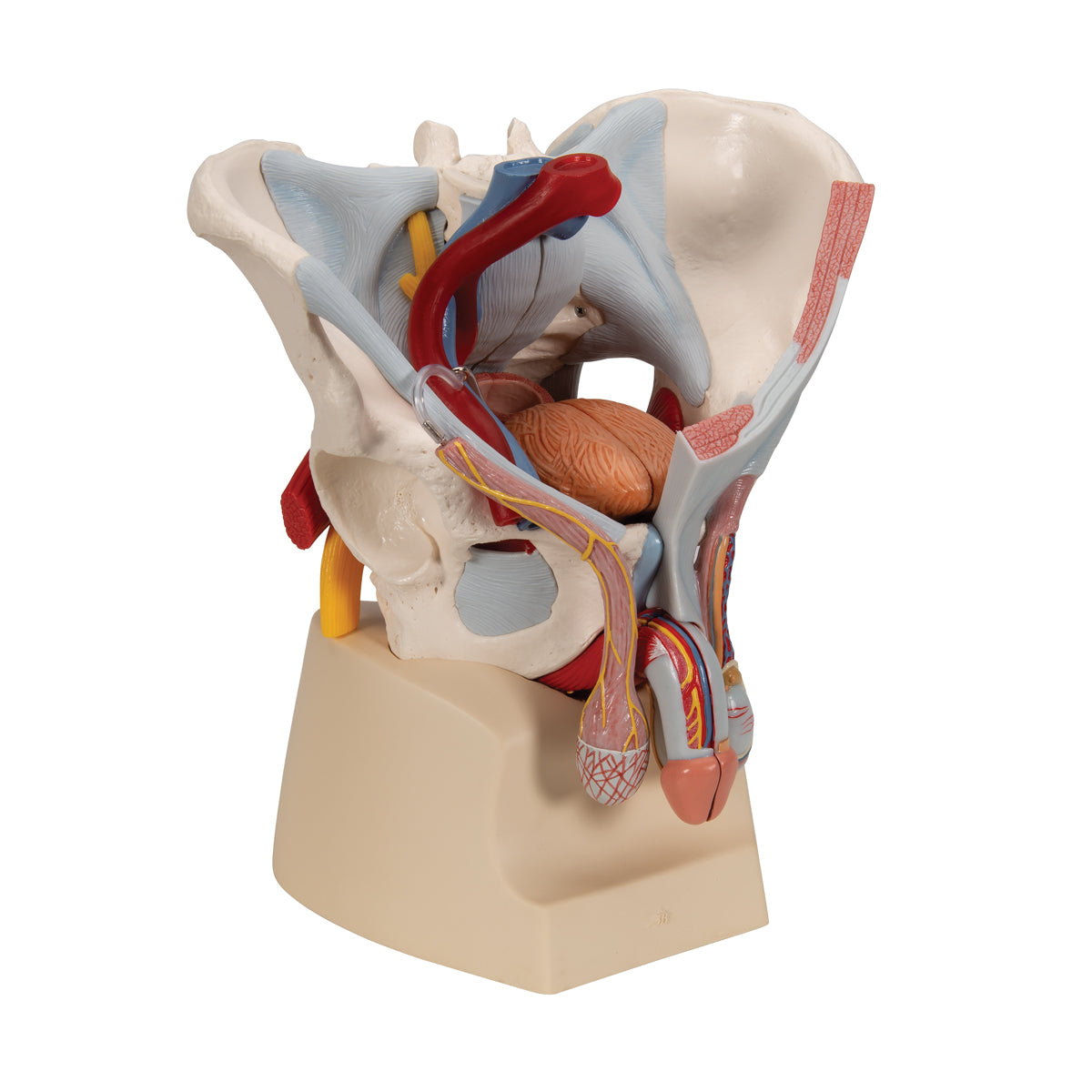
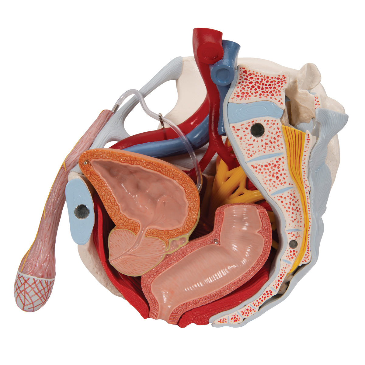
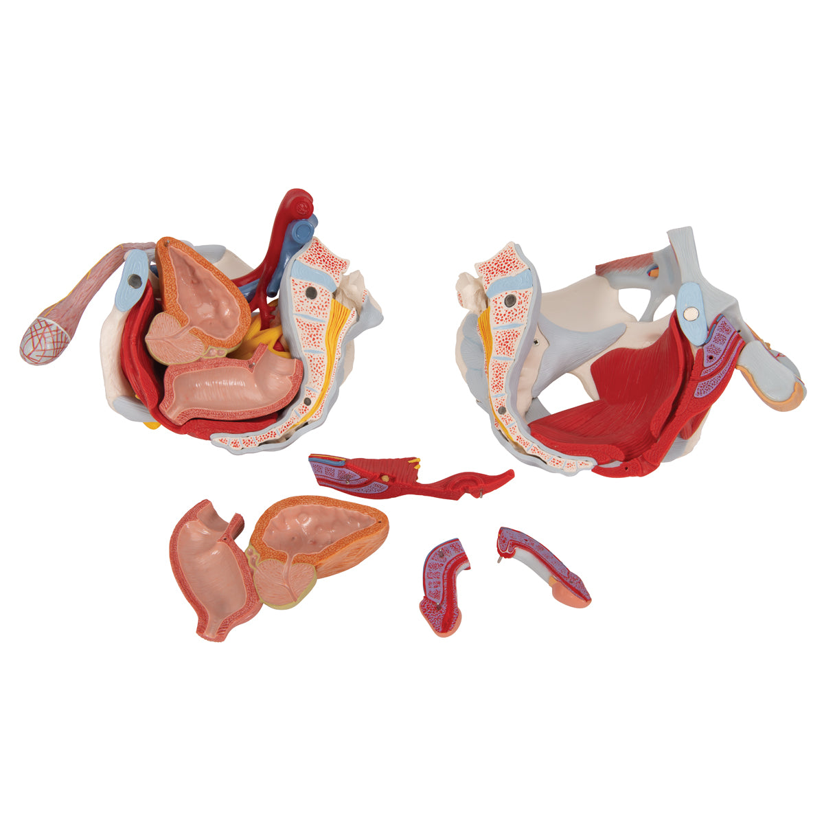
Anatomically speaking, the model shows many different tissues and organs, several of which can be seen in 3 dimensions because the model can be separated into 7 parts.
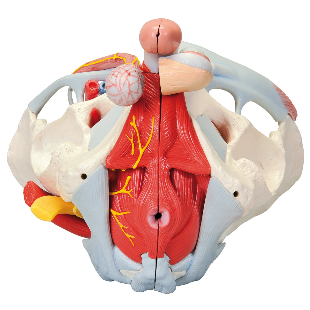
1) The pelvis consists of the 3 building blocks, which are the 2 hip bones and the sacrum. The model also shows the lower lumbar vertebra. In the bone tissue, the most important details can be seen, such as large nodules. Smaller details such as the linea glutaea are omitted. Overall, the model shows the following bones:
Os coxae (the 2 hip bones) of which both are composed
- Os ilium (iliac bone/hip bone)
- Os ischii (seat bone)
- Os pubis (pubic bone)
Us sacrum (sacrum) incl. os coccygis (coccyx)
5th lumbar vertebra (lumbar vertebra)
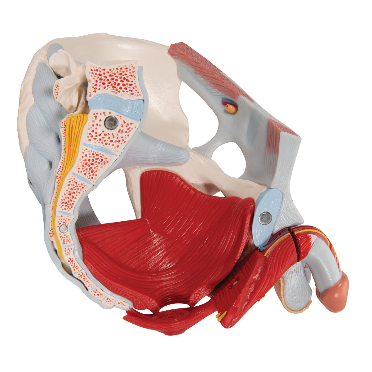
2) The pelvic floor (also called the pelvic floor or diaphragma pelvis in Latin) consists of the following muscles, which are clearly visible:
- M. levator ani (consisting of m. pubococcygeus and m. iliococcygeus)
- M. coccygeus
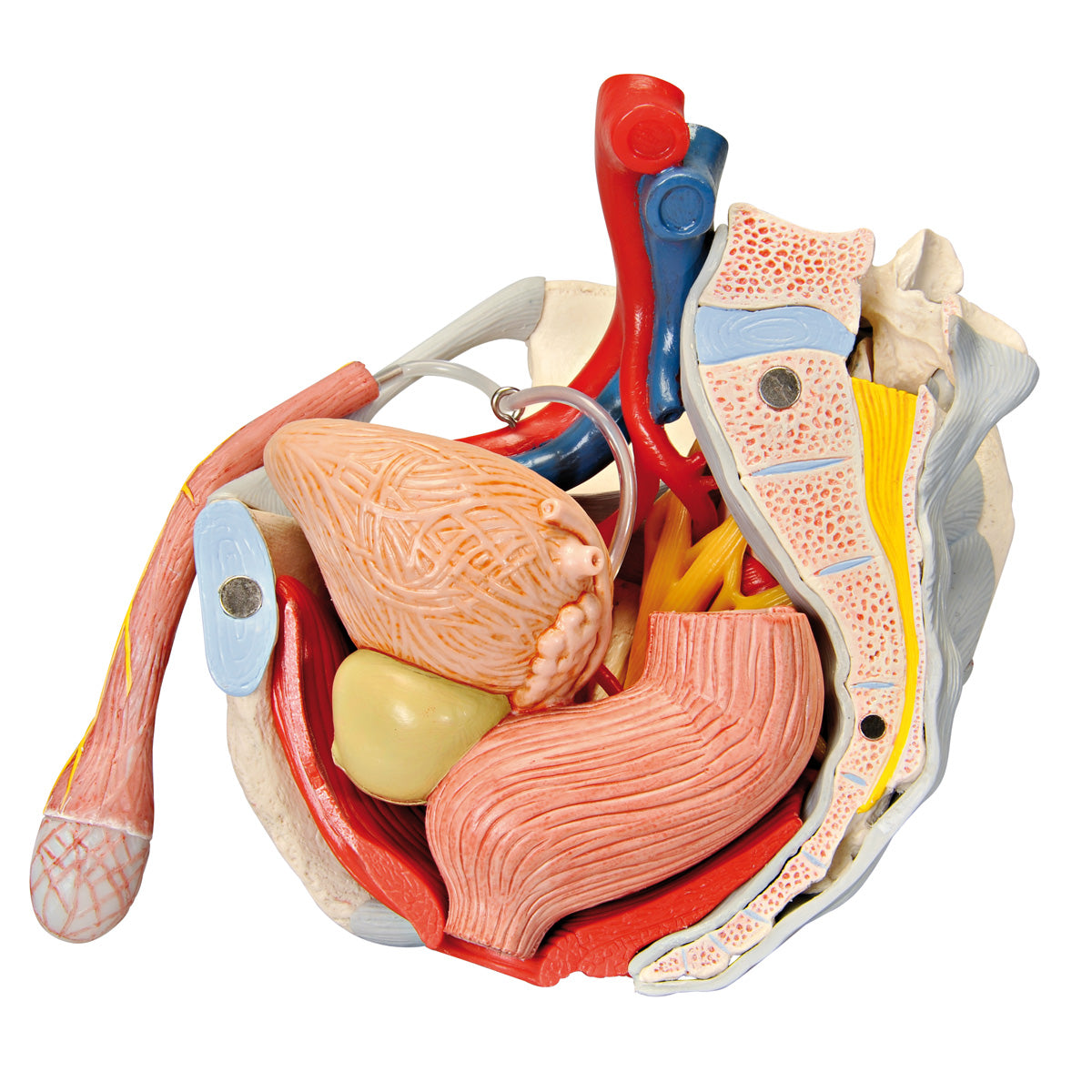
Other muscles are also seen such as the ischiocavernosus muscle, the bulbospongiosus muscle, the obturatorius internus muscle and the sphincter ani externus muscle. In addition, the relationships to the urinary bladder (vesica urinaria) and the rectum (rectum) are seen in detail.
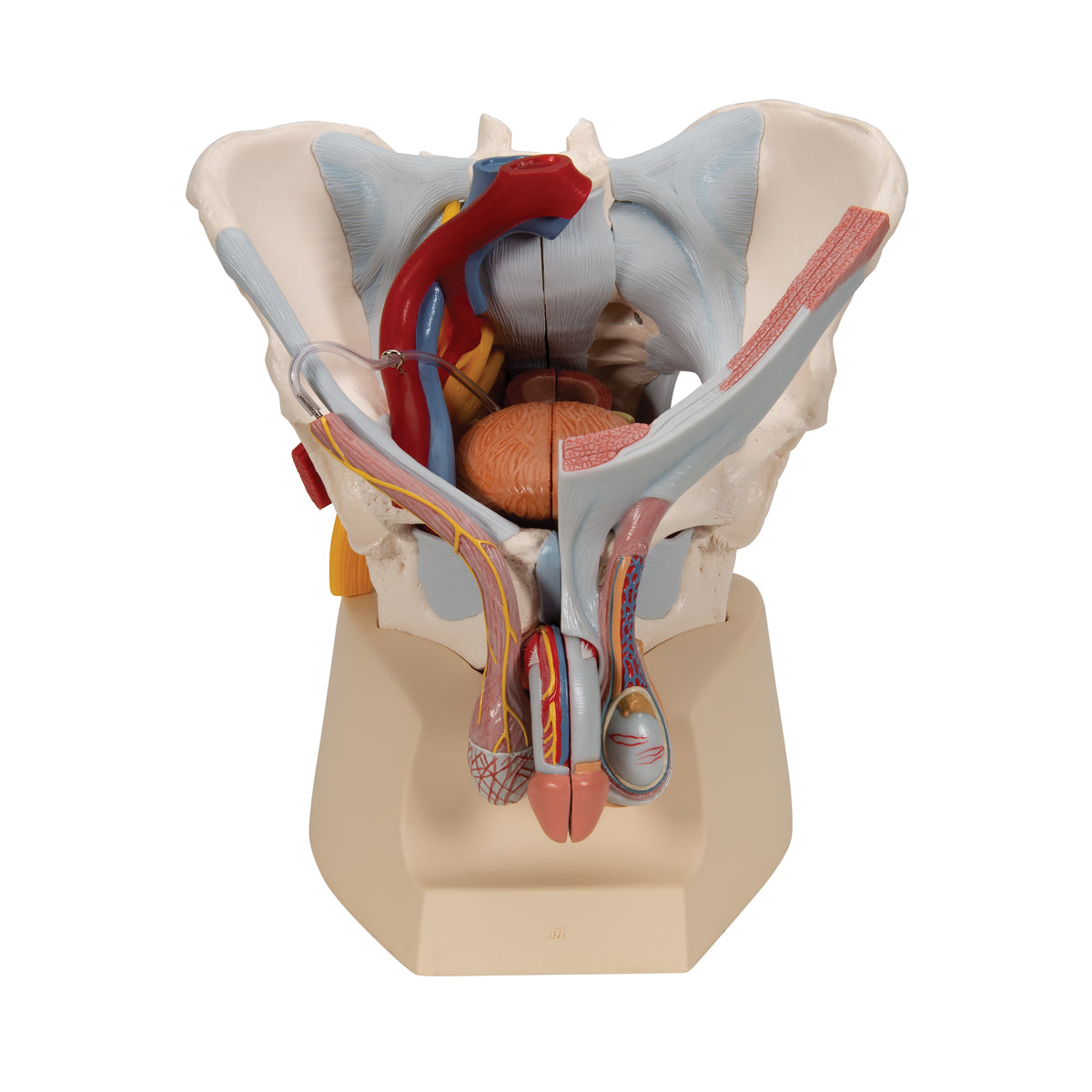
3) The internal genitalia include the testicles (testes) and the epididymides, which are seen in relation to the scrotum and the spermatic cord (funicle). It is not possible to see the internal structures of the testicles (tubules, etc.). Both the contents of the spermatic cord and its barrier/leaves are clearly seen.
NB: In our assortment of pelvic models, we have 2 models that show the internal structures of the testicles: A large model with many details that can be viewed via THIS LINK and a miniature model that can be viewed via THIS LINK
4) The seminal ducts include the ductus deferens (the vas deferens), the ductus ejaculatorius and the urethra masculinum (the urethra), all of which are visible.
5) In relation to the accessory gonads, the prostate (bladder neck gland) and vesiculae seminales (seminal vesicles) are seen, but not gll. bulbourethrales and etc. urethral. The 2 first-mentioned glands are seen in cross-section, and therefore the excretory ducts and their connection are seen in detail.
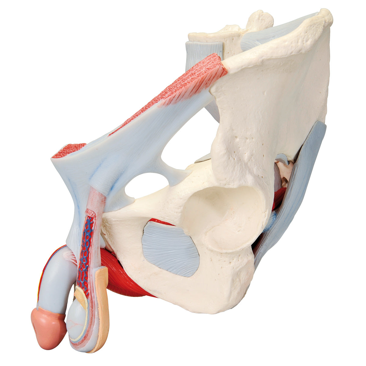
6) The external genitalia include the penis and the scrotum, which are also seen. Opposite the testicles, the internal structure of the penis can be seen.
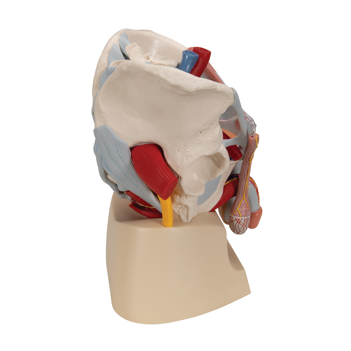
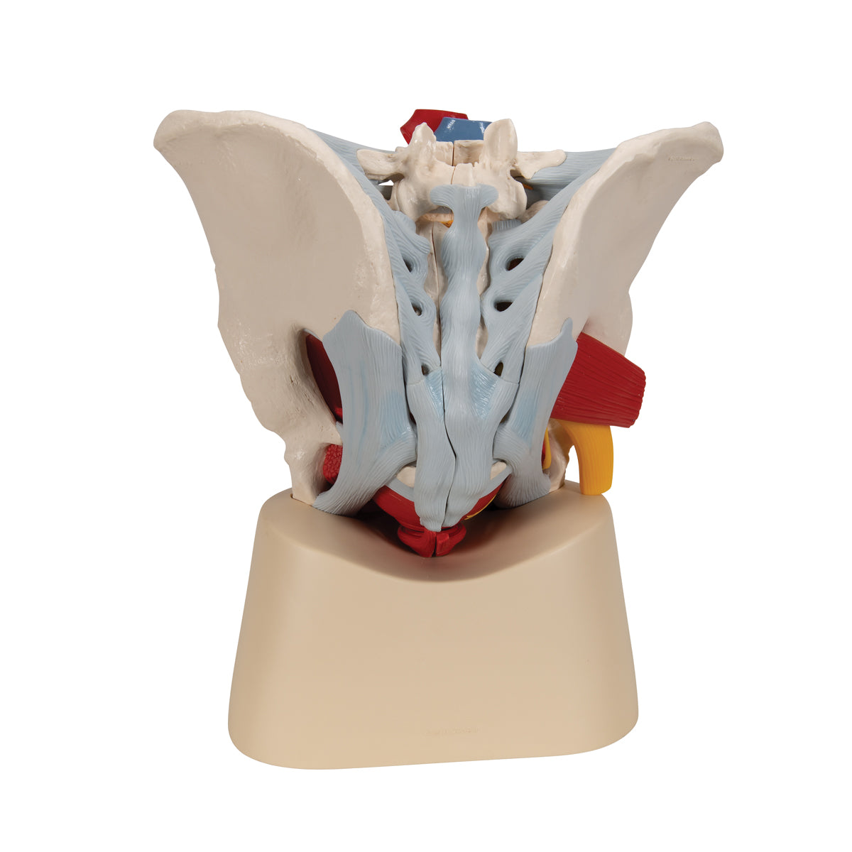
7) Ligaments, blood vessels and nerves are seen in detail. Ligaments are seen on both sides of the pelvic model, while blood vessels and nerves are mainly seen on the right side of the model.
Since the model i.a. can be divided in half, important details can be seen in a median section (including cauda equina). As for nerves, the sacral plexus, sciatic nerve (nervus ischiadicus) and other important nerves are also seen. As for blood vessels, the large artery and vein of the pelvis and parts of their branches are seen.
Many of the pelvic organs function as a channel or a reservoir (e.g. the rectum and the urinary bladder), which is why they are seen in a pedagogical way with an air-filled cavity (cavity).
8) Some of the abdominal muscles can also be seen on the model, so that the connection between the testes, the spermatic cords and the abdominal wall can be better seen.