SKU:EA1-1000054
Skull model showing blood vessels and nerves in the lower jaw as well as colored muscle indications. Can be divided into 3
Skull model showing blood vessels and nerves in the lower jaw as well as colored muscle indications. Can be divided into 3
ATTENTION! This item ships separately. The delivery time may vary.
Couldn't load pickup availability
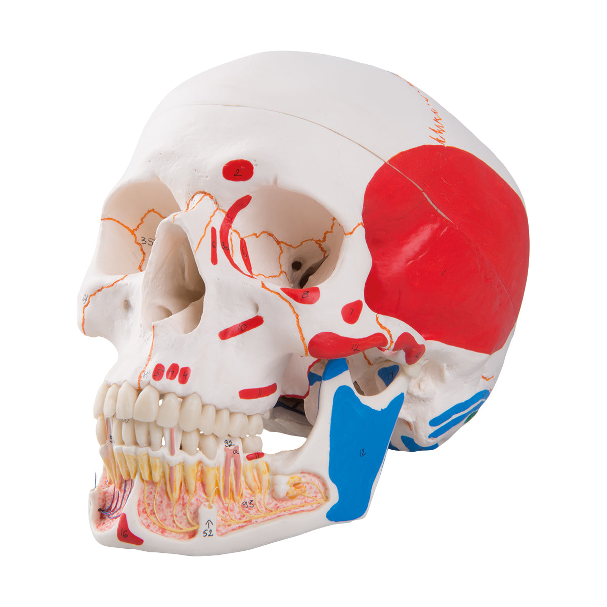
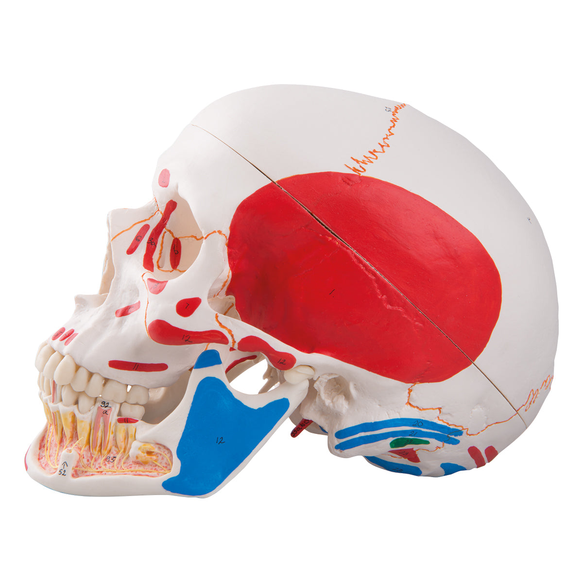
This skull model offers a very educational and detailed insight into the blood vessels and nerves of the lower jaw. Furthermore, the model includes colored muscle markings, colored sutures between the skull bones and colored lines on the inside of the braincase corresponding to the course of blood vessels (arteries and veins).
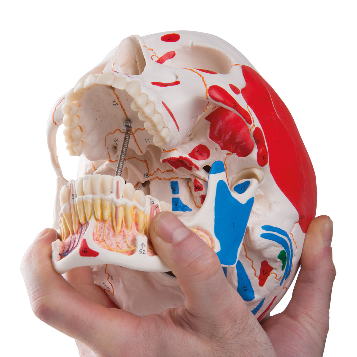
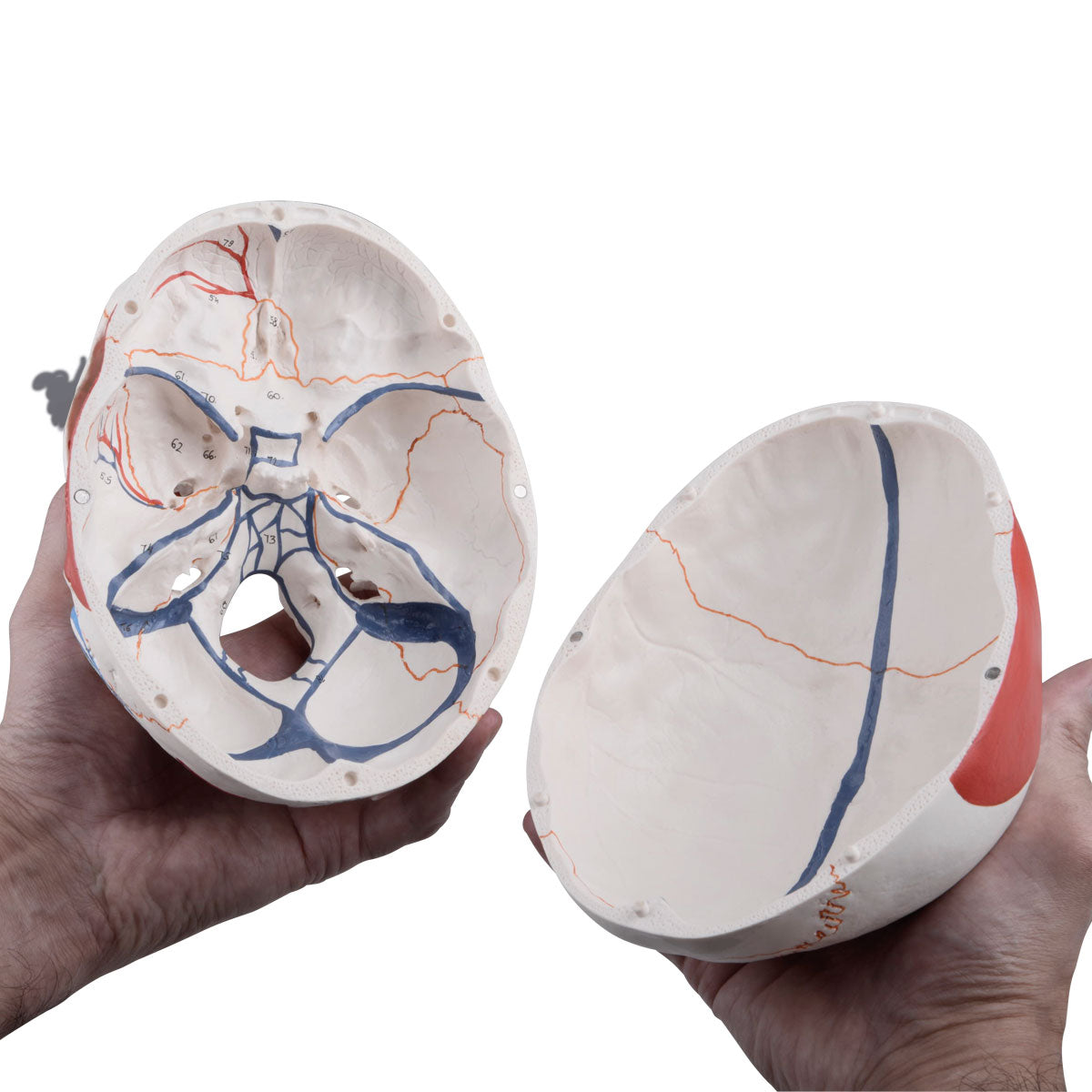
The model is cast in plastic and comes in a size that corresponds to an adult person. The dimensions are 20 x 13.5 x 15.5 cm and the weight is approximately 900 grams. The skull cap ("the top") can be removed, so i.a. the base of the skull (basis cranii interna) can be studied. In addition, the lower jaw bone ("mandible") can be quickly removed. Therefore, the model can be separated into 3 parts.
The skull model has been developed to be able to contain a specific brain model from the same manufacturer.
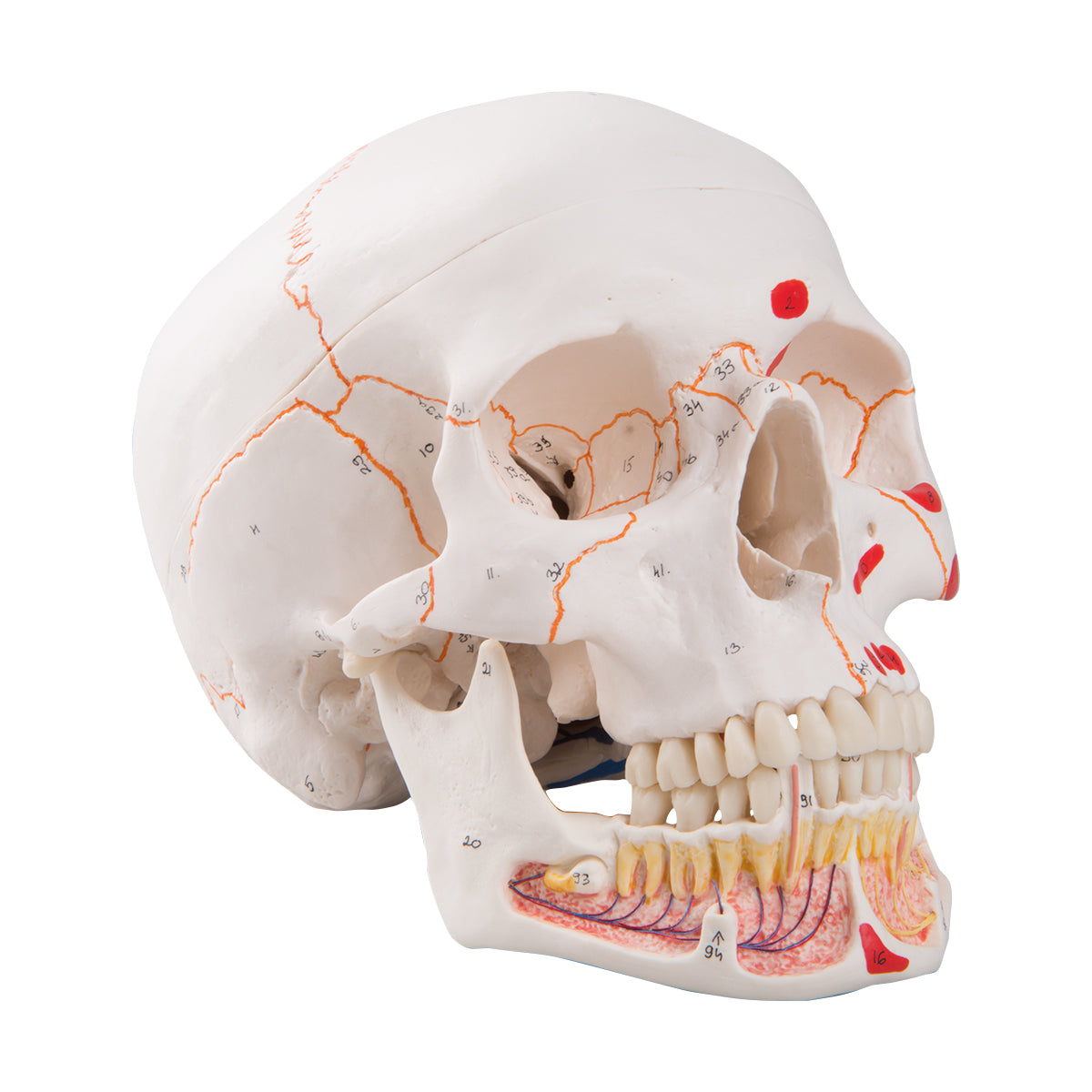
Both bones, the colored muscle markings, the colored sutures and the lines on the inside of the braincase (which symbolize blood vessels) are numbered on the model.
Anatomically speaking
Anatomically speaking
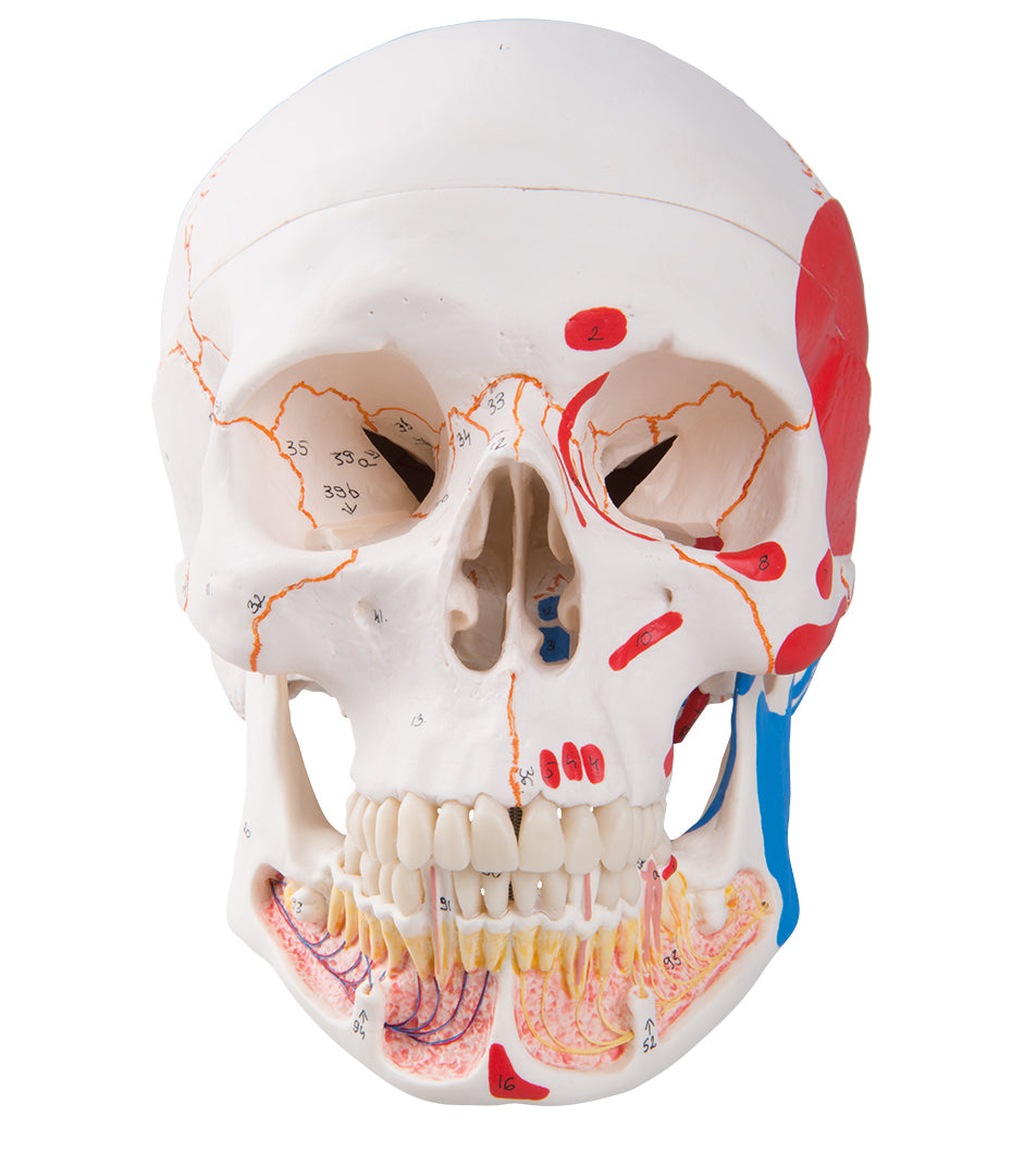
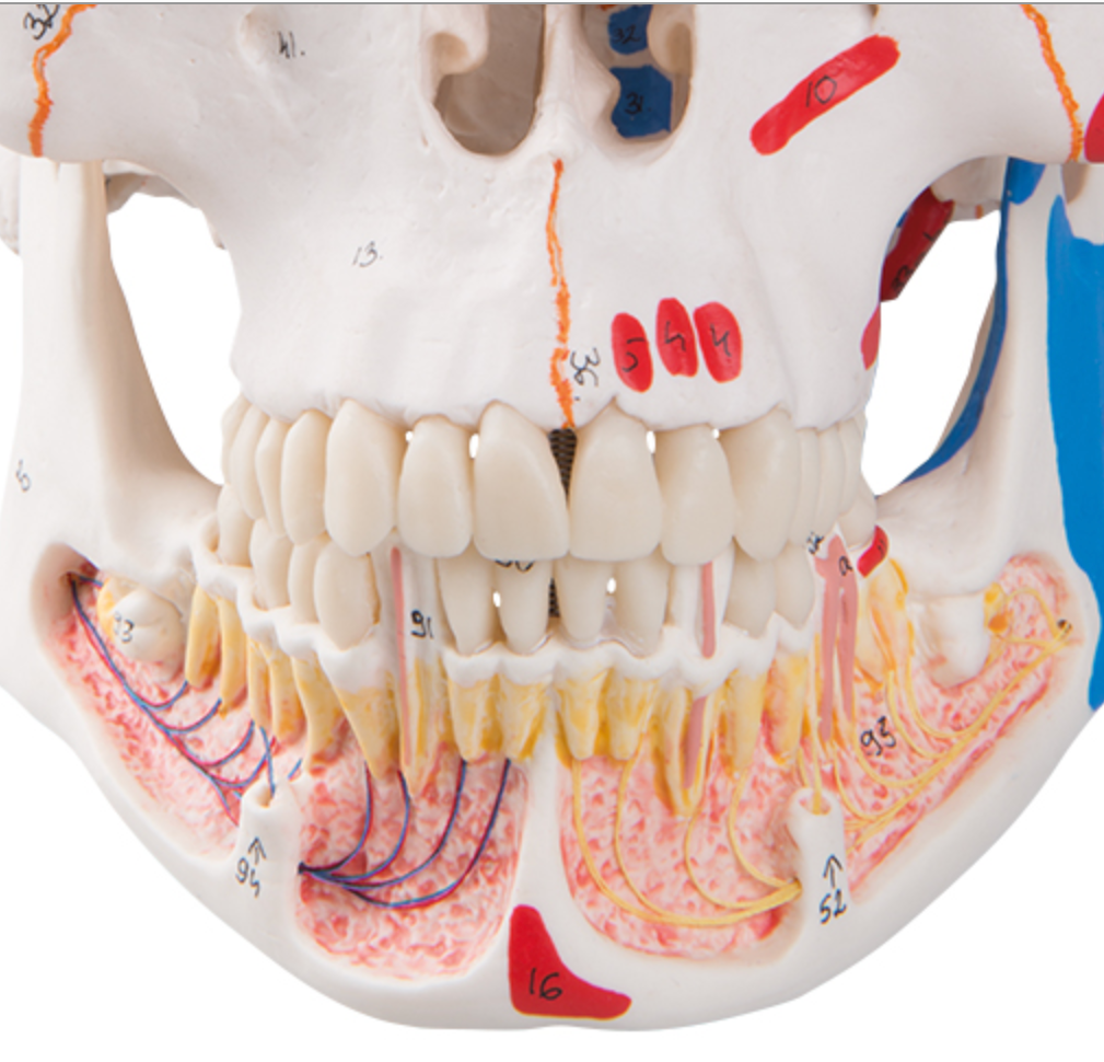
Anatomically speaking, the educational and detailed insight into the blood vessels and nerves of the lower jaw must be emphasised. The insight provides the opportunity to study anatomical details such as the inferior dental plexus and a. and v. mentalis (see the pictures on the left).
Generally speaking, the human skull can be divided into 2 parts, and the skull model therefore shows the following:
1) The braincase (neurocranium), which is intended to enclose the brain and the hearing-equilibrium organ
2) The facial skeleton (viscerocranium) which surrounds the nasal cavity and forms the tooth-bearing framework around the oral cavity. The 32 teeth are also included
The braincase consists of 8 bones. There are 4 unpaired (the frontal, sphenoid, sphenoid and occipital bones) and 2 paired (the occipital and temporal bones). All these bones as well as sutures can be identified on the skull model.
The facial skeleton includes 6 paired bones (the maxilla, palatine, cheekbone, nasal bone, lacrimal bone, and lower conchbone) and 3 unpaired bones called the mandible, the ploughshare, and the zygomatic bone (some do not count the zygomatic bone as part of the facial skeleton). All these bones and sutures can also be identified on the skull model. NB: The hyoid bone is also included in the facial skeleton but cannot be seen on this skull model.
The human skull contains many holes and channels containing vessels and nerves. Overall, there are connections between the braincase and the neck, to and from the eye socket, to and from the pterygo-palatine fossa and to and from the nasal cavity.
This skull model shows many of the most important holes and canals, but not all. Furthermore, the level of detail on the bones is good. As for "osseous landmarks" such as the processus styloideus, many of the most important are seen, while some minor participants are omitted.
As for skeletal muscles, red and blue color markings are seen on the skull model. These colors symbolize/show respectively origin and attachment. There are color markings for facial muscles, masticatory muscles, palate muscles, tongue muscles as well as some deep neck muscles and some back muscles.
Furthermore, the sutures are colored in an orange color, while on the inside of the brain case, respectively, red and blue lines/strokes, which should symbolize/show the "meningeal arteries and veins" as well as the venous sinuses.
Flexibility
Flexibility
In terms of movement, it is only relevant to mention the jaw joint. The lower jaw bone ("mandible") is held in place via a relatively long metal spring, which is attached via screws to the mandible and palatine bone. In addition, rubber is fitted in the joint bowls of the jaw joints (fossa mandibularis), which holds the joint head (caput mandibulae) "like in a rubber bowl" (see the pictures on the left).
The model's jaw joints are not very flexible. Natural movements in the jaw joint require that, among other things, the articular head can slide forward and downward until the articular head is forward on the tuberculum articulare. The latter is the bony part with a weak guiding groove in front of the depression (fossa mandibularis). This can only be demonstrated on the model if the rubber in the joint cups is removed, which the model is not designed for.
If you take a good hold of the mandible, you can however move it up and down and to the sides, although the natural sliding joint mechanism as described above cannot be demonstrated.
Clinically speaking
Clinically speaking
Clinically speaking, the skull model is ideal for understanding diseases, disorders and disorders in this part of the skeleton.
Share a link to this product

A safe transaction
For 19 years I have been managing eAnatomi and sold anatomical models and posters to 'almost everyone' who has anything to do with anatomi in Scandinavia and abroad. When you place your order with eAnatomi, you place your order with me and I personally guarantee a safe transaction.
Christian Birksø
Owner and founder of eAnatomi and Anatomic Aesthetics