SKU:EA1-1013283
Skull model with both masticatory and facial muscles
Skull model with both masticatory and facial muscles
ATTENTION! This item ships separately. The delivery time may vary.
Couldn't load pickup availability
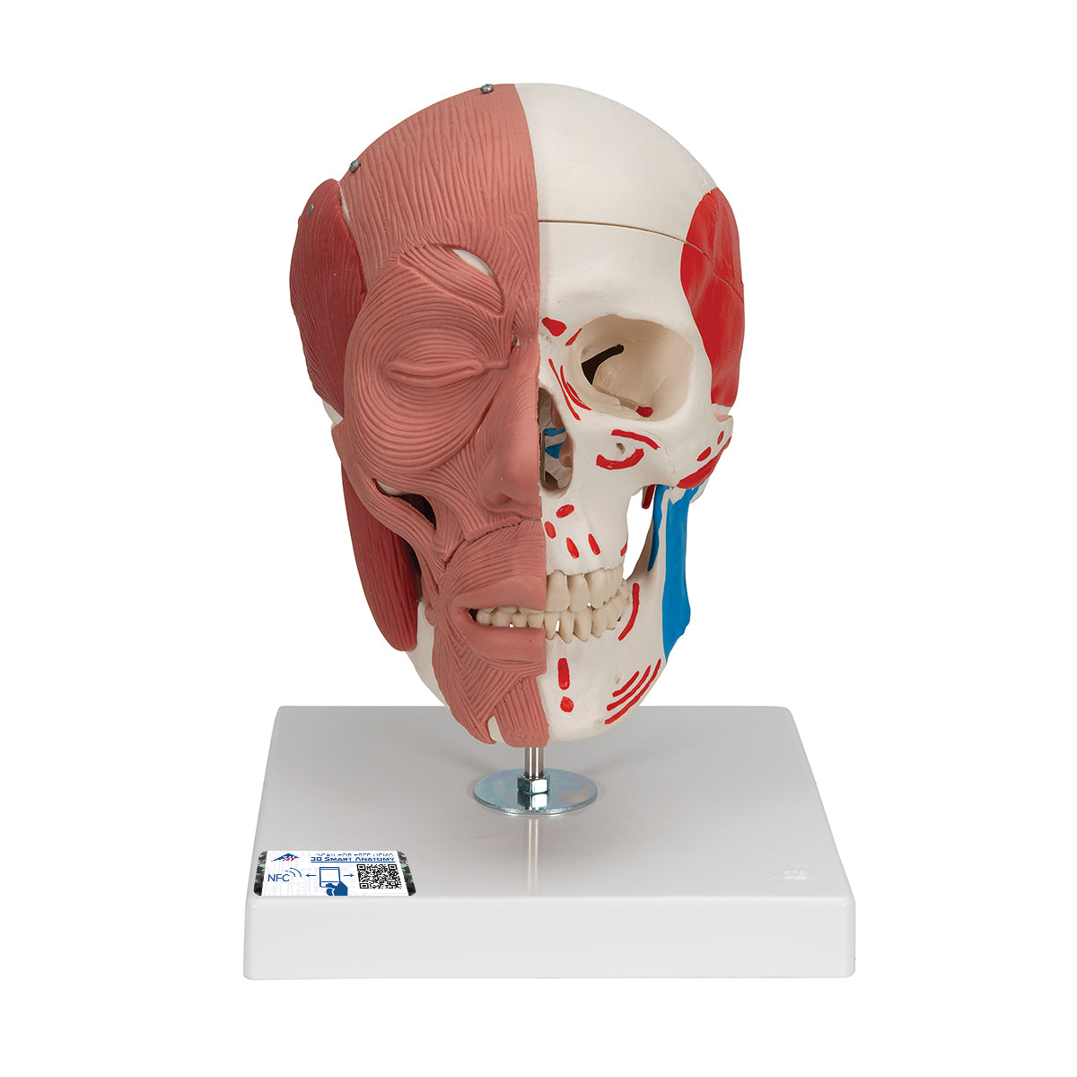
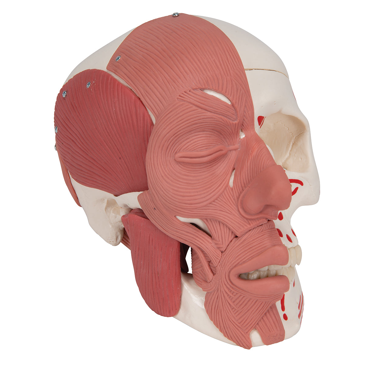
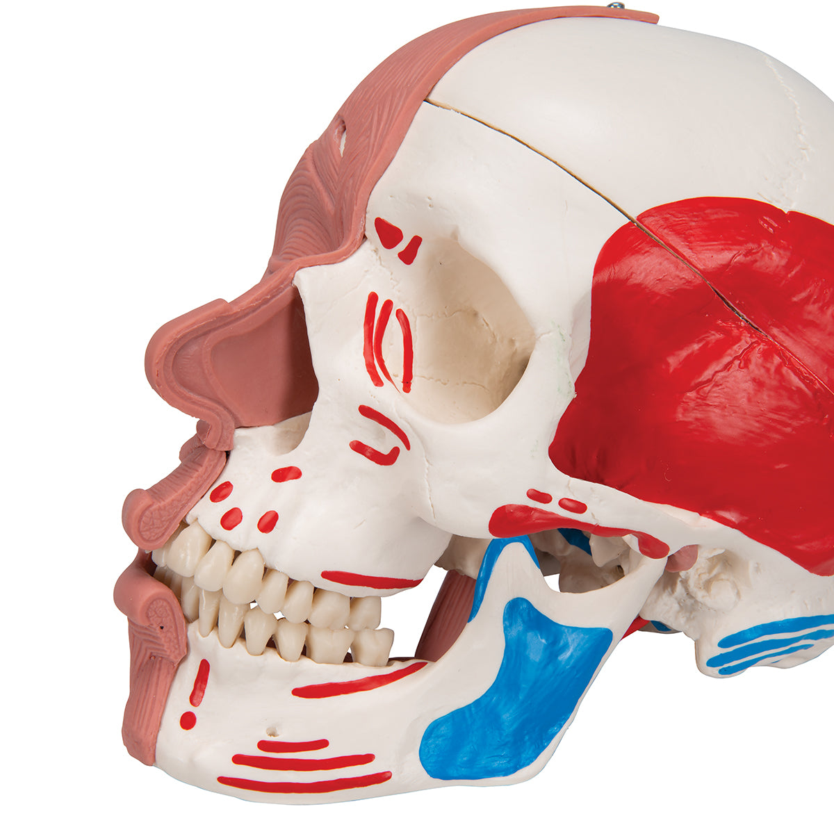
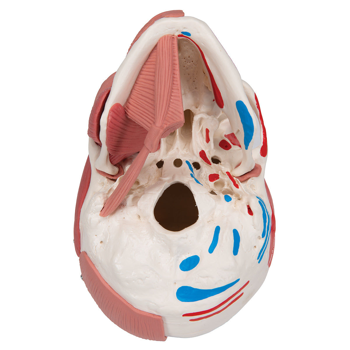
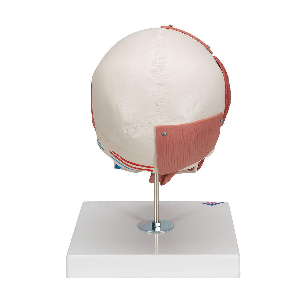
This skull model shows the masticatory and facial muscles visible on the right side, while colored muscle indications are seen on the bone tissue on the opposite side.
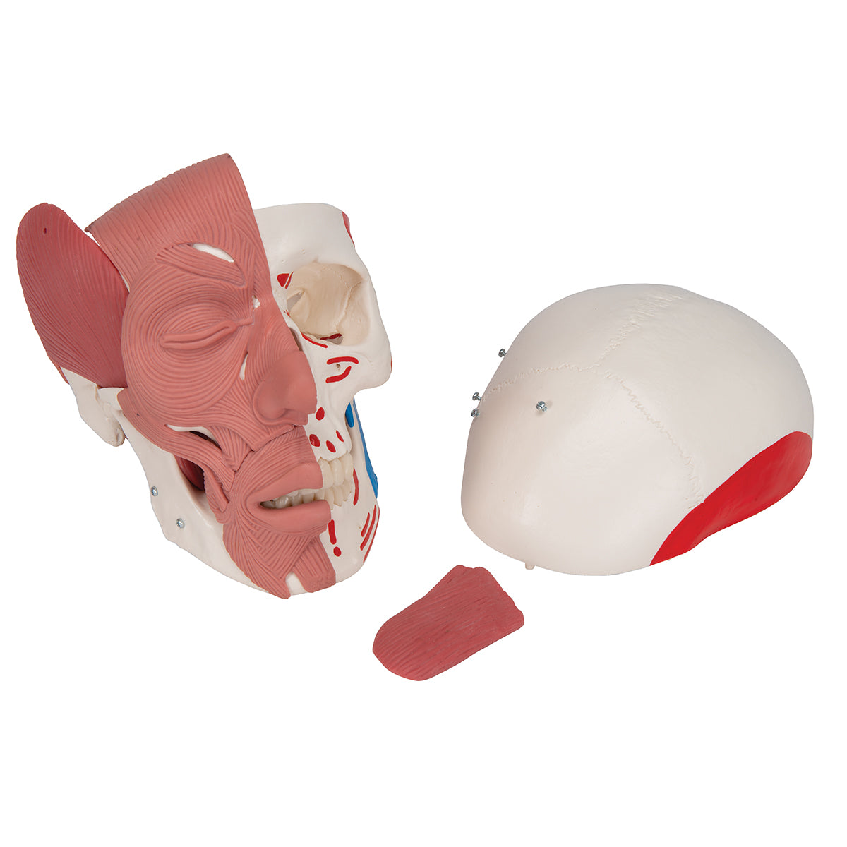
The model is cast in plastic and comes in a size that corresponds to an adult person. The dimensions are 18 x 18 x 25 cm and the weight is approximately 1.1 kg. The skull cap ("the top") can be removed, so i.a. the base of the skull (basis cranii interna) can be studied. Furthermore, one of the chewing muscles (m. masseter) and the lower jaw bone ("mandible") can also be removed relatively easily.
Anatomically speaking
Anatomically speaking
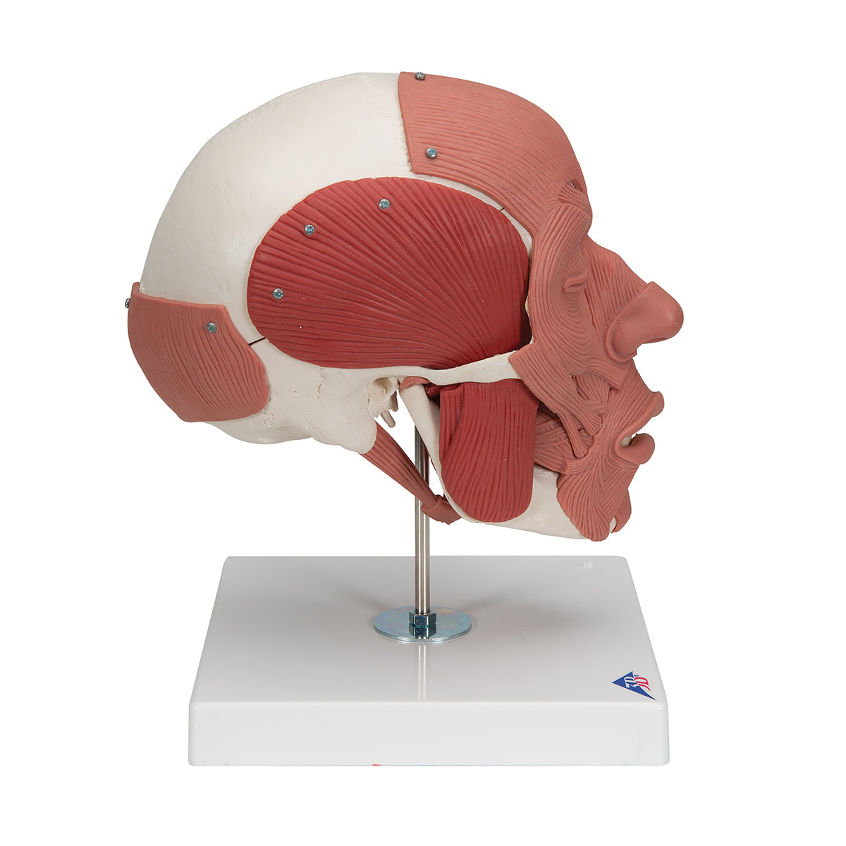
Anatomically, it should be emphasized that this skull model shows visible masticatory and facial muscles on the right side. These 2 muscle groups can be easily separated visually because they each appear in their own shade of brown. Of the many muscles, only the m. masseter can be detached.
Generally speaking, the human skull can be divided into 2 parts, and the skull model therefore shows the following:
1) The braincase (neurocranium), which is intended to enclose the brain and the hearing-equilibrium organ
2) The facial skeleton (viscerocranium) which surrounds the nasal cavity and forms the tooth-bearing framework around the oral cavity. The 32 teeth are also included
The braincase consists of 8 bones. There are 4 unpaired (the frontal, sphenoid, sphenoid and occipital bones) and 2 paired (the occipital and temporal bones). All these bones as well as sutures can be identified on the skull model.
The facial skeleton includes 6 paired bones (the maxilla, palatine, cheekbone, nasal bone, lacrimal bone, and lower conchbone) and 3 unpaired bones called the mandible, the ploughshare, and the zygomatic bone (some do not count the zygomatic bone as part of the facial skeleton). All these bones and sutures can also be identified on the skull model. NB: The hyoid bone is also included in the facial skeleton but cannot be seen on this skull model.
The human skull contains many holes and channels containing vessels and nerves. Overall, there are connections between the braincase and the neck, to and from the eye socket, to and from the pterygo-palatine fossa and to and from the nasal cavity.
This skull model shows many of the most important holes and canals, but not all. Furthermore, the level of detail on the bones is good. As for "osseous landmarks" such as the processus styloideus, many of the most important are seen, while some minor participants are omitted
As for color markings for skeletal muscles on the left side of the skull model, they are seen in red and blue. These colors symbolize/show respectively origin and attachment. There are color markings for facial muscles, masticatory muscles, palate muscles, tongue muscles as well as some deep neck muscles and some back muscles.
Flexibility
Flexibility
In terms of movement, it is only relevant to mention the jaw joint. The lower jaw bone ("mandible") can be moved because the visible masticatory muscles on the right side are flexible. This makes it possible to demonstrate chewing movements with the mandible.
Clinically speaking
Clinically speaking
Clinically, the skull model can be used to understand diseases and disorders in the jaw joint and masticatory muscles, such as jaw tension and temporomandibular dysfunction (TMD). The model can also be used for medical problems related to facial muscles, such as facial paresis.
The model is also ideal for understanding other diseases, disorders and disorders in this part of the skeleton.
Share a link to this product

A safe transaction
For 19 years I have been managing eAnatomi and sold anatomical models and posters to 'almost everyone' who has anything to do with anatomi in Scandinavia and abroad. When you place your order with eAnatomi, you place your order with me and I personally guarantee a safe transaction.
Christian Birksø
Owner and founder of eAnatomi and Anatomic Aesthetics


