SKU:EA1-1020161
Skull model with colors, cervical vertebrae and more
ATTENTION! This item ships separately. The delivery time may vary.
Anatomical model of the skull including the cervical vertebrae including part of the brainstem, spinal cord, the origin of the spinal nerves and the vertebral artery.
This model includes colored skull bones, just as the Atlas and Axis (C1 and C2) differ in color from the other cervical vertebrae. The colors are included to facilitate the explanation of the size, location and identification of the bones.
The skull can have removable skull top, lower jaw and can be detached from the cervical vertebrae. Cervical spine mounted on a white plastic stand measuring 13.5 x 15.5 cm.
Anatomically speaking
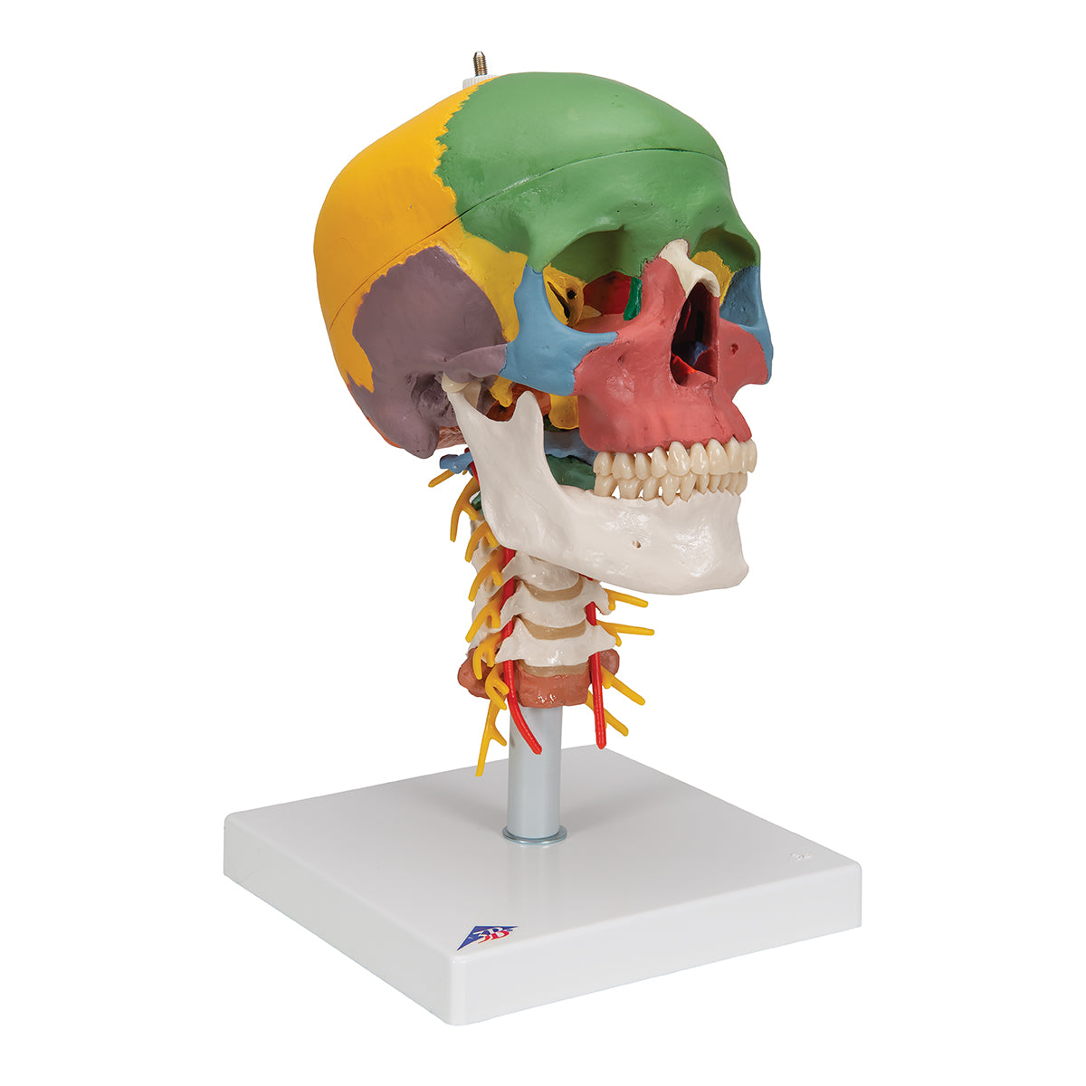
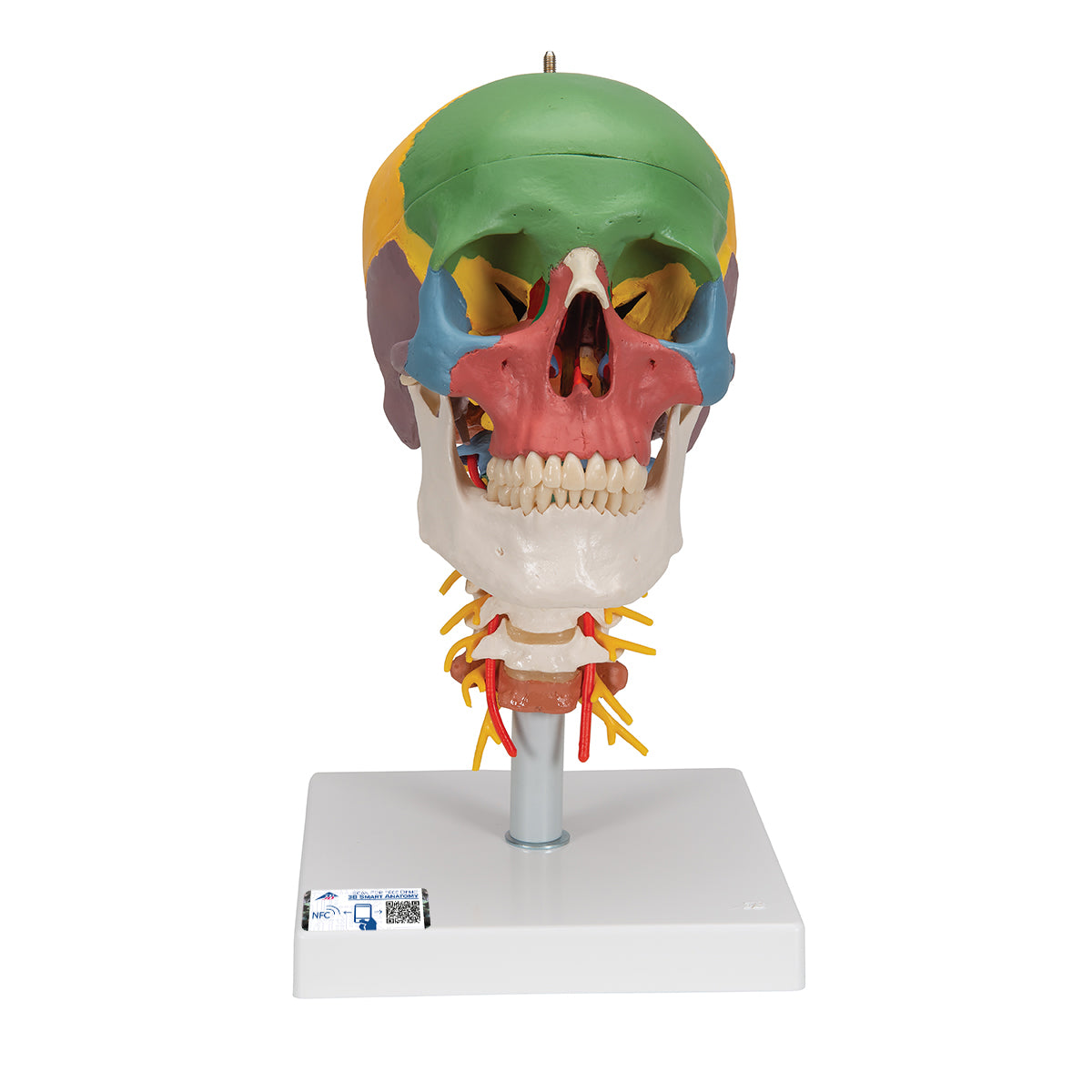
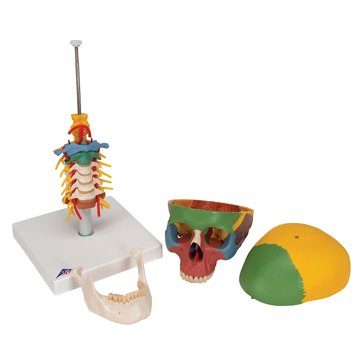
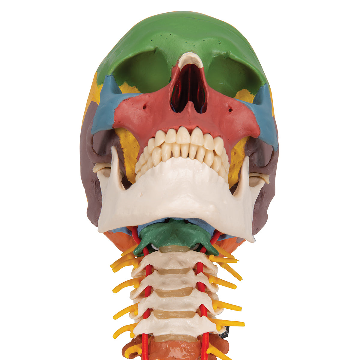
Anatomically, the human skull can be divided into 2 parts, and the skull model therefore shows the following:
1) The braincase (neurocranium), which is intended to enclose the brain and the hearing-equilibrium organ
2) The facial skeleton (viscerocranium) which surrounds the nasal cavity and forms the tooth-bearing framework around the oral cavity. The 32 teeth are also included
The braincase consists of 8 bones. There are 4 unpaired (the frontal, sphenoid, sphenoid and occipital bones) and 2 paired (the occipital and temporal bones). All these bones as well as sutures can be identified on the skull model.
The facial skeleton includes 6 paired bones (the maxilla, palatine, cheekbone, nasal bone, lacrimal bone, and lower conchbone) and 3 unpaired bones called the mandible, the ploughshare, and the zygomatic bone (some do not count the zygomatic bone as part of the facial skeleton). All these bones and sutures can also be identified on the skull model. NB: The hyoid bone is also included in the facial skeleton but cannot be seen on this skull model.
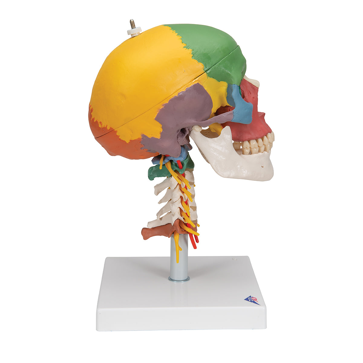
The human skull contains many holes and channels containing vessels and nerves. Overall, there are connections between the braincase and the neck, to and from the eye socket, to and from the pterygo-palatine fossa and to and from the nasal cavity.
On this skull model, most holes and canals are visible, but not all.
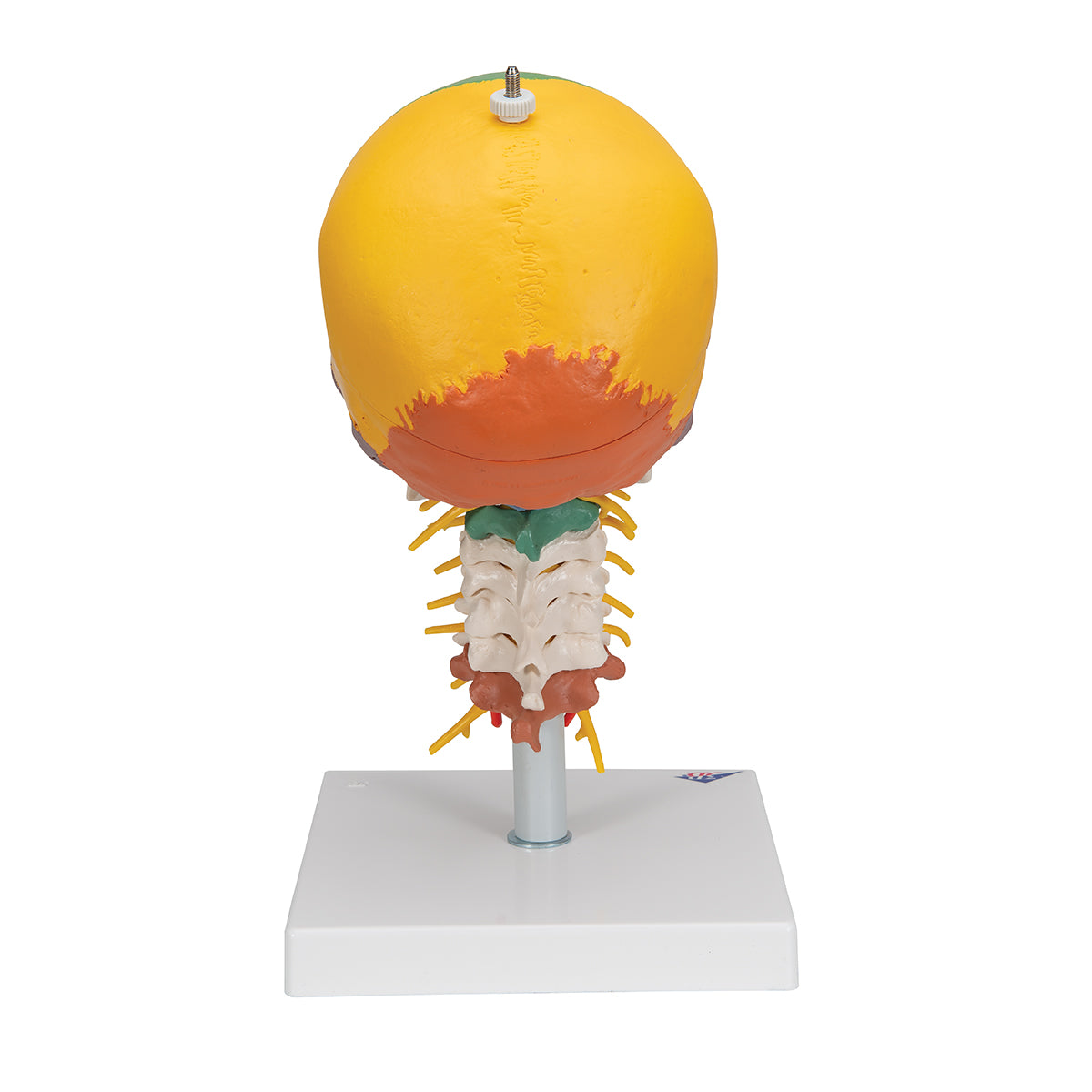
The model also includes the 7 cervical vertebrae (including the intervening discs) as well as a piece of the vertebral artery, nerve roots and spinal cord that runs up to and including the pons in the brainstem.
Flexibility
Flexibility
In terms of movement, the model includes many joints because it shows the cervical spine.
The lower jaw bone ("mandible") is held in place via a spring in the oral cavity and it is possible to illustrate the mobility of the jaw joint. The lower jaw can also be completely removed.
It is possible to demonstrate movements in the upper and lower cervical joints (or in the cervical spine as a whole) as the vertebrae are assembled on a flexible copper stick. That is flexion or extension of the neck joint, as well as rotation is possible.
Clinically speaking
Clinically speaking
Share a link to this product
A safe transaction
For 19 years I have been managing eAnatomi and sold anatomical models and posters to 'almost everyone' who has anything to do with anatomi in Scandinavia and abroad. When you place your order with eAnatomi, you place your order with me and I personally guarantee a safe transaction.
Christian Birksø
Owner and founder of eAnatomi and Anatomic Aesthetics