Skip to product information
1 of 5

SKU:EA1-DS 1

Enlarged ear model with auricula of the highest quality

Enlarged ear model with auricula of the highest quality

Regular price 12.800,00 DKK
Regular price Sale price 12.800,00 DKK
Sale Sold out
Not including taxes if ordering from outside EU. Shipping calculated at checkout.

Out of stock

this product is made to order. To place an order please call or write us.

This model shows in an extremely high level of detail the structure of both the outer ear, the middle ear and the inner ear. Since the model can be separated into several parts, it is possible to examine even the smallest structures, thereby gaining an understanding of the ear's complex anatomy.


The model is produced in SOMSO plastic and is enlarged approximately four times compared to the size of a natural ear. The model has the dimensions 26 x 44 x 41 cm (length x width x height) and weighs 3.7 kg.


In total, the model can be separated into 8 separate parts and it is delivered on a green plastic stand.


The individual structures on the model are numbered and the names appear in the product manual, which is included with the purchase of the model.

Anatomical features

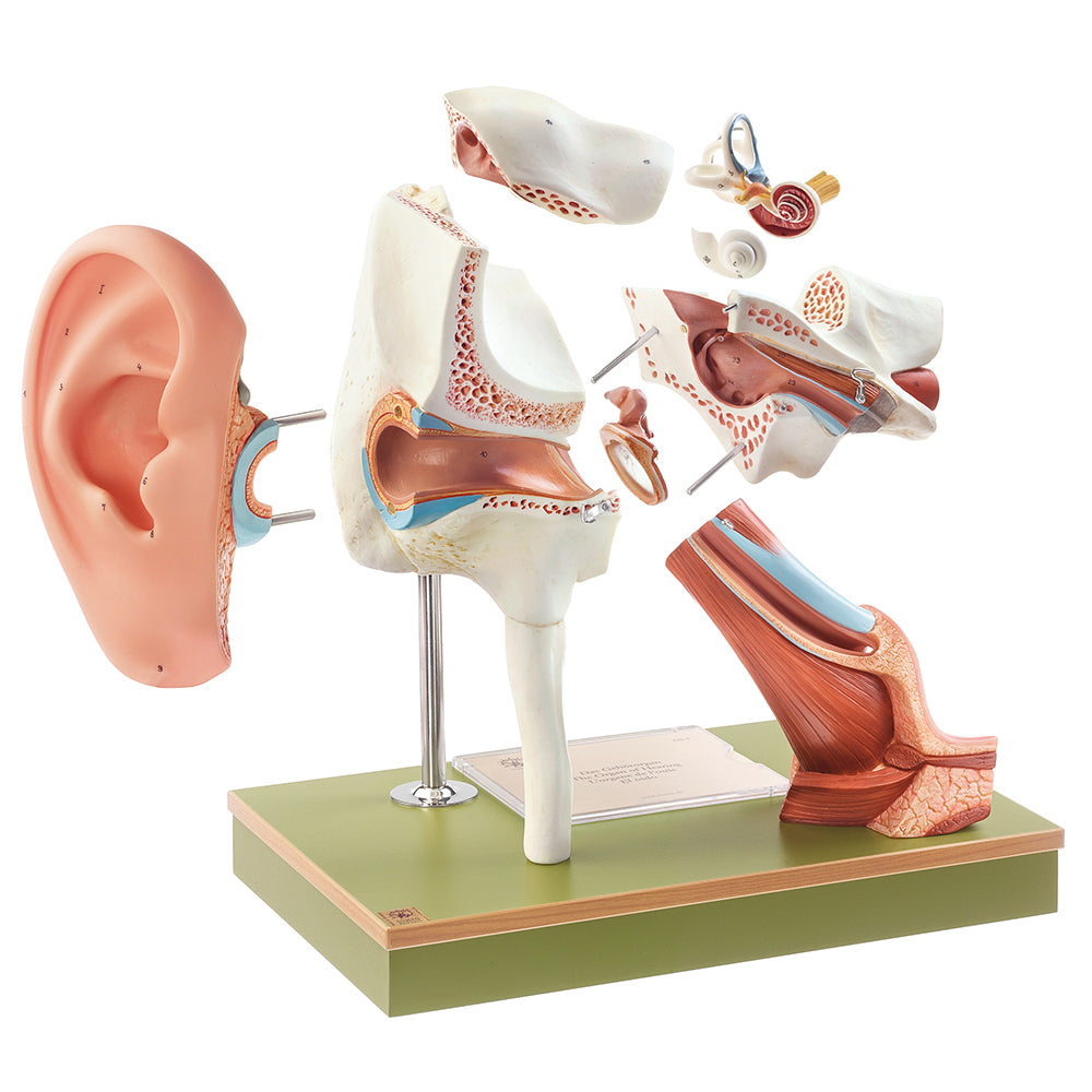
Anatomically speaking, the model can be used to understand the structure of the entire ear, consisting of the outer ear with the auricle and the external ear canal, the middle ear and the inner ear. In addition, the eustachian tube (tuba auditoria) is seen, which communicates with the nasopharynx, as well as the location of the ear in the temporal bone (os temporale).


The model can be separated into 8 separate parts as follows:


The auricle (pinna) can be removed
The eardrum (membrana tympanica) with the three middle ear bones - the hammer (malleus), the anvil (incus) and the stapes - can be removed and examined
Part of the pars petrosa of the temporal bone can be removed
Where the eardrum separates the outer ear from the middle ear, the model can be separated into two larger parts
The lower part of the Eustachian tube can be separated from the middle ear


In the inner ear, the organs of hearing and equilibrium are illustrated with associated nerves (N. vestibulocochlearis - N. cochlearis and N. vestibularis). The model shows how the osseous labyrinth (pictured in white) surrounds the membranous labyrinth (pictured in blue) in the arch canals (canales semicirculares), while this is not the case in the vestibulum and in the cochlea.

Product flexibility

Clinical features

Clinically speaking, the model can be used to understand ear diseases such as otitis media, cholesteatoma (bone eater), perforated eardrum and bullous myringitis.


Furthermore, the ear model can be used to understand examination methods and treatments such as the installation of a drain (tympanic membrane drain).

View full details

A safe deal

For 19 years I have been at the head of eAnatomi and sold anatomical models and posters to 'almost' everyone who has anything to do with anatomy in Denmark and abroad. When you shop at eAnatomi, you shop with me and I personally guarantee a safe deal.

Christian Birksø
Owner and founder of eAnatomi ApS

Read more about eAnatomi