Skip to product information
1 of 3

SKU:EA1-HS 26

Heart model in life size of the highest quality

Heart model in life size of the highest quality

Regular price 6.695,00 DKK
Regular price Sale price 6.695,00 DKK
Sale Sold out
Not including taxes if ordering from outside EU. Shipping calculated at checkout.

Out of stock

this product is made to order. To place an order please call or write us.

This model shows the heart's anatomy in an unusually fine degree of detail, both on the surface, but also inside. The front surface of the heart can be removed, whereby both atria and ventricles become visible. The valve system of the heart is also seen.


The model is produced in life size from a cast of a young, fit person. The model measures 18 x 18 x 30 cm (length x width x height), weighs 0.7 kg and is delivered on a green plastic stand.

Anatomical features

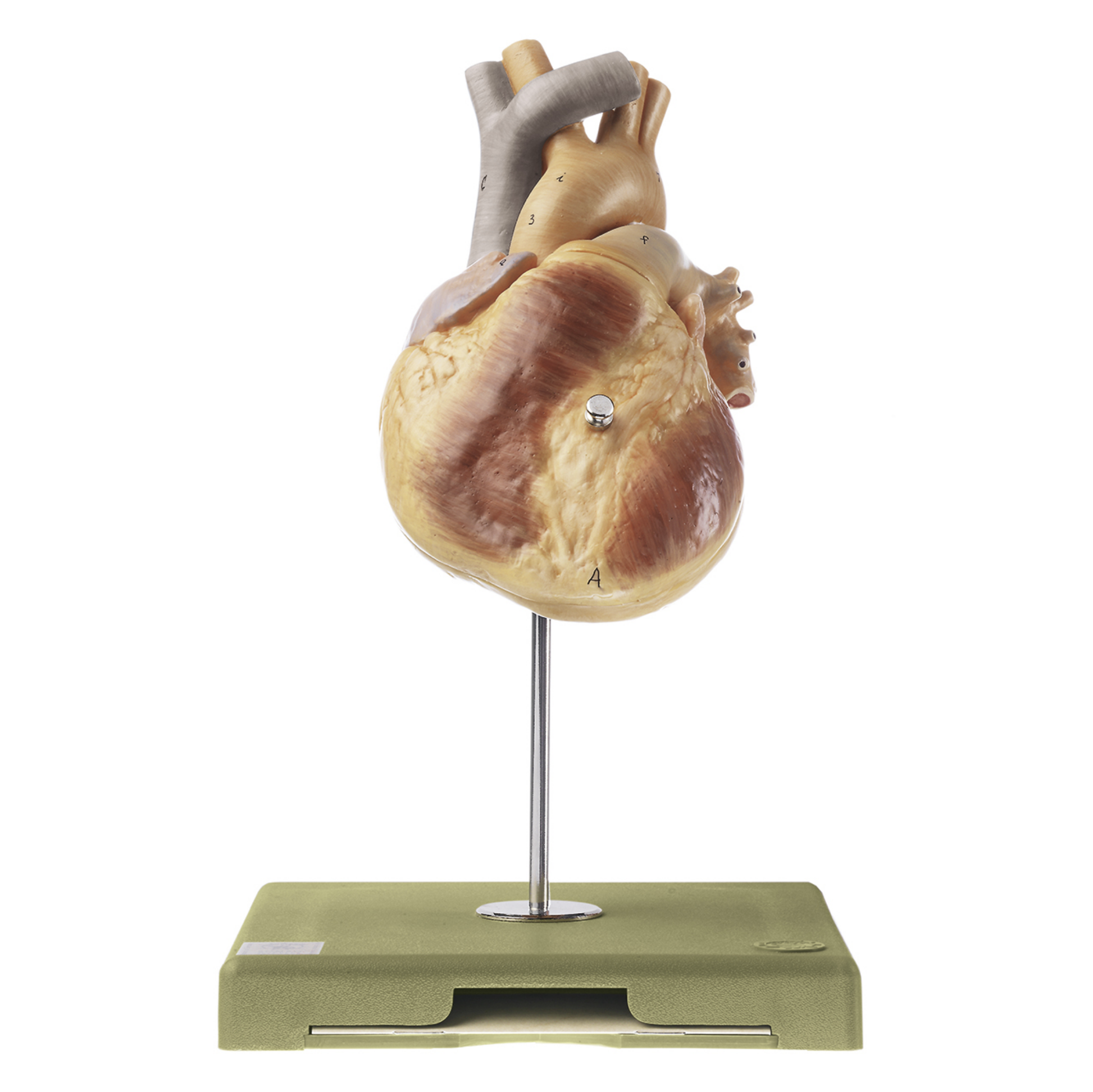
Anatomically speaking, the model can be used to gain an understanding of both the surface anatomy of the heart, but also the structure of the individual heart chambers and the valves between them.


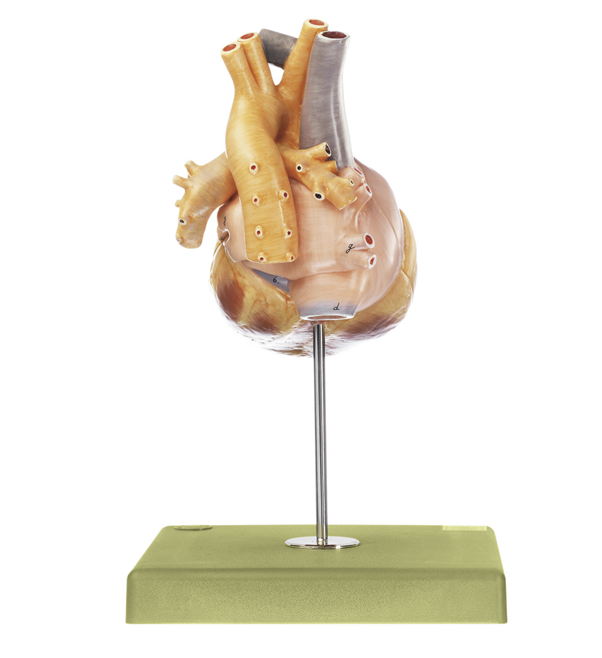
The model shows both atria, with right and left auricles respectively, and both ventricles. In addition, the large arteries and veins that run to and from the heart are seen:


Aorta
Ah. pulmonales (pulmonary arteries)
Vv. pulmonales (pulmonary veins)
Superior and inferior vena cava


In addition, the heart's own blood supply is seen, consisting of the right and left coronary arteries and their branches. On the back of the heart, the coronary sinus is seen, where the veins that drain the heart empty.


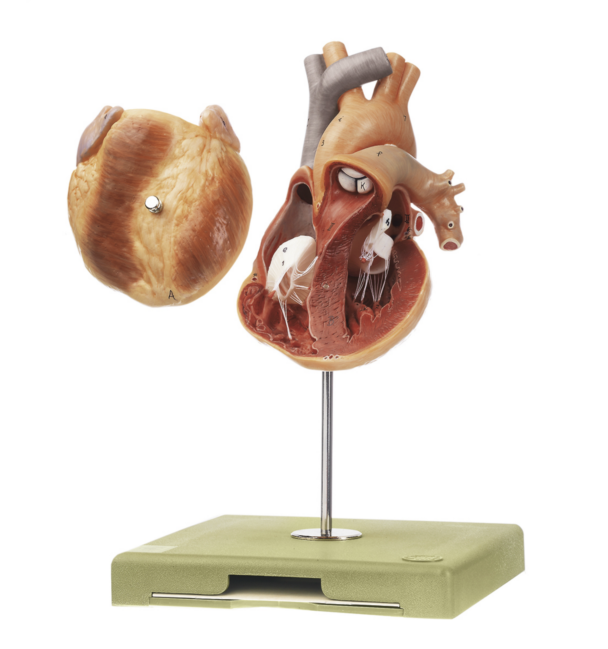
The valve system of the heart is also seen. Between the atria and ventricles are the two lobed valves of the heart (the tricuspid valve and the mitral valve) and between the right ventricle and truncus pulmonalis, and the left ventricle and the aorta, are the two sacral valves of the heart (the aortic valve and the pulmonary valve).


When the front surface is removed, you get a sense of the three layers of the heart wall - the endocardium, the myocardium and the epicardium. Furthermore, it can be seen how the wall thickness in the left ventricle is significantly greater than in the right ventricle.


The heart's impulse conduction system is not depicted.

Product flexibility

Clinical features

Clinically speaking, the model does not show anything pathological, but can be used to understand diseases and disorders affecting the various structures of the heart.


It could be, for example, leaky valves (valvular insufficiency), stenoses (narrowed heart valves), heart failure or blood clots in the heart (myocardial infarction).

View full details

A safe deal

For 19 years I have been at the head of eAnatomi and sold anatomical models and posters to 'almost' everyone who has anything to do with anatomy in Denmark and abroad. When you shop at eAnatomi, you shop with me and I personally guarantee a safe deal.

Christian Birksø
Owner and founder of eAnatomi ApS

Read more about eAnatomi