SKU:EA-1000127
Flexible model of the spine with nerves, muscle indications and other bones without stand
Flexible model of the spine with nerves, muscle indications and other bones without stand
ATTENTION! This item ships separately. The delivery time may vary.
Couldn't load pickup availability
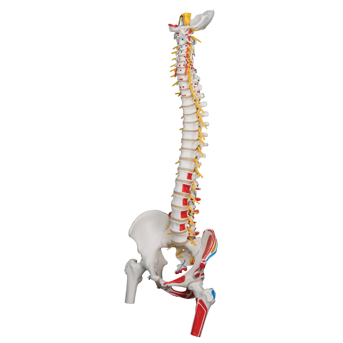
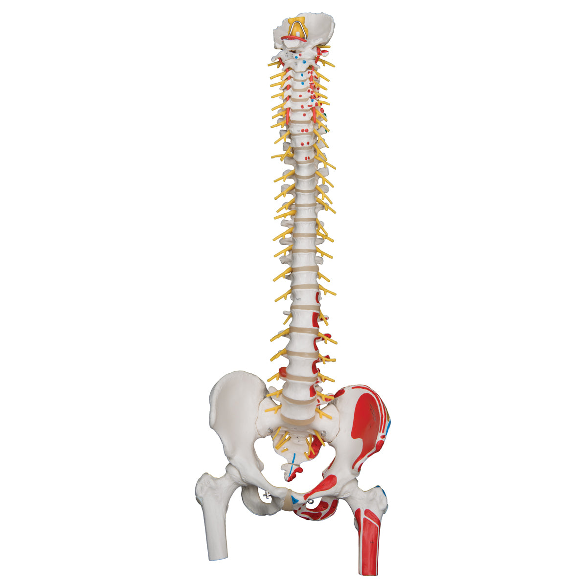
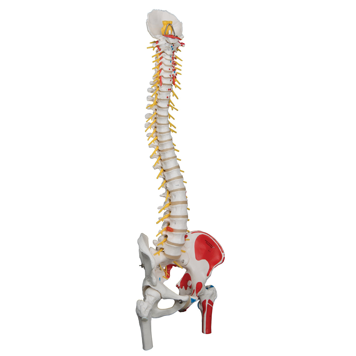
This model of the spine is very flexible and illustrates the origin and attachment of muscles using color. In addition, it includes the brainstem, spinal nerves, a. vertebralis, a herniated disc in the lower back, the neck bone (os occipitale) and the pelvis and hip joint.
The model measures 83 cm in height. All the vertebrae are held together by a copper wire, and in addition an elastic system has been inserted in the upper cervical vertebrae. The model is delivered without a stand.
Anatomically speaking
Anatomically speaking
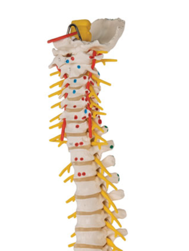
Anatomically speaking, the model shows the entire vertebral column, which consists of a neck part, a chest part, a lumbar part, a pelvic part and the coccyx. The neck bone is also included. Therefore, the model shows the two most important joint types in the spinal column, which are the symphyses (disci/band discs) between the vertebral bodies and the facet joints, which are sliding joints between the vertebrae's pivots. The lower neck joint and the sacrococcygeal joint are also seen. Since the neck bone is included on this model, the upper neck joint is also visible.
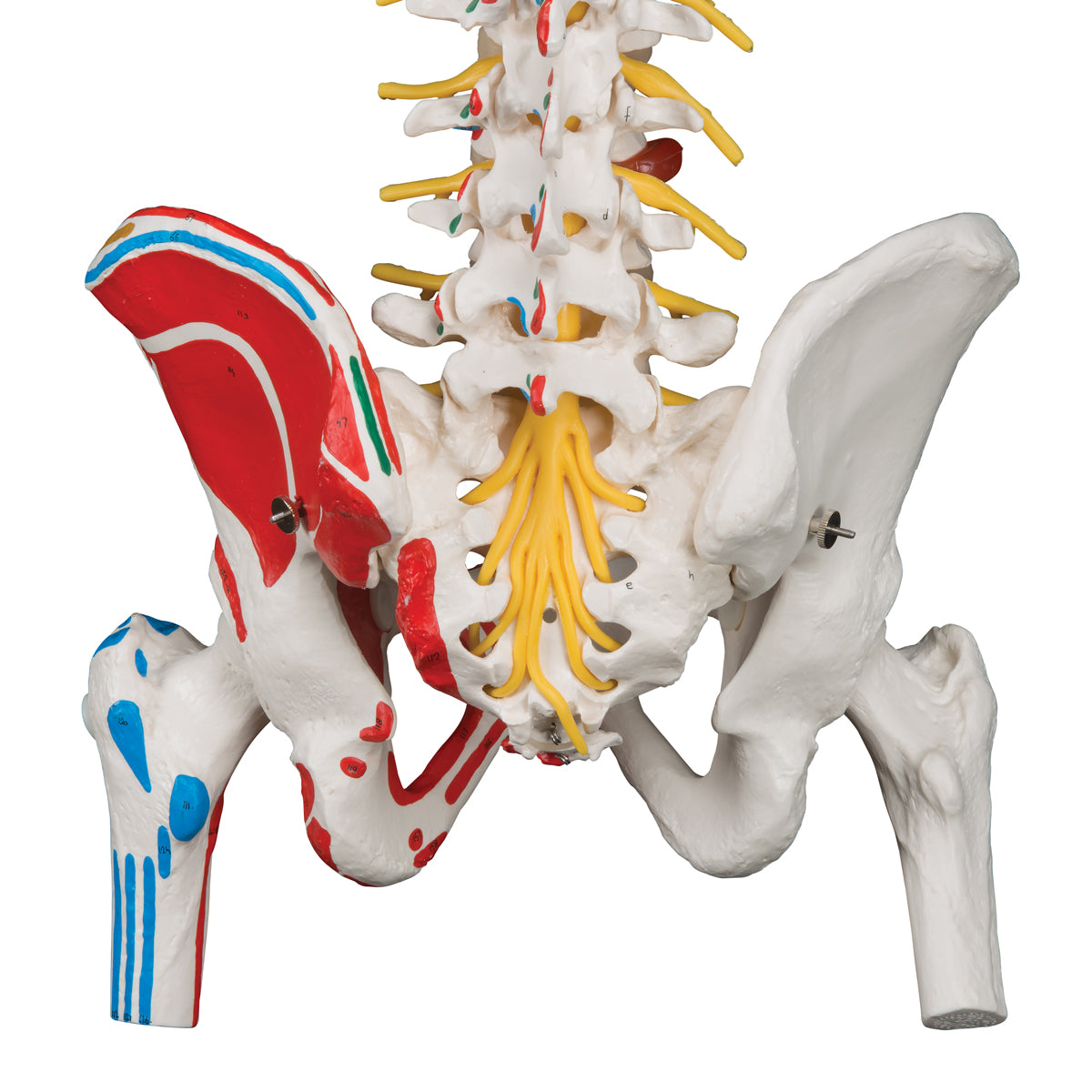
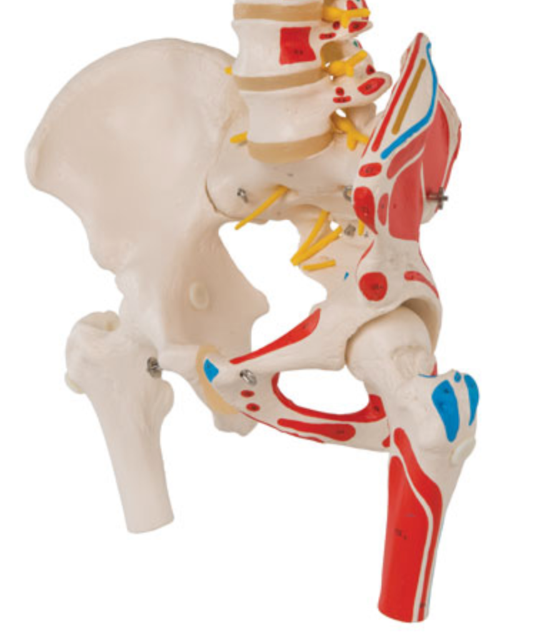
In addition, the model shows the pelvis in its entirety as a ring structure as well as both femoral heads with the femoral neck and part of the femur. Therefore, the model also shows the SI joint between the sacrum and the pubic bone, the symphysis between the pubic bones and the hip joint.
Color markings can be seen on one side of the model. Red color symbolizes the origin of muscles, while blue color symbolizes the attachments of muscles. The model can therefore also be used for muscle training.
Finally, the model shows the brainstem, the 31 pairs of spinal nerves (with both the anterior and posterior radix), the vertebral artery and a herniated disc in the lower back. Overall, the bone quality is very good, and therefore, for example, knots and depressions in the bone tissue are clearly visible.
All the most important anatomical structures on the model are numbered at a high level of detail, and a comprehensive overview with their names on e.g. Latin and English. You can download and print the overview via our website - look under the product images on the left.
Flexibility
Flexibility
In terms of movement, the model is very flexible and produced for active use. The mobility of the human spine is greatest in the cervical and lumbar regions (which are lordotic), while the thoracic region (which is kyphotic) is less mobile. This model can be used to demonstrate the most important movements of the spine, which are flexion-extension, lateral flexion and rotation in the neck and lumbar region.
The neck bone (os occipitale), upper cervical vertebra (atlas) and second upper cervical vertebra (axis) are assembled with elastic string, which causes a special elastic effect, where the vertebrae both follow each other during movement and come together again in a natural way. You can thus demonstrate the most important movements in the neck joints, which are the nodding and rocking of the head, which takes place in the upper neck joint and the shaking of the head, which takes place in the lower neck joint.
In the hip joints, the head of the femur is held firmly in the socket via elastic. Therefore, different movements of the hip joint, such as abduction and rotation, can be demonstrated. However, the femoral head cannot be removed.
Clinically speaking
Clinically speaking
Clinically, this model is ideal for understanding disc herniation, root involvement and spondylolisthesis. It can also be used to understand disorders such as back pain, scoliosis, spondylosis, spinal stenosis and osteoarthritis.
The model can of course also be used to understand fractures, a. vertebralis dissection and many different disorders related to the hip joint and the pelvis.
Share a link to this product

A safe transaction
For 19 years I have been managing eAnatomi and sold anatomical models and posters to 'almost everyone' who has anything to do with anatomi in Scandinavia and abroad. When you place your order with eAnatomi, you place your order with me and I personally guarantee a safe transaction.
Christian Birksø
Owner and founder of eAnatomi and Anatomic Aesthetics