Skip to product information
1 of 6

SKU:EA1-1000333

The pelvis with a fetus in the 40th week of pregnancy and an additional fetus in the 3rd month of pregnancy. Can be separated

The pelvis with a fetus in the 40th week of pregnancy and an additional fetus in the 3rd month of pregnancy. Can be separated

Regular price 5.030,00 DKK
Regular price Sale price 5.030,00 DKK
Sale Sold out
Not including taxes if ordering from outside EU. Shipping calculated at checkout.

ATTENTION! This item ships separately. The delivery time may vary.

This model shows the woman's pelvis in a median section with a fetus in the 40th week of pregnancy as well as a small supplementary model of a fetus in the womb corresponding to the 3rd month of pregnancy.


The dimensions of the model are 38 x 25 x 40 cm, and the weight is 2.5 kg. The large fetus in the pelvis can be removed. The model is delivered on a stand, where the small supplementary model of a fetus in the womb is fixed.

Anatomically speaking

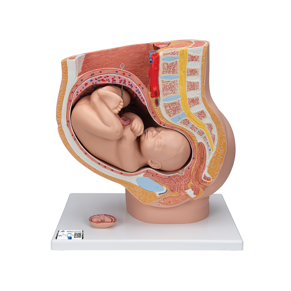
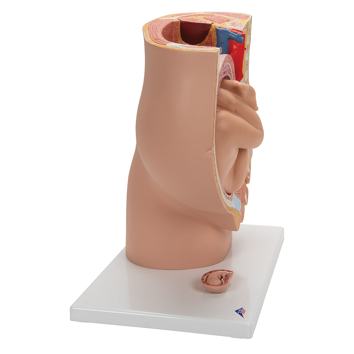
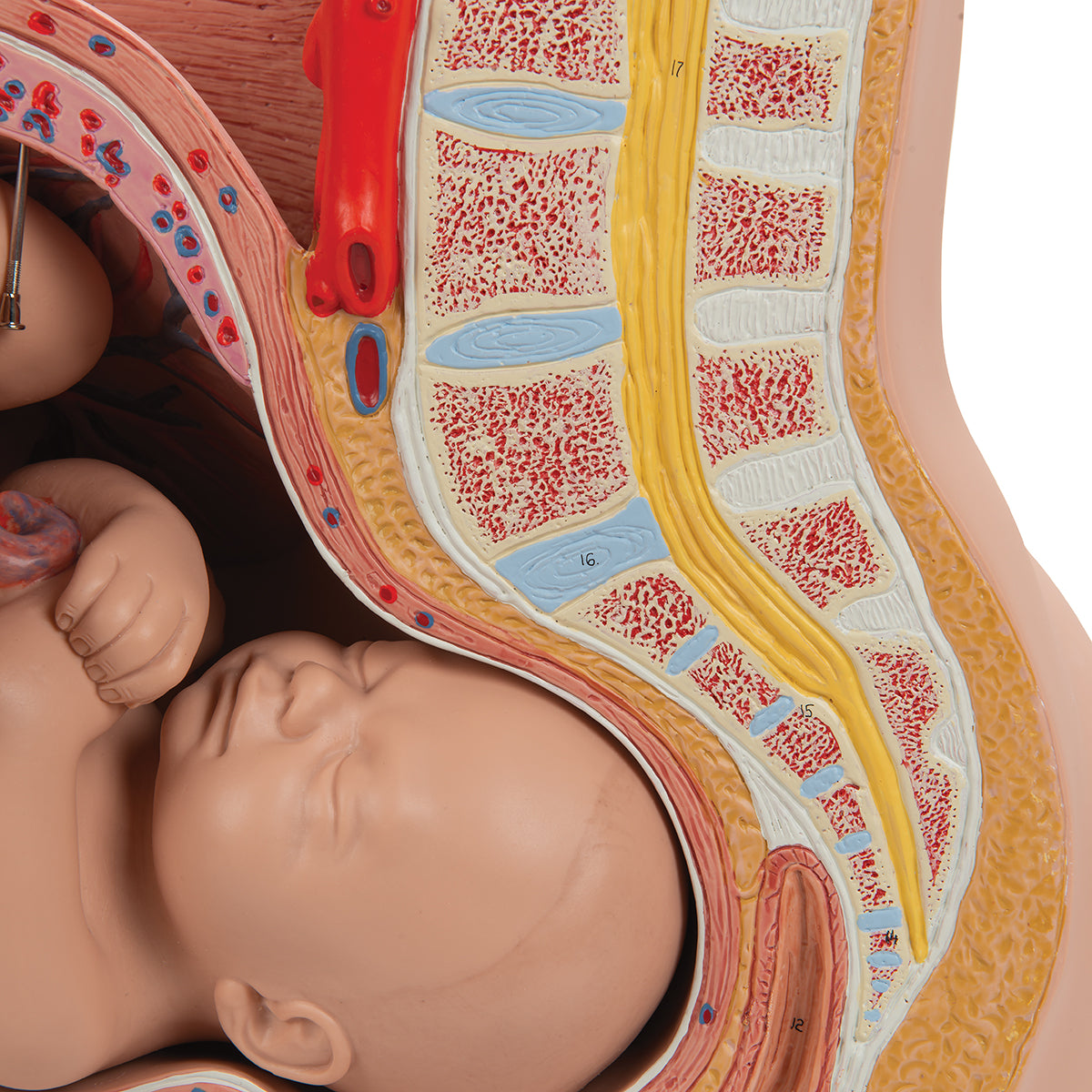
Anatomically speaking, the model primarily shows a median section of the woman's pelvis with a fetus lying in a normal position (posterior head position).


The model shows how the large fetus in the womb lies in relation to other organs and tissues. These include the front joint of the pelvis (symphysis/symphysis pubica), the urinary bladder (vesica urinaria), the vagina (vagina), the rectum (rectum), the lower back incl. the spinal cord (medulla spinalis) and the largest blood vessels which are the aorta abdominalis and inferior vena cava.


Some tissues/organs are also seen in the horizontal plane (also called the transverse plane) when viewing the model "from above". Above all, the back and abdominal muscles and the kidney tissue in the right kidney are seen here.


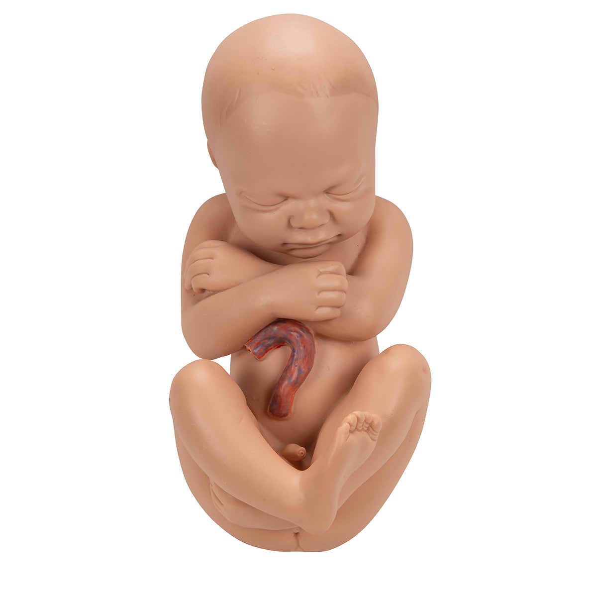
When the large fetus is removed, the inner (fetal) side of the placenta is visible. You see the attachment of the umbilical cord and that the umbilical cord vessels radiate beyond the placenta. The cross-section of the umbilical cord shows the umbilical vein and the 2 umbilical arteries.


The amnion (amniotic membrane) encloses the amniotic fluid in which the fetus lies. Although it is not named on the model, it is clearly visible when the large fetus is removed.


On the model's stand, you can also see a small fetus in the womb corresponding to the 3rd month of pregnancy, which can for example be used for comparison.

Flexibility

Clinically speaking

Clinically speaking, the model can be used, for example, in connection with birth preparation and complications such as placenta previa, placental abruption (abruptio placenta) and fetuses that face the wrong way (eg with the rear down).

View full details

A safe transaction

For 19 years I have been managing eAnatomi and sold anatomical models and posters to 'almost everyone' who has anything to do with anatomi in Scandinavia and abroad. When you place your order with eAnatomi, you place your order with me and I personally guarantee a safe transaction.

Christian Birksø
Owner and founder of eAnatomi and Anatomic Aesthetics

Read more about eAnatomi