SKU:EA-ProLXH
Dynamic spine model of 2 lumbar vertebrae showing a herniated disc. Most learning friendly
Dynamic spine model of 2 lumbar vertebrae showing a herniated disc. Most learning friendly
Out of stock
this product is made to order. To place an order please call or write us.Couldn't load pickup availability
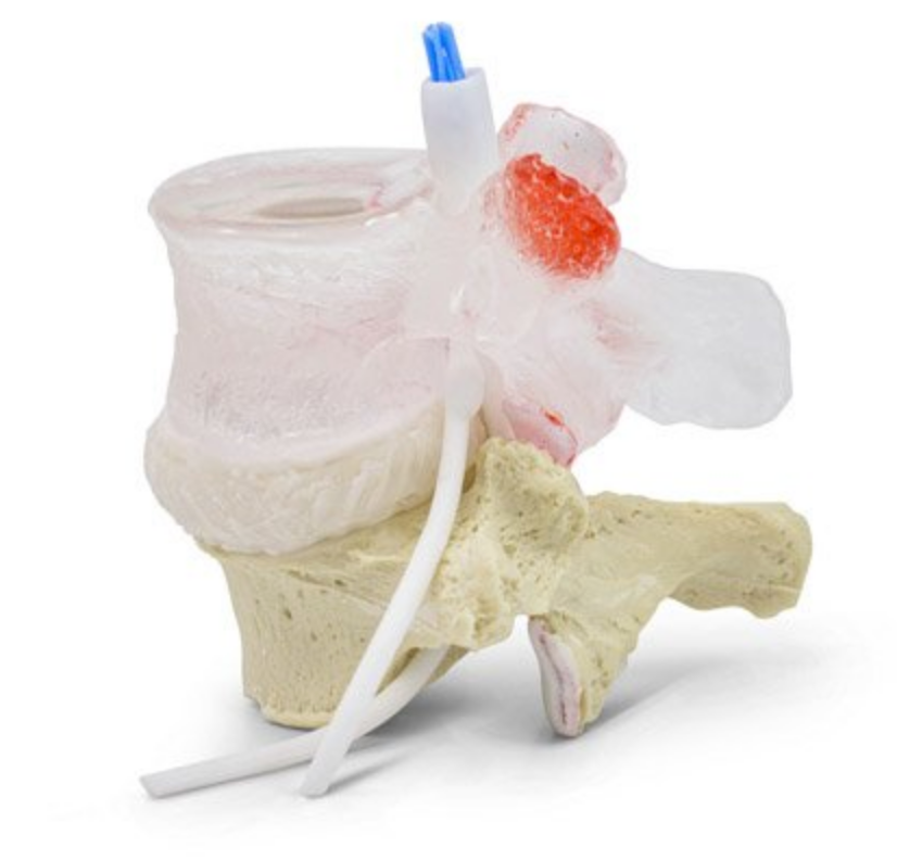
This dynamic spinal joint model consists of 2 lumbar vertebrae, an intermediate disc (disc) and nerves. It can primarily be used to demonstrate a herniated disc in the lower back. As something extraordinary, the upper lumbar vertebra is transparent. This easy-to-learn and stylish design offers additional and cutting-edge features such as visualization of blood vessels and lesions within the transparent lumbar vertebra interior as well as intervertebral disc architecture and lesions within it.
Compared to our comparable spine model called "extra learner friendly" where the upper lumbar vertebra is also transparent, this model offers the same details PLUS
the following properties:
- Nerve supply to the intervertebral disc
- Nerve ingrowth into damaged parts of the disc
- A crack at the front of the disc's connective tissue ring
- Complete view of the cauda equina (the collection of spinal nerve roots surrounded by the dura mater spinalis and arachnoid mater spinalis in this region of the lower back)
Both in terms of size and weight, the model corresponds to 2 lumbar vertebrae in an adult person. The model is not designed to be disassembled. It is possible to purchase a stand.
NB: This model is part of our unique series of dynamic and innovative spine models . The models are handmade in Canada, cast after human vertebrae and can demonstrate all the movements in the joints of the back. The materials are extremely lifelike in very high quality with components from the USA (and therefore not China etc.).
The models are developed with a focus on dynamics, interaction and simplicity in their use. The purpose is that the professional with the spinal joint model in hand can accurately and quickly explain the mechanisms of degenerative changes in the spine as well as their relationship to pain, function and the biomechanics of the spine. Furthermore, the models are developed to be able to withstand active and repeated use.
Anatomical features
Anatomical features
Anatomically, the model shows 2 lumbar vertebrae (L4-L5) and therefore the two most important joint types in the spinal column, which are the symphysis (disc/disc) between 2 vertebral bodies and the facet joints, which are sliding joints between the vertebrae's pivots. In relation to the articular pins, asymmetry is seen at L5 (bottom).
Overall, the bone quality is extremely good, and therefore, for example, knots and depressions in the bone tissue are clearly visible.
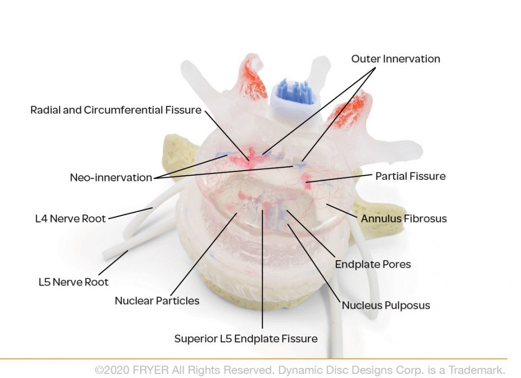
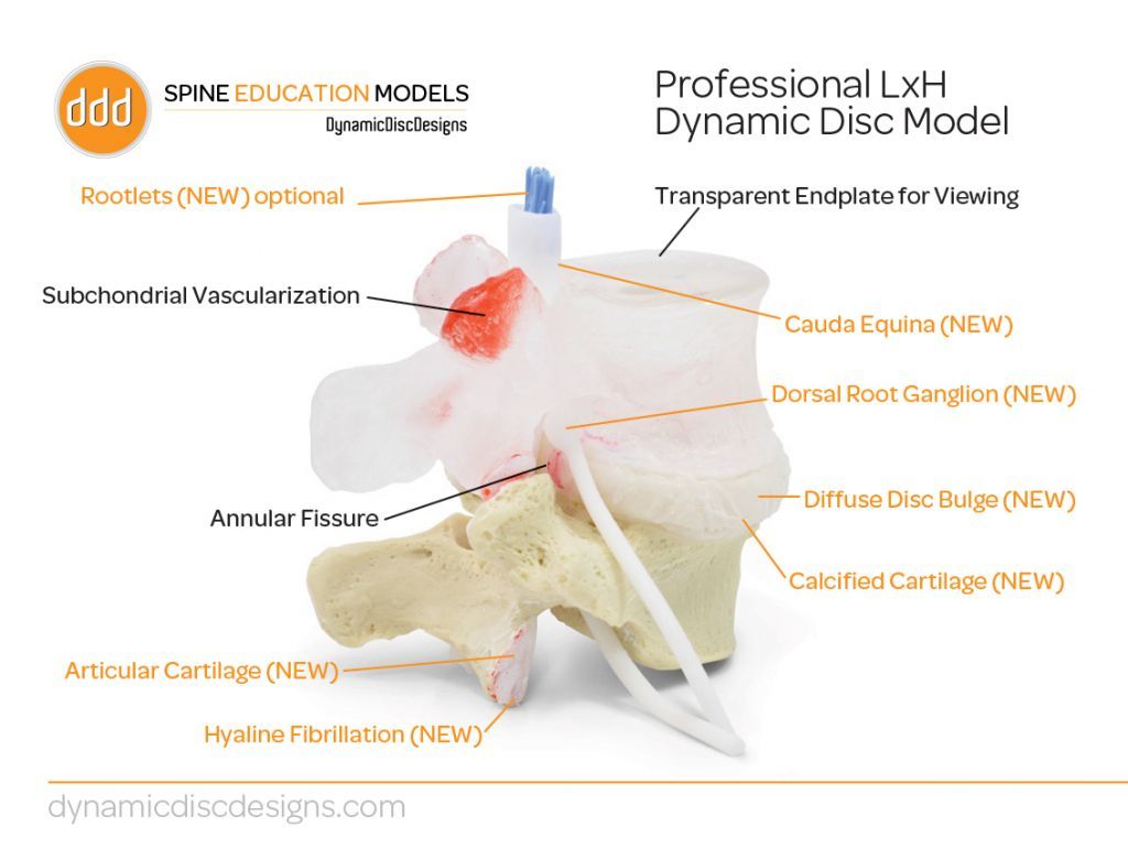
L4 (the model's "top" lumbar vertebra) is transparent as part of the learner-friendly design. The model and in particular the transparency of L4 offers possible to see the following:
The special shape of the tape disc
Calcified endplate (peripheral)
Other cartilage change
Subchondral bone with "hyaline fibrillation"
Right posterolateral, "radial" and "circumferential" tear in the connective tissue ring of the disc
Blood vessels in L4 (in the vertebral body)
Openings (pores) in black color in the end plate at the top of L5
Lesions in red color in the endplate at the top of L5
Subchondral blood vessels in synovium (at L4)
Randomly scattered black structures in the disc core targeted demonstration of dynamic changes in the disc core
Compared to our comparable spine model called "extra learner friendly" where the top lumbar vertebra is also transparent, this model offers the same details (as mentioned above) PLUS the following features:
Nerve supply to the intervertebral disc
Nerve ingrowth into damaged parts of the disc
A "circumferential" anterior tear in the disc's connective tissue ring
Complete view of the cauda equina (the collection of spinal nerve roots surrounded by the dura mater spinalis and arachnoid mater spinalis in this region of the lower back)
The view of the cauda equina is complete on this model because the inserted nerve bundle in the vertebral canal shows a mass of loose spinal nerve roots in blue color surrounded by a whitish membrane. This visual and educational construction thus shows the accumulation of spinal nerve roots surrounded by dura mater spinalis and arachnoid mater spinalis in this region of the lower back. See also the pictures and the video on the left.
Departing from the nerve bundle, lumbar spinal nerves are seen in the same way in white through the foramen intervertebrale ("the hole between the vertebrae"). On the spinal nerves, bulges (ganglion) are seen. The nerves can be moved in relation to the rest of the model (e.g. by pulling on them).
A tear in the disc can be used to demonstrate a herniated disc - read more below.
Product flexibility
Product flexibility
In terms of movement, the model is in a special class, just like the other models in this unique series of dynamic spine models. The model's 2 types of joints (the disc and the facet joints) can be moved in different natural ways.
The model's movements include flexion-extension, lateral-flexion, rotation and compression incl. decompression.
The model has been developed with a tear in the dynamic band disc (discus intervertebralis). If you compress the model, you can realistically demonstrate a herniated disc, where red material bulges out into the spinal canal and affects the nerve bundle (see the pictures and the video on the left). This corresponds to a pouching out of the gel-like disc core, which breaks through the disc's connective tissue ring backwards towards the spinal canal and its contents.
If, on the other hand, you simply perform flexion-extension and lateral flexion on the model without compressing it, you can also demonstrate how the disc's connective tissue ring can affect both the nerve bundle in the vertebral canal and the spinal nerves without the disc core breaking through the connective tissue ring (i.e. protrusion ).
Besides the fact that the nerves can be moved relative to the rest of the model (e.g. by pulling on them), it must be emphasized that the complete view of the cauda equina (the collection of spinal nerve roots in the vertebral canal) is deformable. As an example, you can "squeeze the nerve bundle" and see how it is all affected. See also the video on the left.
Clinical features
Clinical features
Clinically, this model has been developed both for learning about lumbar disc herniation as well as general learning about lesions in both the spine and the intervertebral disc based on degenerative changes.
Disc herniation most often occurs in the lower back. The model is both ideal for demonstrating disc herniations in this region, where the gel-like disc core breaks through the disc's connective tissue ring, as well as protrusion where the disc's connective tissue ring remains intact but affects nerves.
As L4 is transparent, the model offers the opportunity to learn about the anatomy of the lumbar vertebra and disc, which is particularly relevant to degenerative changes that lead to lesions. An example is the end plate of the vertebral body, where openings (pores) are seen in a significantly greater number centrally, which makes the end plate weaker in this area.
The nerve bundle (cauda equina) in the vertebral canal on this model is complete. Therefore, the model is particularly suitable for learning about cauda equina syndrome and procedures such as lumbar puncture as well as spinal and epidural anaesthesia.
As part of our unique series of dynamic spine models, which offer an exclusive opportunity to demonstrate all the movements of the spine, the model can be applied to many aspects of spine biomechanics.
Finally, the model can also be used for learning about other degenerative changes such as spinal stenosis and facet joint osteoarthritis. However, in this series of spinal models we have models specifically developed to demonstrate the characteristics of these diseases.
Share a link to this product

A safe deal
For 19 years I have been at the head of eAnatomi and sold anatomical models and posters to 'almost' everyone who has anything to do with anatomy in Denmark and abroad. When you shop at eAnatomi, you shop with me and I personally guarantee a safe deal.
Christian Birksø
Owner and founder of eAnatomi ApS