Collection: Microscopic anatomy
In the large and wide selection, you will primarily find models that show tissues and cells. These include liver tissue, kidney tissue, bone tissue, a model of blood vessels, tongue tissue as well as a model of specific tissues and cells of the eye
-
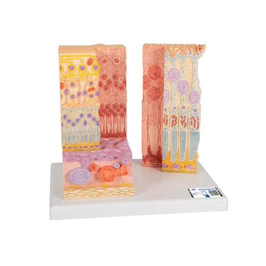
Detailed model of the cells in the eye's retina, choroid and sclera in a microscopic perspective
Regular price $294.00 USDRegular priceUnit price / per -
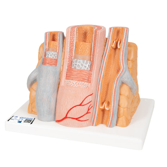
Detailed model of 1 artery and 2 veins which is greatly enlarged
Regular price $294.00 USDRegular priceUnit price / per -
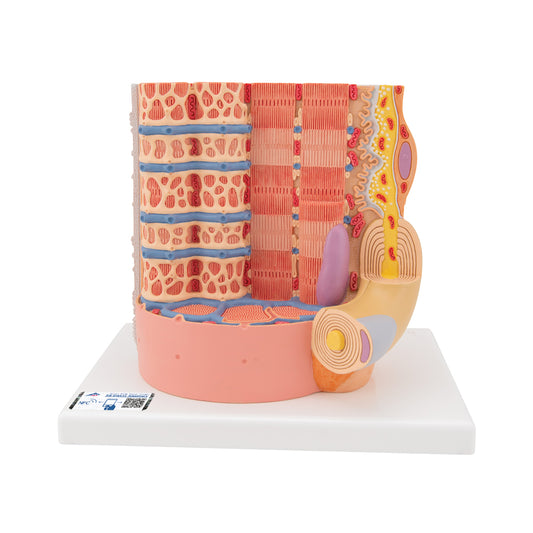
Detailed model of a muscle fiber from skeletal muscle magnified well over 10,000 times
Regular price $278.00 USDRegular priceUnit price / per -
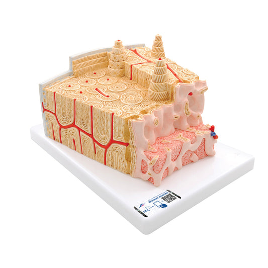
Detailed model of bone tissue incl. blood vessels in a microscopic perspective
Regular price $160.00 USDRegular priceUnit price / per -
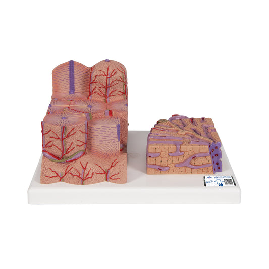
Detailed model of the liver tissue and blood vessels in a microscopic perspective
Regular price $304.00 USDRegular priceUnit price / per -
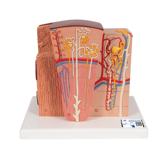
Detailed model of the different tissues and cells of the kidney in a microscopic perspective
Regular price $297.00 USDRegular priceUnit price / per -
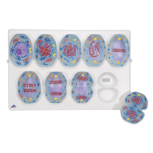
Flexible model of the phases of mitosis
Regular price $455.00 USDRegular priceUnit price / per -
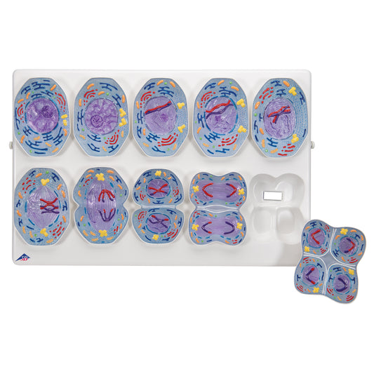
Flexible model of the phases of meiosis
Regular price $455.00 USDRegular priceUnit price / per -
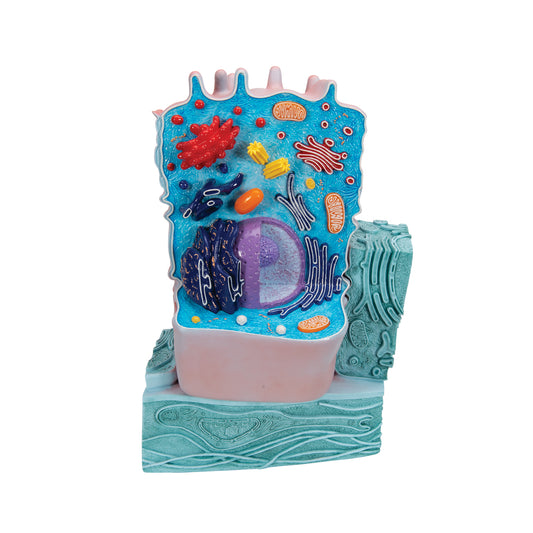
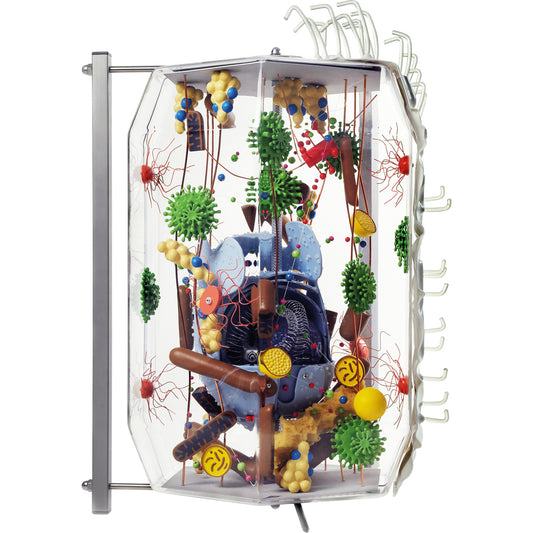
Highly detailed model of a cell in an electron microscopic perspective
Regular price $345.00 USDRegular priceUnit price / per -
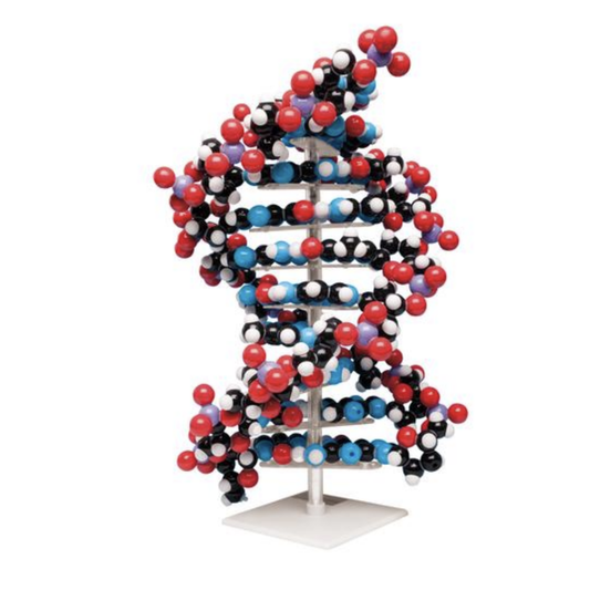
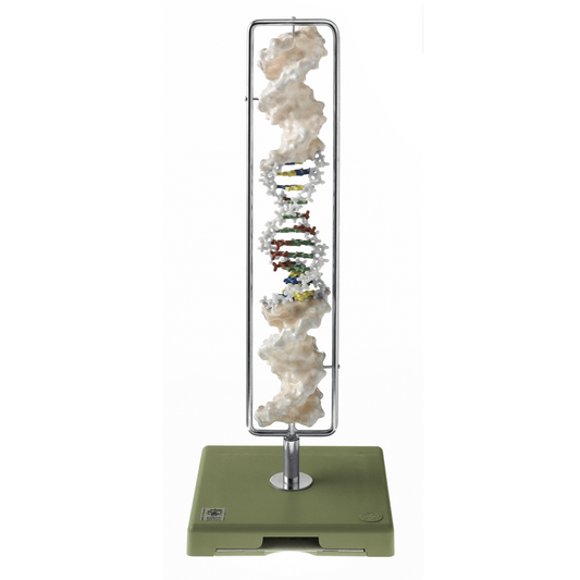
Large model of DNA as a kit made up of educationally colored atoms
Regular price $579.00 USDRegular priceUnit price / per -
Giant model of an undifferentiated human cell in an electron microscopic perspective
Regular price $4,079.00 USDRegular priceUnit price / per -
Exclusive and complete model of DNA presented on a rotating stand
Regular price $1,056.00 USDRegular priceUnit price / per
Collapsible content
Read more about the product category here
We call this selection microscopic anatomy. The microscopic anatomy is about structures that are examined by optical microscopy or electron microscopy.
In the large and wide selection, you will primarily find models that show tissues and cells. These include liver tissue, kidney tissue, bone tissue, a model of blood vessels, tongue tissue and a model of specific tissues and cells in the eye.
You will also find models of individual cells as well as structures in the cell. These include a muscle fiber from skeletal muscle, a general animal cell, an undifferentiated human cell, models of the phases of meiosis and mitosis, and DNA models.
Previously, we called the selection microanatomy. The models in the selection are used both for histological purposes (the doctrine of tissue), biological purposes and for understanding diseases.
Anatomically speaking, the models of tissue and cells show the structure/architecture that is characteristic of the tissue of the organ. With the model of liver tissue in hand, you can e.g. see liver lobules, liver cells, bile ducts, central vein and the Glissonian triads (portal triads).
With the model of kidney tissue in hand, you can see the beginning of the ureter (ureters), the renal cortex (renal cortex), the renal medulla (medulla renalis) in the form of pyramids and the entire renal pelvis (with calices). Furthermore, you can see many details regarding small and physiologically important structures such as the renal corpuscle (corpusculum renis), the nephron with Henle's loop and tubule and collecting duct.
The model of bone tissue shows the clear differences between the compact bone tissue (substantia compacta/cortical bone) and the spongy bone tissue (substantia spongiosa/trabecular bone). With the model in hand, you can see that the former consists of bone matrix in the form of lamellae, which are organized into Haversian systems (cortical osteons). One can also see the cancellous bone tissue's spongy network of trabeculae with communicating cavities filled with bone marrow. Barriers such as the periosteum and endosteum are also shown.
The model of blood vessels primarily shows 2 arteries and 1 vein. With the model in hand, you can see details such as the venous pump (valve and muscle pump system) and the different layers of the blood vessel walls (tunica intima, tunica media and tunica adventitia).
The model of the tongue shows the many different tissues. With the model in hand, you can see details such as taste buds, nerve supply (via the cranial nerves), tongue muscles and the 4 papillae (papillae vallatae, papillae filiformes, papillae fungiformes and papillae foliatae).
The model of specific tissues and cells in the eye shows the retina (retina), choroid (choroid) and sclera (tendon) as well as the branching of related nerve cells. With the model in hand, you can e.g. see the photoreceptors (rod and cone layer/stratum photosensorium) in great detail.
As for the models, which show individual cells and structures in the cell, you will find, for example, the model of a muscle fiber from skeletal muscle. With the model in hand, you can see the neuromuscular end plate and details such as the sarcolemma, Transverse Tubules/T-tubules, sarcomeres, myosin and actin filaments, the cell nucleus and mitochondria.
The models of cells primarily show organelles. With one of the models in hand, in addition to the cell membrane (plasmalemma), you can see structures such as the nucleus with chromatin and nucleolus, ribosomes, rough and smooth ER, the Golgi apparatus, mitochondria, lysosomes and peroxisomes.
With a model in hand of mitosis or meiosis, you can see the different phases of cell division in an educational way.
DNA models offer different insights into the chemical structure of DNA strands, which form the basis of the heredity/genome/genes.
Clinically, the models of tissues and cells can be used to understand diseases in the respective tissues. These can be, for example, liver diseases such as cirrhosis ("cirrhosis of the liver"), acute and chronic hepatitis as well as NASH and liver tumors. It can also be kidney diseases such as diabetic nephropathy and glomerulonephritis.
The model of bone tissue can be used to understand e.g. osteoporosis and multiple myeloma. The model of blood vessels is ideal for understanding atherosclerosis, blood clots, DVT and the use of support stockings, while the model of the tongue can be used to understand disorders such as fissured tongue, infections, cancer and problems related to the cranial nerve supply to the tongue.
The model of tissue and cells in the eye can be used to understand disorders such as retinal detachment, AMD, uveitis, scleritis as well as metastases and primary tumors.
The models of individual cells and the cell's structures can also be used for clinical purposes. The model of a muscle fiber from skeletal muscle can be used to understand hypertrophy and the importance of protein. The other models of cells can be used for understanding e.g. mitochondrial diseases and lysosomal diseases.
The DNA models can also be used to understand disease - eg sickle cell anemia due to a mutation.

A window to a world of anatomy
Whatever you're looking for
Then we can procure or produce it. eAnatomi is more than just a retailer of existing products. We have our own development department, where we create unique and original products that are used for training, guidance and inspiration.

19 years of anatomy
A safe transaction
For 19 years I have been at the head of eAnatomi and sold anatomical models and posters to 'almost everyone' working with anatomy in Denmark and abroad. When you do business with eAnatomi, you do business with me and I personally guarantee a safe transaction.
Christian Birksø
Owner and founder of eAnatomi
Latest blog news
View all-

2 meter tall anatomy figures - we call it anato...
For more than a decade, eAnatomy has produced our own anatomical illustrations, performed by the best illustrators. Where most illustrations are used on classic printed media such as posters, selected...
2 meter tall anatomy figures - we call it anato...
For more than a decade, eAnatomy has produced our own anatomical illustrations, performed by the best illustrators. Where most illustrations are used on classic printed media such as posters, selected...
-

Anatomical Chart Company - changes the format
Anatomical Chart Company har i 2023 besluttet af udfase papirvarianten for fremover kun at levere den let laminerede version med ringhuller. Dette betyder at det ikke længere bliver muligt, at...
Anatomical Chart Company - changes the format
Anatomical Chart Company har i 2023 besluttet af udfase papirvarianten for fremover kun at levere den let laminerede version med ringhuller. Dette betyder at det ikke længere bliver muligt, at...
-

The heart model's place in teaching & expla...
Teachers, professionals and other mediators can easily be challenged when they have to explain the anatomy and diseases of the heart.
The heart model's place in teaching & expla...
Teachers, professionals and other mediators can easily be challenged when they have to explain the anatomy and diseases of the heart.


