SKU:EA-MMC
Dynamic vertebra model of the cervical spine showing various consequences of degeneration
Dynamic vertebra model of the cervical spine showing various consequences of degeneration
Out of stock
this product is made to order. To place an order please call or write us.Couldn't load pickup availability
This spine model shows the entire cervical spine, which consists of 7 cervical vertebrae.
The following characteristics must be highlighted (read more about the characteristics in the text below):
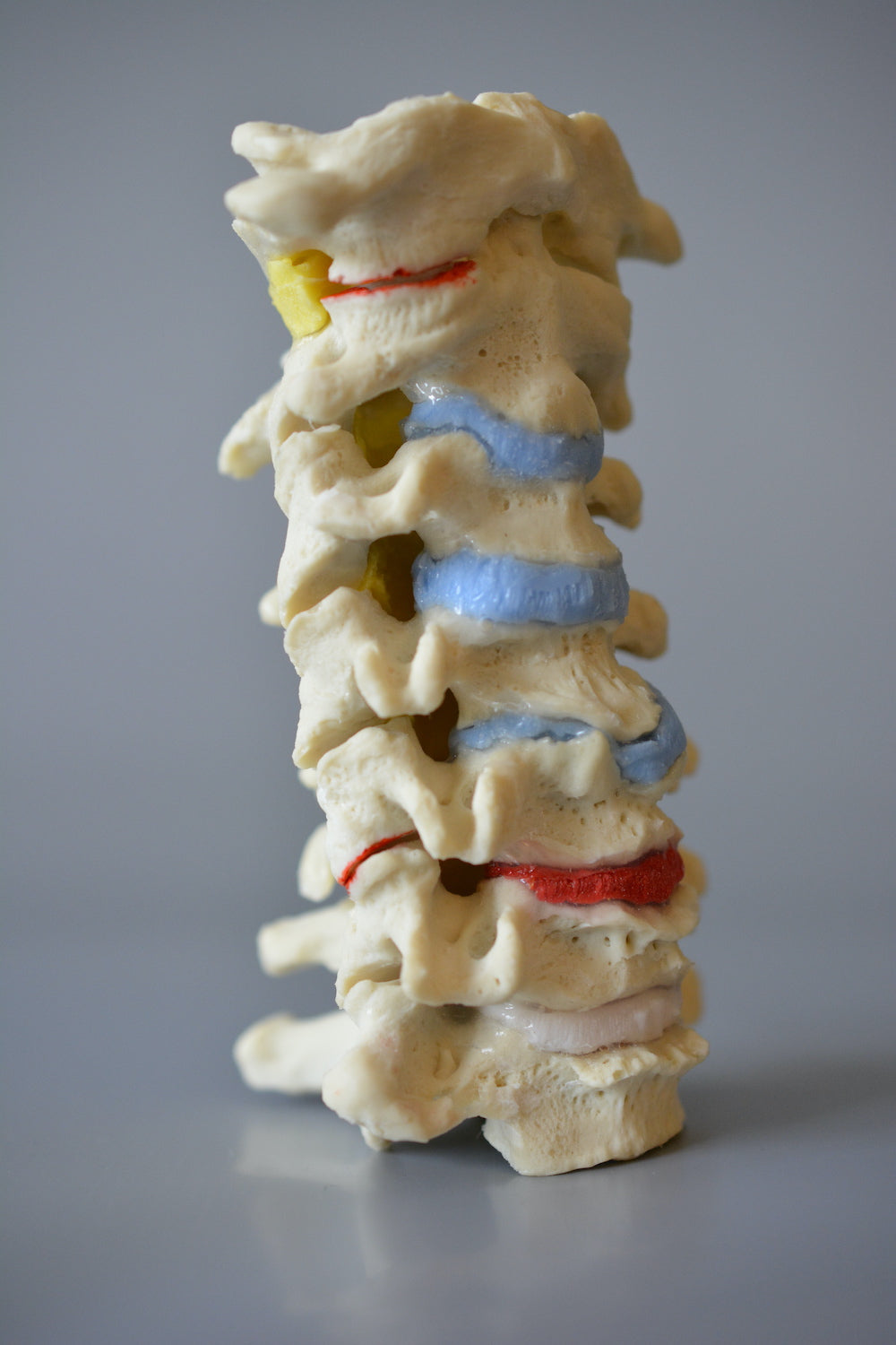
- Reduced curvature (lordosis)
- Degenerative changes such as inflammation (via colour), osteophytosis and a herniated disc
- Reduced joint gap between the joint surfaces in the facet joints
- Hyperelastic band disc between C5 and C6 (to demonstrate hypermobility)
- An exceptional range of motion in the joints, which includes rotation
- Realistic tissue casting
Both in terms of size and weight, the model corresponds to the cervical spine of an adult. The model is not designed to be disassembled. It is possible to purchase a stand.
NB: This model is part of our unique series of dynamic and innovative spine models. The models are handmade in Canada, cast after human vertebrae and can demonstrate all the movements in the joints of the back. The materials are extremely lifelike in very high quality with components from the USA (and therefore not China etc.).
The models are developed with a focus on dynamics, interaction and simplicity in their use. The purpose is that the professional with the spinal joint model in hand can accurately and quickly explain the mechanisms of degenerative changes in the spine as well as their relationship to pain, function and the biomechanics of the spine. Furthermore, the models are developed to be able to withstand active and repeated use.
Anatomical features
Anatomical features
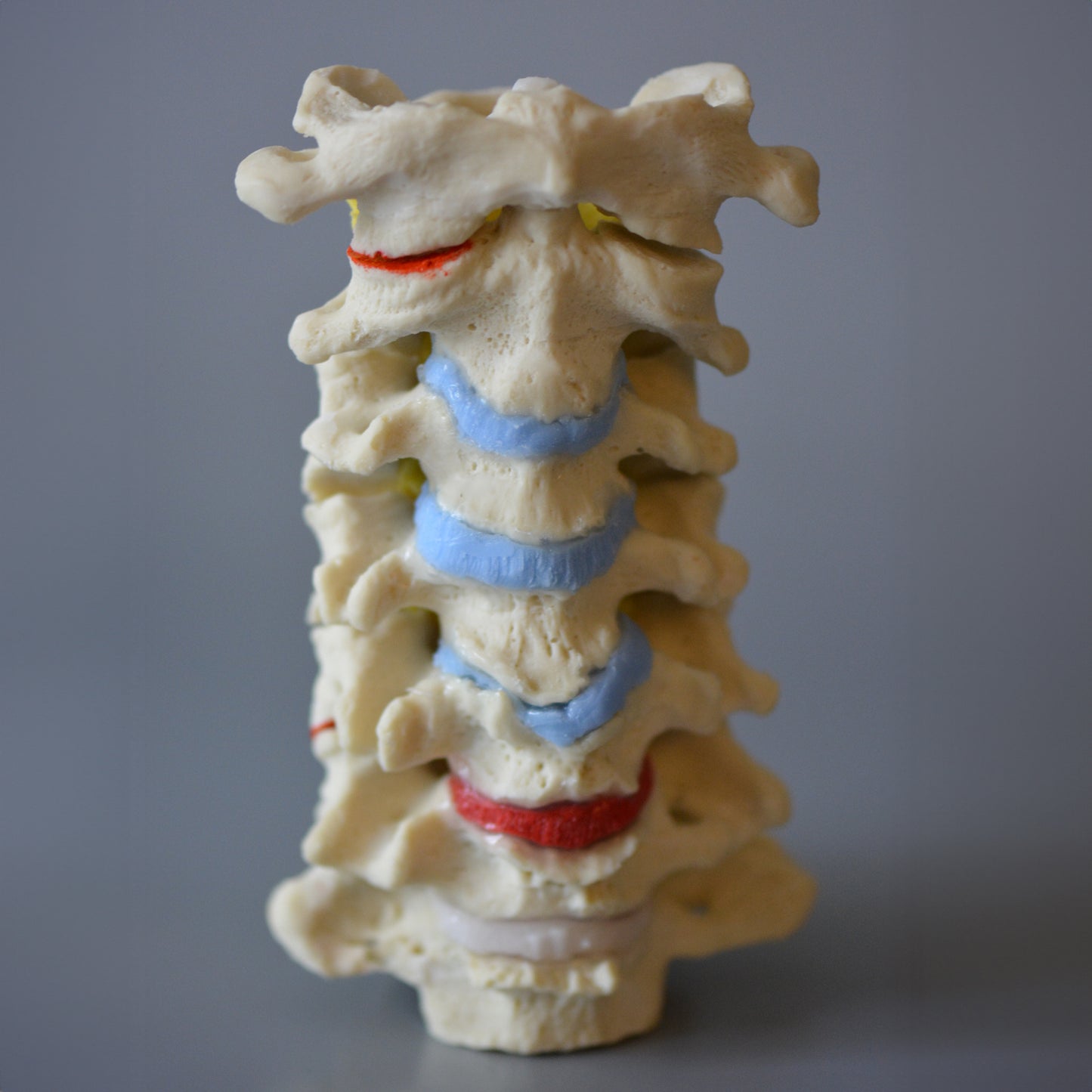
Anatomically, the model shows the entire cervical spine (C1-C7) and therefore the two most important joint types in the spinal column, which are the symphyses (disci/band discs) between the vertebral bodies and the facet joints, which are sliding joints between the vertebrae's pivots. Bodies are seen throughout the vertebral canal. flavum, and at C1-C2, bodies are seen. cruciform Atlantis.
Overall, the bone quality is extremely good, and therefore, for example, knots and depressions in the bone tissue are clearly visible.
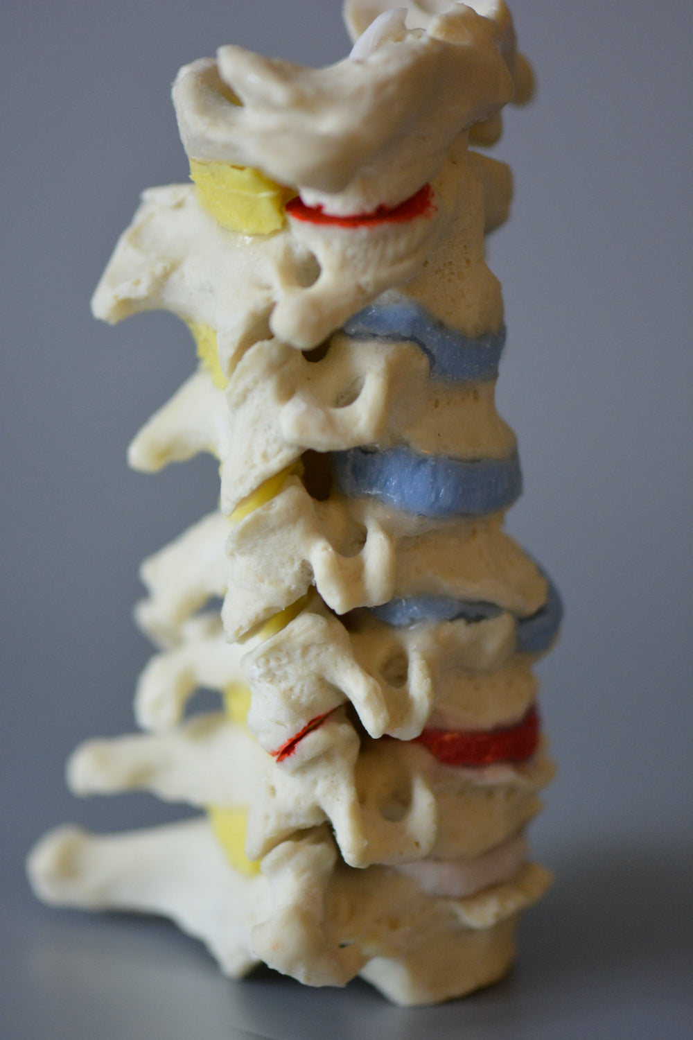
Degenerative changes on the model include i.a. osteophytosis (edge expansion), reduced curvature (lordosis/backward bending) and inflammation in some of the facet joints using educational red color in combination with reduced joint space.
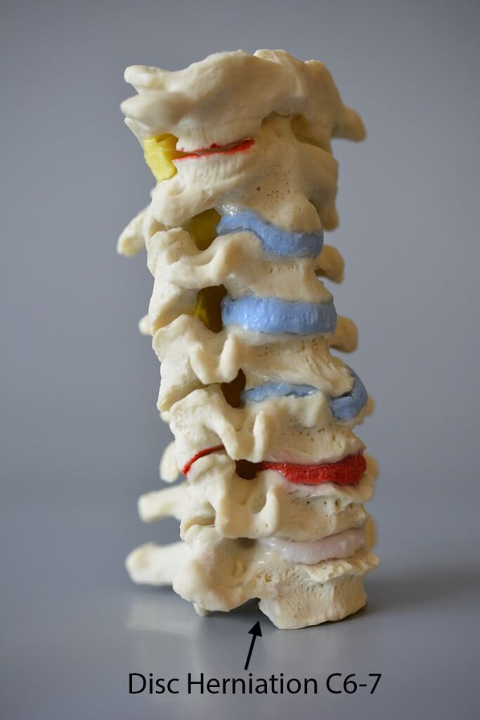
Furthermore, the model has been developed with a hyperelastic disc between C5 and C6 as well as a tear centrally in the outer, fixed part of the disc between C6 and C7 (ie centrally in the annulus fibrosus), where a disc herniation can be demonstrated. Read more about these 2 conditions below.
Product flexibility
Product flexibility
In terms of movement, the model is in a special class, just like the other models in this unique series of dynamic spine models. The model's 2 types of joints (the disc and the facet joints) can be moved in different natural (and unnatural) ways.
The discs can be compressed and the cervical vertebrae can be moved naturally (see the video among the images on the left). Both by compression (compression) and dynamic extension of the cervical vertebrae, one can realistically demonstrate a herniated disc between C6 and C7, where red material bulges into the vertebral canal (centrally) from a tear in the outer, fixed part of the intervertebral disc (i.e. centrally in annulus fibrosus).
A hyperelastic disc is inserted between C5 and C6, which can be used to demonstrate the importance of this in the facet joints compared to the other natural discs on the model (see the video among the images on the left).
As the model consists of 7 cervical vertebrae, rotation in the cervical spine can also be demonstrated. Thereby, the "opening and closing" of the IVF (intervertebral foramen) can be demonstrated - for example in relation to learning about degenerative changes that can affect the neurovascular content of the opening.
Clinical features
Clinical features
Clinically, the model is developed for learning about degenerative changes that cause pain. The model is ideal for learning about disc herniation, spinal stenosis and osteoarthritis of the cervical vertebrae.
Based on the model's color markings of inflammation in facet joints and altered joint space, it is also ideal for learning about cascades of changes in the joints that cause altered mechanics and pain. As a special feature, the hyperelastic disc between C5 and C6 can be used to demonstrate the importance of this in the facet joints.
Since rotation can be clearly demonstrated, it is also ideal for learning about degenerative changes that can affect the neurovascular content of the intervertebral foramen.
Share a link to this product

A safe deal
For 19 years I have been at the head of eAnatomi and sold anatomical models and posters to 'almost' everyone who has anything to do with anatomy in Denmark and abroad. When you shop at eAnatomi, you shop with me and I personally guarantee a safe deal.
Christian Birksø
Owner and founder of eAnatomi ApS