SKU:EA1-MS 8/1
Complete model of the female pelvis and genitals in extremely high quality
Complete model of the female pelvis and genitals in extremely high quality
Out of stock
this product is made to order. To place an order please call or write us.Couldn't load pickup availability
If you are looking for a complete model of the woman's pelvis, which shows both the pelvic ring of bones, the pelvic floor, the internal and external genitalia as well as other tissues such as muscles, ligaments, blood vessels and nerves, this is ideal.
It is produced in SOMSO plastic by the manufacturer SOMSO, which is world-renowned for very high quality. This means quality materials, a sense of accuracy and longevity. In other words: World-class craftsmanship.
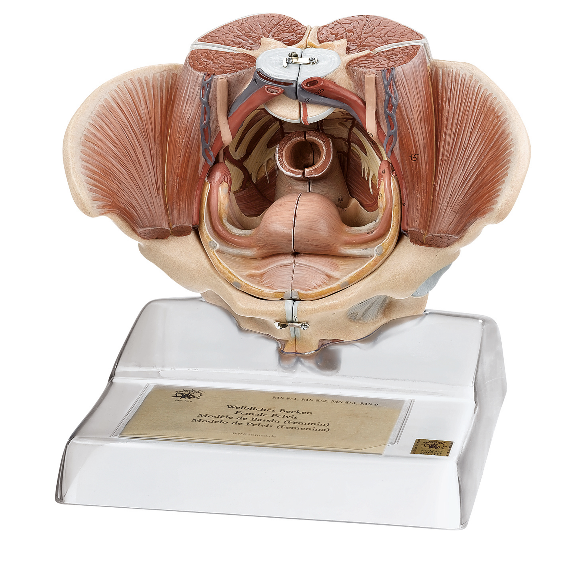
The model is developed in natural adult size. It weighs 1.9 kg and the dimensions are
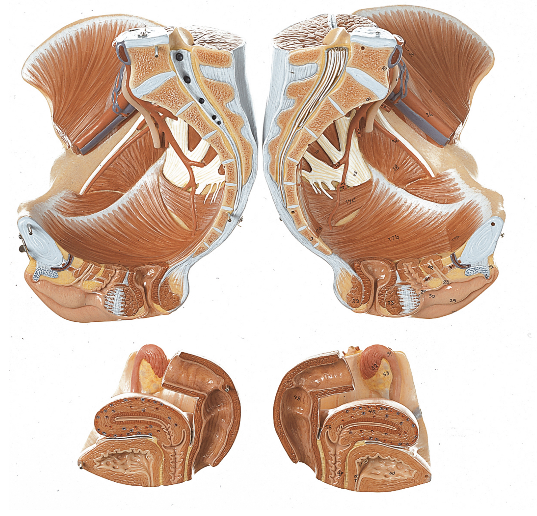
23 x 28 x 17 cm. It can be separated into 4 parts (see the pictures on the left), which are held together using metal pins on the inside and practical metal hooks on the outside. There is no movement in the pelvic joints, although it i.a. can be split in half. The model is delivered on a stand.
Important anatomical structures are numbered on the model. With the purchase, an overview with naming is included, cf. the mentioned numberings. The nomenclature is prepared in Latin and English.
Anatomical features
Anatomical features
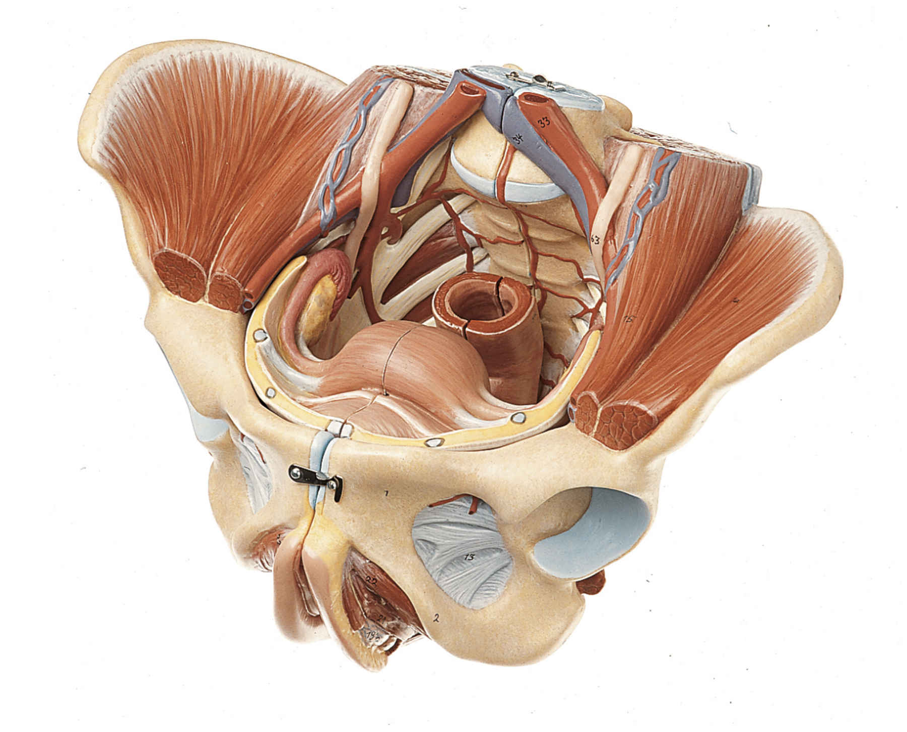
Anatomically, it must be emphasized that this model shows many different structures. Most often only selected structures are seen on a pelvic model, but this complete pelvic model includes:
1) The pelvis (pelvic ring) incl. ligaments
2) Pelvic floor (pelvic floor) and other muscles (skeletal muscles)
3) The internal genitalia
4) The external genitalia
5) Relationships to other tissues/organs such as the ureters, bladder and rectum
6) The lower part of the spinal cord and sacral plexus
7) Blood vessels
1) The pelvis (pelvic ring) consists of the 3 building blocks, which are the two hip bones and the sacrum. The model also shows the lower lumbar vertebra. In the bone tissue, only the largest details are visible, such as large nodules. Smaller details such as the linea glutaea are omitted. Overall, the model shows the following bones:
Os coxae (the 2 hip bones) of which both are composed
- Os ilium (iliac bone/hip bone)
- Os ischii (seat bone)
- Os pubis (pubic bone)
Us sacrum (sacrum) incl. os coccygis (coccyx)
5th lumbar vertebra (lumbar vertebra)
Many ligaments are visible, and some of the above bones are partially covered by these. In addition to the pelvic ligaments such as sacrotuberale can also be seen in some of the back (e.g. lig. interspinale) and the attachment of m. erector spinae. Ligaments, ligament discs (disci) and cartilage in e.g. the hip socket are seen in blue.
2) The pelvic floor (also called the pelvic floor or diaphragma pelvis in Latin) consists of the following muscles, which are clearly visible:
- M. levator ani (consisting of m. pubococcygeus and m. iliococcygeus)
- M. coccygeus
Other muscles such as m. obturatorius internus, m. iliacus and m. psoas major are also seen.
3) The internal genitalia include the ovaries (ovaries), the fallopian tubes (tubae uterinae), the uterus (uterus) and the vagina (vagina), all of which are seen in detail. Anatomically, it is located in the small pelvis.
4) The external genitalia are shown with the largest and most important details, which are anatomically located in front of and below the symphysis.
5) Relationships to other tissues/organs primarily include the ureters, the urinary bladder (vesica urinaria) and the rectum (rectum). Since the model can be split in the middle, structures are seen in a median section. Therefore, the regions where the excavatio vesicouterina and excavatio rectouterina are formed are also seen. On the model, however, you cannot see that they are formed by the peritoneum (peritoneum).
6) Nerves comprise the lower part of the spinal cord (medulla spinalis), which in this region is called the cauda equina. This is seen in the cisterna lumbar, and one can also sense the fibrous string (filum terminale). The sacral plexus is also shown, but the further course of the nerves is not seen.
7) Blood vessels are seen with some important ramifications. In addition to the common iliac artery, the internal and external iliac arteries with some smaller branches (and the corresponding veins), for example, the median and lateral sacral arteries as well as the a. and v. ovarica (and corresponding veins) can be seen.
Many of the pelvic organs function as a channel or a reservoir (e.g. the rectum and the urinary bladder), which is why they are seen in a pedagogical way with an air-filled cavity (cavity).
Product flexibility
Product flexibility
In terms of movement, the pelvis is not flexible. The few joints that are visible are not movable.
Clinical features
Clinical features
Clinically speaking, the model can be used to understand many diseases and disorders in the woman's pelvis - for example in the genitals, the urinary bladder or the rectum. Examples are anal fistula, anal fissure, urinary tract infection and tumors.
Furthermore, the model can be used to understand disorders and diseases such as disc herniation, cauda equina syndrome, stones in the ureter and deep vein thrombosis (DVT) in the pelvis.
Share a link to this product


A safe deal
For 19 years I have been at the head of eAnatomi and sold anatomical models and posters to 'almost' everyone who has anything to do with anatomy in Denmark and abroad. When you shop at eAnatomi, you shop with me and I personally guarantee a safe deal.
Christian Birksø
Owner and founder of eAnatomi ApS